What is Pes Cavus?
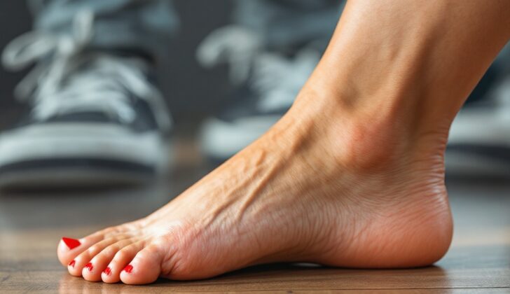
Pes cavus, also known as a high-arched foot, is a foot condition that affects both children and adults. This condition is often the result of an underlying neurological disorder, but can also be seen in individuals without any known health issues. The affected foot typically has an elevated arch, a toe region that points downwards (first ray plantar flexion), and the foot twists inwards (forefoot pronation). Due to its close link with neurological issues, it’s important for doctors to thoroughly understand a patient’s medical history before starting treatment.
In terms of structure, a healthy foot typically forms a ‘tripod’ with contact points at the heel and the heads of the first and fifth metatarsals (bones in the mid-foot region). However, in a high-arched foot, the front part of the foot points downwards, causing the other two ‘points’ of the ‘tripod’ to bear extra weight.
This condition can be driven by issues in the forefoot (the front part of the foot), often due to muscular imbalances caused by neurological diseases. Because of this imbalance, the stronger muscles overpower the weaker ones, causing the front part of the foot to point downwards and twist inwards. Over time, as the shape of the foot changes, the Achilles tendon (found at the back of the ankle) may shorten and the mid-part of the foot may become stiff and less capable of absorbing shock.
On the other hand, a high-arched foot can also be a result of injuries to the ankle or foot. For example, remnants of fractures or issues with long-term ankle stability can lead to this condition. When this happens, the foot attempts to restore its balance by twisting at the subtalar joint (the joint below the ankle), which may result in further deformation with time.
At the joints between the foot and the toes (metatarsophalangeal joints), the toes might be lifted upwards, often due to imbalances in the muscles controlling these regions. This ‘cock-up’ deformity, as it is called, could lead to dislocation of these joints over time. This condition may progress slowly and it is commonly seen to start before puberty. In kids, this condition gradually becomes worse with time and can affect the growth and shaping of foot bones.
The most commonly observed cause of high-arched foot is Charcot-Marie-Tooth (CMT) disease, a neurological disorder affecting the peripheral nerves. In patients with this disease, foot deformities usually worsen over time and surgery is often required to prevent progression to a fixed deformity.
If left untreated, this condition can also lead to instability in several parts of the foot. In the long term, this instability can cause advanced arthritis of the ankle joint. The pattern of the spread of high-arched foot is usually associated with the underlying cause. It can be genetic in nature, where it’s passed down within a family through genes. Some people without any evident neurological disorder may also inherit this foot condition, although the genetic pattern isn’t clear yet.
What Causes Pes Cavus?
Pes cavus, a foot condition seen in both adults and children, is typically caused by a hereditary or genetic issue if it affects both feet. If only one foot has this condition, it’s often due to an injury. If no injury has happened, an MRI of the brain and spinal cord should be performed to get rid of the possibility of serious conditions like a brain tumor or tethered spinal cord, which is a condition where the spinal cord is attached to the spine in a way that limits its movement.
There can be many other causes of cavovarus foot, which is another name for the pes cavus condition. These include:
– Neurologic conditions: These are diseases that affect the nervous system, such as hereditary motor and sensory neuropathies, cerebral palsy, stroke symptoms, spinal cord diseases, Parkinson’s disease, and lesions.
– Traumatic causes: This includes injuries or conditions caused by accidents, like compartment syndrome (a painful condition caused by pressure buildup from internal bleeding or swelling of tissues), ankle or knee dislocations, scar tissue formation, burns, and fractures.
– Post-traumatic bone deformities or imbalance in the ligaments: These could lead to this condition if there was a deformity or injury that didn’t heal correctly.
– An incorrectly treated clubfoot: Clubfoot is a birth defect where the baby’s foot is twisted out of shape or position.
– Other causes: These can include a tarsal coalition (an abnormal connection of two bones at the back of the foot), rheumatoid arthritis, plantar fibromatosis (a benign growth on the bottom of the foot), and diabetic foot syndrome among others.
Some people have a less-obvious version of pes cavus; this is usually grouped in with the ‘idiopathic’ causes, which means the cause is unknown.
Risk Factors and Frequency for Pes Cavus
The actual number of people with a high-arched foot, also called a cavus foot, is not known. This is likely due to the fact that it’s somewhat loosely defined. However, several studies have attempted to identify how common it is in certain groups. The results differ based on factors like where the study was done, who comes to the practice, the health care system, and how a high-arched foot is defined.
- In one study, it was found that if a person has high-arched feet on both sides, there is a 78% chance they have a condition called Charcot-Marie-Tooth disease (CMT). If there’s a family history of high-arched feet, the chance of having CMT goes up to 91%.
- A study in southern India, which looked at 1846 healthy adults without any nervous system conditions, found that about 10% of them had a high-arched foot.
- Another study found that around 25% of male diabetes patients had high-arched foot deformities.
- A review of a foot specialist’s work with eight foot and ankle surgeons showed that more than half of all patients were fitted with a high-arched foot orthosis, a kind of shoe insert to help with the condition.
- In another study, about two-thirds of patients who came in for evaluation of their high-arched foot symptoms had an underlying neurological problem.
It’s important to note that high-arched feet occur in both males and females without any known difference.
Signs and Symptoms of Pes Cavus
Diagnosing an underlying condition often requires a detailed medical history and physical exam. A common cause of concern can be frequent ankle sprains, foot arch pain, and sometimes even knee pain. Pain can be located around the heel, cuboid region, lateral foot, and first metatarsal head due to the increased stress cause by the deformity. Patients may also mention that their shoes no longer fit or wear out quickly, painful calluses (especially under the first and fifth metatarsal heads), foot pain, or pain on certain parts of the foot.
A family history of similar foot deformities might suggest a genetic cause and patients should be asked about the family’s medical history. If a patient suddenly develops a unilateral pes cavus (a high arch) along with other neurological symptoms, it might hint at a spinal issue and needs further examination.
Patients with a subtle cavus foot deformity could present less severe symptoms over a long duration, including lateral foot pain, stress fractures, ankle instability, or issues with the peroneal tendon.
The physical examination should entail a comprehensive foot and ankle check. Besides taking note of the shape and symmetry of the feet, doctors need to check the tightness of the heel cord and the triceps surae, strength of lower extremity muscles, and motion of the subtalar joint. A fixed and rigid subtalar joint could indicate conditions like a coalition or cavovarus. The skin and soft tissues need to be inspected for any abnormalities or corns and calluses. Neurological examination including checking reflexes, sensation, proprioception, and vibratory sensation is also key.
To determine the rigidity of the deformity, the Coleman block test can be used. During this test, a 1-inch block or a book is placed under the outside edge of the foot and heel. If the hindfoot switches from inward bending (varus) to outward bending (valgus) once the influence of the first metatarsal is removed, it means the deformity is flexible. If the varus position does not correct, however, it indicates a rigid and fixed deformity.
The doctor will also need to palpate along the lesser metatarsals and fifth metatarsal for signs of stress fractures, and check the ankle for stability, joint tenderness and issues with the peroneal tendon. The ankle might also have anterior pain due to the dorsiflexed talus impinging.
The patient’s entire body should be evaluated including a physical examination of the spine for signs of curvature (scoliosis) which could point to Charcot-Marie-Tooth disease, and any unusual signs like hairy patches or dimples which might suggest spinal dysraphism. A detailed neurological examination of reflexes, sensation, and vibratory sensation should also be conducted.
One major clinical sign of the subtle cavus foot is the “peek-a-boo” heel, wherein the heel pad becomes easily visible from the front when the patient stands with both feet pointing forward. In a normal foot, this does not occur as the natural alignment of the hindfoot is valgus.
Testing for Pes Cavus
When a doctor suspects a foot condition known as cavus foot, the first step is usually to take X-rays. Specifically, they will look for things like broken bones, dislocations, and signs of wear and tear. These X-rays can also show a bunch of more detailed information, based on specific measurements and angles, which can tell the doctor more about how your foot is holding itself.
One of the simple ways doctors check for cavus foot is to compare the position of two bones in your foot – the medial cuneiform and the fifth metatarsal base – on a side-view X-ray. If the fifth metatarsal base is lower down (closer to the ground), it can indicate cavus foot.
Additionally, doctors might use a handful of other measurements. For example, they might measure a thing called Meary’s Angle, which is the angle between the talus and the first metatarsal bones in your foot. If this angle is greater than usual, that could hint at cavus foot. Similar measurements that doctors might look at include the Hibb angle and the Djian-Annonier angle, both of which can suggest cavus foot if they’re larger than normal.
Apart from X-rays, your doctor might also order a CT scan or an MRI. These imaging techniques can give a more detailed look at your foot and help the doctor plan for any potential surgery. An MRI could be particularly useful for looking at specific issues like damage to the lateral ligaments, problems with the peroneal tendons, joint surface injuries, and certain types of fractures in the foot.
Sometimes, if you have cavus foot in only one foot and there’s no obvious explanation why, doctors might also order an MRI of the brain and spinal cord. They might also refer you to a neurologist if they suspect a condition called hereditary motor sensory neuropathies (HMSNs).
Treatment Options for Pes Cavus
Non-surgical interventions for foot deformities often include changing physical activities, taking anti-inflammatory medications, wearing comfortable shoes, and using custom orthotics (specially designed shoe inserts). Those with mild foot deformities and symptoms can often successfully manage their condition with custom orthotics, which aim to realign the foot and distribute weight more evenly. For more severe deformities, more restrictive orthotics may be used. In a study of 154 patients with painful foot deformities, those who used custom orthotics reported significant improvements in foot pain and function, as well as their overall quality of life, compared to those who used placebo.
Medications such as baclofen, dantrolene, and diazepam may be used to treat underlying muscle stiffness. Botulinum toxin injections have also been used. However, a recent trial for children with a specific type of nerve disorder found no significant decrease in foot deformity progression due to Botulinum toxin A. To help prevent muscle tightening, stretching programs can be beneficial. One patient who underwent a 12-week lower body strengthening program saw significant increases in muscle strength, which in turn, improved physical function. Strengthening and stretching remain reasonable short-term options, although more research is needed to determine their influence on long-term outcomes or prevention of further treatments.
Many experts encourage early surgical treatment for foot deformities due to the development of severe imbalances or deformities that can lead to irreversible damage with degenerative joints. Surgical procedures can include various tendon transfers, tendon lengthenings, and specific types of surgeries to reshape bones (osteotomies). Which procedure(s) is/are used depends greatly on each patient’s individual case and the treating physician’s expertise.
Effective procedures for the flexible foot deformity can include transferring the posterior tibial tendon (a major tendon in the lower leg) to the top of the foot to augment a weak tibialis anterior (muscle in the shin). And transferring the peroneus longus (a tendon that runs along the outside of the calf) to the peroneus brevis (another tendon in the calf) to reduce the pull on the foot’s first bone and assist foot eversion (outward roll).
Soft tissue releases such as lengthening the calf muscle or Achilles tendon are sometimes recommended if the back of the foot is tight, but many surgeons prefer to reduce the tension in the Achilles by changing the foot’s position through various surgeries. When it comes to the more rigid sections of the foot, procedures target each section differently: forefoot-driven foot deformities often require a specific reshaping surgery, while lateralizing calcaneal osteotomies (surgery to reposition the heel bone) are common procedures to correct varus hindfoot deformities (inward-angled heel).
Most patients prefer bone-reshaping surgeries that spare the joints unless they have advanced degenerative disease. Even in cases where a joint fusion is performed, achieving a balance in muscle forces is recommended to avoid failure of the bone fusion. The most extreme of these surgeries, known as “triple arthrodesis,” is typically viewed as a last-resort operation, often as a result of significant disease in nearby joints.
In subtle cases of foot deformity, surgical treatment often occurs while the surgery is addressing other issues such as stress fractures or instabilities. The sequence of surgery usually starts with reshaping the forefoot, followed by modifying the heel. The release of plantar fascia (a band of tissue that runs along the bottom of your foot) is not commonly included in the treatment plan for mild foot deformities.
What else can Pes Cavus be?
The main issue doctors face isn’t just recognizing a cavus foot, but correctly diagnosing what’s causing it. This is vital in determining the best treatment for the patient. ‘Pes cavus’ is a term that describes a high arch in the foot, which can be a sign of numerous different diseases and conditions. So, it’s important for the doctor to realize that most patients who come in with a noticeably high arch in their foot have an underlying disorder.
With this in mind, doctors need to be able to tell the difference between a cavovarus foot (a specific type of high-arched foot where the heel tilts inward) and an equinovarus foot (another type of high-arched foot, but this one is caused by different issues). While these conditions might look similar, they are caused by different things and need different surgical approaches.
Some causes of cavus foot include:
- Charcot–Marie–Tooth disease
- Congenital hypomyelinating neuropathy
- Dejerine–Sottas neuropathy
- Distal hereditary motor neuropathies
What to expect with Pes Cavus
The future outcome, or prognosis, of a cavovarus foot – a condition where the foot has a higher arch and turns inward – strongly depends on the specific underlying health condition that caused it.
Some diseases, such as Charcot-Marie-Tooth disease (CMT), a hereditary nerve disorder, can lead to an ongoing deformity. Conversely, other causes like poliomyelitis (a disease caused by a virus that affects the nerves and can lead to partial or full paralysis), generally do not lead to further deformity.
The treatment and future outlook for these conditions can greatly differ. For instance, CMT type 1 typically begins during teenage years and progresses sooner than CMT type 2, which usually commences in the third and fourth decades of life. Furthermore, in CMT, the hands are generally affected after the feet, which can obviously contribute to the disability. Certain rare types of CMT can result in further neurological problems affecting the cranial nerves (the nerves that come directly from your brain).
Possible Complications When Diagnosed with Pes Cavus
If a foot deformity diagnosis is delayed or orthotics are used for too long even as the deformity worsens, it can result in permanent and rigid deformity that might only be treated with a surgical procedure known as arthrodesis.
However, the surgery may fail to correct or stabilize the underlying bone deformity, and if the nerve condition is progressive (like Charcot-Marie-Tooth or CMT), no surgery can fully prevent it from recurring.
While arthrodesis is a surgical option, its long-term results haven’t been as promising, and complications such as adjacent joint disease are known to occur in foot and ankle surgeries. Based on a study, even after performing a triple arthrodesis for various foot deformities, a significant number of patients had adjacent joint degeneration after the surgery.
The subtle cavus foot condition can also lead to complications such as:
- Ankle instability
- Varus ankle arthrosis
- Peroneal tendon disorders
- Stress fractures in the 5th metatarsal on the outer foot
- Impingement syndromes in the anteromedial ankle
- Plantar fasciitis
- Claw toe deformities
- Knee derangement
Cavovarus alignment and chronic ankle instability can also increase the risk of developing ankle joint arthritis. One study showed that cavovarus alignment increases pressure on the medial joint in cadaver studies, but the abnormal contact pressures could be improved with a surgery known as lateralizing calcaneal osteotomy.
Preventing Pes Cavus
The education for patients and their caregivers should focus on understanding the benefits of physical therapy and using supportive devices for milder conditions. It’s important to understand that these non-surgical treatments may not be able to completely stop the worsen condition of the high-arched, inward-turned foot, commonly referred to as a ‘cavovarus deformity’. There might be a need for further procedures to keep the foot flat when standing, also known as a ‘plantigrade’ foot.
Like many bone-related health problems, the initial step towards treatment usually doesn’t involve surgery. However, discussions need to be held with the patient and their caregiver about potential surgical methods if the deformity continues to worsen. This allows everyone to make an informed decision regarding the future treatment plan.


