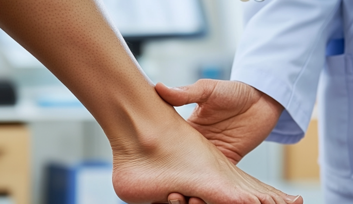
What is Posterior Tibial Tendon Dysfunction?
Posterior tibial tendon dysfunction (PTTD), which is now known as progressive collapsing foot deformity (PCFD), is the main reason for flatfoot deformity in adults. This happens when the tendon at the back of your ankle, known as the posterior tibial tendon, fails to work properly. This failure affects the surrounding ligaments, the fibrous tissue connecting bones and cartilages, leading to an abnormal change in the shape and size of the bone.
PTTD is an increasing condition that can greatly affect patients as it results in movement limitations, severe pain, and weakness. There are various risk factors for this condition, which include high blood pressure, obesity, diabetes, past injuries, or exposure to steroids – a type of medicine used for different conditions. This condition is something we need to understand because of how it progresses and affects overall health and mobility.
What Causes Posterior Tibial Tendon Dysfunction?
Historically, a classified system for PTTD (Posterior Tibial Tendon Dysfunction), a condition that affects your foot’s ability to support its arch, was proposed by Johnson and Strom. It was based on three factors: the condition of the PTT (Posterior Tibial Tendon), the position of the back of the foot, and how flexible the twisted foot shape was. The most common reason for the deterioration of the PTT is repetitive pressure leading to minor injuries and progressive failure.
There is a specific area behind the bony bump of your ankle (retromalleolar region) that may contribute to the condition. It was found that this area in the samples from human bodies (cadaver specimens) has less blood supply.
The PTT’s shape and path seem to also play a role in this condition. This tendon takes a sharp turn around the inside of the ankle (medial malleolus), which can put a lot of tension on it, especially back towards the rear it and away from the ankle bone. The nearby tendons, known as the flexor hallucis longus and the flexor digitorum longus, don’t make such a sharp turn.
The effects of diabetes and genetic traits should also be considered. Diabetes can cause changes in the tendon’s structure, causing it to thicken and harden, all of which increase the risk of tendon thinning. Patients with high fat levels in the blood (hyperlipidemia) have shown to produce inflammation causing substances and increase tendon-damaging proteins, which make the tendon stiffer and decrease its healing ability. Moreover, recent studies have shown that genetic inheritance can predetermine the likelihood of a damaged tendon. The likelihood of PTT damage has been linked to naturally high levels of certain types of collagen and proteins that change the tendon’s structure.
Even though these theories of PTT degeneration are still seen as potential causes of PTTD, it is now understood that the condition can involve many factors in the midfoot, rearfoot, and ankle that weren’t previously known. Also, the weakening or tearing of the plantar fascia (the band of tissue that runs along the bottom of your foot), spring ligament (the ligament that supports the foot’s arch), and deltoid ligaments (the strong ligaments on the inside of the ankle) can lead to the condition. These factors have led to a change in terminology from PTTD to PCFD (Peripheral Compartment Fluid Dynamics).
Studies have shown that an injury to the spring ligament alone, or a combination of injuries to the spring and deltoid ligaments, even without PTT damage, can cause PCFD. These conditions have only really been identified in small studies, where patients showed normal strength in foot inversion (the action of turning the sole of the foot inward), tenderness in front of the medial malleolus (the bony bump on the inside of your ankle), and an MRI proving a tear in the spring ligament. Other possible factors include a constriction under the flexor retinaculum (the band of fibrous tissue at your ankle), abnormal shape of the talus bone (the bone in your ankle), degeneration due to osteoarthritis, and pre-existing flat foot.
Risk Factors and Frequency for Posterior Tibial Tendon Dysfunction
The prevalence of a disease known as PCFD hasn’t been determined by any extensive studies, but estimates suggest that between 3.3% and 10% of people may have it, and this can vary depending on a person’s age and sex. A typical patient with PCFD is a woman in her 60s who is overweight. In fact, a body mass index (BMI) of 25 or above is found in around 81.5% of patients with PCFD, and is more common in women.
- Shorter leg length was significantly more common in patients with PCFD than in other individuals in a case-control study, indicating that it may play a role in developing the disease.
- PCFD is also associated with adult-acquired flatfoot deficiency, which might lead to people being wrongly diagnosed with other conditions.
- This misdiagnosis might mean that the actual number of people with PCFD is higher than what’s been reported.
- Furthermore, the underreporting of PCFD could be due to the disease not showing any symptoms in its early stages.

resonance imaging (MRI) of posterior tibial tendon dysfunction, also known as
progressive collapsing foot deformity. The MRI demonstrates extensive
tenosynovitis of the posterior tibial tendon (PTT); no tears were noted. The PTT
is over double the size of the flexor digitorum longus tendon (FDL).
Signs and Symptoms of Posterior Tibial Tendon Dysfunction
People with PCFD typically first experience pain in the middle parts of their ankle and foot. As the disease progresses, they may start to feel pain on the outer side of their foot due to either pressure on the fibula bone or injury to the peroneal tendon.
Detecting how severe the disease is and its stage can be best done through a thorough physical examination. It’s very important to carefully look at the feet while the person is standing. This is because the foot may not show the true extent of the deformity when the person is not putting any weight on it. The collapse of the arch of the foot causes the foot to become flat, and this can easily be seen. The “too many toes” sign is another common observation. From the back, you can see more than two toes sticking out to the side because of the slanting position of the foot. The foot might also show that it can’t fully flex upward because of a condition known as equinus contracture.
The single-limb heel raise test is a useful method to tell the stage of the disease. If it’s stage I, the person should be able to lift their heel off the ground without any pain. If it’s stage II, they might still be able to do the lift, but it will likely cause them discomfort. In more advanced stages, a rigid deformity may stop them from being able to do the lift. It’s also necessary to assess the flexibility of the foot during the evaluation.
Testing for Posterior Tibial Tendon Dysfunction
Imaging is very important for understanding how serious Posterior Tibial Tendon Dysfunction (PTFD), or flatfoot, is and how to treat it. Two types of x-rays, anteroposterior (AP), which is a straight on view, and lateral, or side view, are the basic requirements.
On the straight on x-ray, your doctor will be looking for two particular signs of flatfoot. One is called talonavicular uncoverage, which is how much of one bone in your foot (the talus) is not touching another bone (the navicular). Too much space here suggests that the forefoot has drifted sideways, a key feature of flatfoot. Doctors typically consider anything more than 30-40% to be too much.
The other thing your doctor will look at is the so-called Simmons angle, or talo-first metatarsal angle. This is the angle between your talus and the first metatarsal (one of the long bones in your foot). Normally this angle is around 7 degrees. If it’s more than 16 degrees, that’s another clear sign of flatfoot.
On the side view x-ray, your doctor will be looking at a different angle, this time between the talus and the first metatarsal, sometimes referred to as the Meary angle. Normally, this angle can vary from 4 degrees in one direction to 4 degrees in the other (in other words, around 0 degrees). If the angle is more than 20 degrees, it’s another signal that flatfoot is present.
Other types of imaging, like weight-bearing computed tomography (WBCT) – a kind of 3D x-ray that you can do while standing up – and magnetic resonance imaging (MRI) – which uses magnets to create very detailed images – can provide additional information. They are particularly useful for getting a better view of how the bones are misaligned, and for checking the condition of the affected soft tissue. Even though there isn’t general agreement about using MRI for classifying the severity of the condition, it’s still a very useful tool for understanding the specifics of a person’s condition.
Treatment Options for Posterior Tibial Tendon Dysfunction
If you have been diagnosed with a condition called Posterior Tibial Tendon Dysfunction (PTTD) or the newly termed Planovalgus Complex Foot Deformity (PCFD), your doctor might choose one of several treatment approaches depending on the stage of your condition.
At all stages, the initial approach often includes conservative management. This typically involves the use of nonsteroidal anti-inflammatory drugs (NSAIDs) to reduce inflammation and pain, along with modifications to your routine activities. This type of treatment is especially helpful for older patients and those who are not suitable for surgery.
In the first stage (Stage I), you may be given a walking boot or a cast to wear for up to 3 to 4 weeks. This is to let the tendon in your foot that is affected by PTTD – called the posterior tibial tendon (PTT) – heal. After the healing phase, you may begin physical therapy to strengthen the muscles in your foot.
If your foot gets better with these treatments, the next step could be to transition to orthotics or an ankle-foot orthosis (AFO). These wearables are designed to provide support and maintain relief for your foot, and are customized just for you. Your doctor will typically advise you to continue with this therapy for 3 to 4 months. If these measures don’t help, you may need surgery, which might include a procedure to remove inflammation from your tendon and reshape it.
In Stage IIA, your doctor might recommend the same treatments as in Stage I, with the addition of a surgical procedure that involves repositioning your heel bone (medial calcaneal osteotomy), along with repairing your posterior tendon. Additional procedures might be carried out, depending on your specific condition.
At stages IIB and III, the treatments suggested for Stage IIA could still be relevant. There are situations where more advanced procedures, like lengthening the side of your foot or fusing some of the bones in your feet (arthrodesis) could be considered. Stage III and IV often require surgical treatment because of changes and deformities in your rear foot and ankle.
For these advanced stages (III and IV), surgical options might include a mix of fusing some of your foot bones, lengthening your calf muscle (Achilles tendon), repairing ligaments in your feet, or replacing damaged parts with prosthetics (total ankle arthroplasty).
The new classification system (PCFD) doesn’t specify a clear-cut treatment plan like the former PTTD, but experts agree that treatments should focus on preserving the range of motion and mobility in your feet. They also stress the importance of considering joint fusion for patients with severe deformities or stiffness.
They note that people with a high Body Mass Index (BMI above 30) generally have poorer outcomes when they undergo reconstructive surgery compared to other procedures. Also, they encourage trying procedures that preserve the joint, especially for younger patients.
What else can Posterior Tibial Tendon Dysfunction be?
Even though Posterior Tibialis Tendon Dysfunction (PTTD), also known as adult-acquired flatfoot, is the most common reason for this condition in adults, there are also many other similar conditions. These other conditions can look very much like PTTD, so it’s important to think about them when trying to figure out what is causing the flatfoot:
- Tarsal coalition (an abnormal connection between two bones in the foot)
- Inflammatory arthritis (a group of conditions involving damage to the joints)
- Charcot arthropathy (a condition of the foot making it to change shape and become deformed)
- Neuromuscular disease (conditions that affect the nerves that control your voluntary muscles)
- Traumatic disruption of midfoot ligaments (ligaments in the middle of the foot being damaged in an injury)
What to expect with Posterior Tibial Tendon Dysfunction
PCFD, or progressive collapsing foot deformity, is a condition that worsens over time if not treated. Detecting and treating it early is crucial to slow down its worsening. Patients who use specially made shoe inserts (orthotics) and go through physical therapy usually see noticeable improvements. In a recent study by Alvarez and his team, about 89% of patients with early to mid-stage PCFD saw positive effects from using orthotics and doing physical therapy, with almost all of them regaining their full strength within four months.
Using orthotics has also been effective in treating injuries to the spring ligament, which is a part of the foot. However, when it comes to surgeries, the results are not as predictable. There is no guarantee that patients will return to the state they were in before the disease started. Additionally, patients may still have some lingering effects even after undergoing reconstructive surgeries.
Possible Complications When Diagnosed with Posterior Tibial Tendon Dysfunction
Common complications that could occur after surgical reconstruction include blood clots, infection, wound reopening, nerve injury, and uncomfortable surgical equipment or implants. These issues can appear alone or concurrently. Studies suggest that nearly one-third of patients who undergo flatfoot reconstruction may face problems with wound healing. This highlights the essential need for careful and consistent wound care.
Likely Complications from Surgical Reconstruction:
- Blood clots
- Infection
- Wound reopening
- Nerve injury
- Uncomfortable surgical implants
- Problems with wound healing
Recovery from Posterior Tibial Tendon Dysfunction
Looking after patients after an operation is very important for the procedure to be successful. Normally, patients are given a cast or splint that doesn’t support weight for about 6 to 8 weeks. It’s also important to arrange appointments 10 to 14 days after the surgery to remove stitches and check on the patient’s progress. Physical therapy might be recommended for some patients to help them recover.
Preventing Posterior Tibial Tendon Dysfunction
Educating the patient and taking a cautious approach can make the treatment more successful:
* Limiting and modifying activities is a vital first step in treatment without surgery.
* Studies have proven that cautious treatment in the early stages of the condition, like using over-the-counter anti-inflammatory drugs (such as Ibuprofen), can be beneficial.
* In lots of cases, patients may need prescribed insoles that support the inner foot arch or custom-made shoe inserts to aid their recovery.
* If the non-surgical treatments don’t work and the recovery takes a long time, then surgery is often considered as a final option.
* When it comes to surgery, it’s crucial to manage patients’ expectations. There may still be some symptoms following surgical reconstruction.


