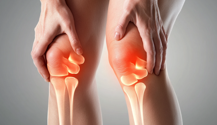
What is Prepatellar Bursitis?
Bursitis is a condition where a small sac in the body, known as a bursa, becomes swollen or inflamed. The bursae, filled with a small amount of lubricating fluid and situated close to your bones, muscles, tendons, and ligaments, help to reduce friction between these structures. Inflammation can occur around large joints such as the shoulder, knee, hip, and elbow and often leads to doctor visits.
Within the knee alone, there are four significant bursae: the suprapatellar, infrapatellar, pes anserine, and prepatellar. The bursa situated between your kneecap (or patella) and the skin above it is known as the prepatellar bursa. This bursa is the most frequently affected in the knee and the second most commonly affected in the body, with the olecranon bursa in the elbow taking the top spot.
Because of its location, the prepatellar bursa is especially prone to irritation during activities that involve a lot of kneeling. Hence, bursitis in this area is often referred to as “housemaids’, carpet-layers’, or carpenter’s knee.”
What Causes Prepatellar Bursitis?
The thin walls of a part of your body called the bursae make it prone to inflammation, especially when it experiences direct harm or repeated stress from activities like frequent kneeling. Furthermore, certain existing conditions like gout, rheumatoid arthritis, or infections can contribute to the development of prepatellar bursitis, which is an inflammation of the bursa at the front of your kneecap.
People with conditions that weaken their immune system, such as diabetes, regular use of steroids, undergoing a specific treatment for kidney failure called hemodialysis, are more at risk of developing bursitis.
Chronic bursitis, a long-term inflammation of the bursa, can also develop from repeated trauma. But this happens less often in the prepatellar bursa (front of the kneecap) than in the olecranon bursa (pointy bone at the tip of your elbow).
Risk Factors and Frequency for Prepatellar Bursitis
Prepatellar bursitis is difficult to measure because many people only seek medical help when it becomes severe, while others with mild symptoms may not seek treatment. Despite this, it’s estimated that 1 in every 10,000 people experiences prepatellar bursitis each year. The majority of these cases—more than 80%—are men between the ages of 40 and 60. Most of these cases are not infectious, although up to a third can become so, which increases the risk of further health problems. While anyone can get prepatellar bursitis, children are more likely to develop the infectious type. People with long-term conditions that weaken the immune system, like diabetes, are also at a higher risk.
Signs and Symptoms of Prepatellar Bursitis
Bursitis is a condition that can come on suddenly or develop over a long period of time, producing a wide range of symptoms. The doctor will need to know about your medical history and your lifestyle to make a diagnosis. Things like having a condition that weakens your immune system (for example, diabetes or long-term use of steroids), or a hobby or job that requires lots of physical activity (for example, housekeeping, carpentry, roofing, gardening) can be important factors. Acute bursitis often results from an injury, infection, or joint disease, while chronic bursitis is usually caused by conditions that cause joint inflammation and repetitive pressure or overuse.
The physical signs of bursitis can also vary. Acute bursitis generally shows up as redness, warmth, tenderness when the bursa is touched, and possibly a limited range of motion due to discomfort. Chronic bursitis, on the other hand, is often painless; the bursa has had time to expand and accommodate the increased fluid. In either case, it’s important to check the affected area for signs of injury, redness, and warmth. One study suggests that a temperature increase of just 2.2°C between the skin over the bursa and the same area on the other side of the body may be a sign of septic bursitis. The bursa may be warm during an episode of acute bursitis, which highlights the need for additional tests, especially in people who have not had similar episodes before.
Testing for Prepatellar Bursitis
In simple terms, diagnosing bursitis, which is inflammation of a fluid-filled sack near your joints, mostly relies on a doctor’s examination and understanding of your symptoms. Routine tests on your blood or other bodily fluids might not be necessary. However, some imaging methods could be really helpful to go alongside a thorough history of your symptoms and a physical check-up. This can help the doctor rule out other possible conditions.
For instance, if your doctor is worried about a fracture or a foreign object in your body after an injury, they might decide to take an x-ray. Ultrasound can be helpful too, it can differentiate between inflammation of a bursa (fluid-filled sac around a joint) and conditions like a skin infection. It can also track changes in the range of motion which might suggest an injury to a tendon (connects muscle to bone). An ultrasound can also give extra information when extracting fluid with a needle from the inflamed area.
Although it’s not commonly used, a magnetic resonance imaging (MRI) scan can rule out other potential conditions if needed. Prepatellar bursitis (inflammation of the bursa in front of the kneecap) can show up as an oval-shaped fluid-filled sac between the skin and the kneecap on an MRI image.
Another important technique in diagnosing bursitis involves extracting fluid from the bursa with a needle. This fluid is then sent to a lab for testing. They’ll check for types of cells, crystals, the glucose level, and germs. If they find crystals that don’t bend light the same way in all directions (negative birefringent), it might mean gout. On the other hand, if they find crystals that do (positively birefringent), it might suggest pseudogout, which is a disease caused by the buildup of calcium pyrophosphate crystals in the joints.
Additionally, if they find more white blood cells than normal that have irregular shapes (polymorphonuclear leukocytes), it might suggest an infection, while more white blood cells that have single nuclei (mononuclear cells) might suggest non-infectious causes. Interestingly, a high white blood cell count in the fluid is not specific, but if it’s above 2000/mm3, that’s a reasonable indication of infectious bursitis. They’ll also check the glucose level in the fluid, as lower levels might suggest an infection. The presence of certain types of bacteria (germs) when staining the fluid varies a lot, it can range from a 15% to 100% chance of finding them.
But the best method to diagnose infectious bursitis is culturing, or growing, the bacteria from the bursa fluid in a lab. This can tell whether an infection is present and also what kind of organism is causing it.
Treatment Options for Prepatellar Bursitis
Prepatellar bursitis, an inflammation in the small, fluid-filled sac in front of your kneecap, can occur suddenly or could build up over time. It’s important to manage the underlying causes such as gout or an infection. Bursitis is typically treated with rest, cold compresses, changing your activities to avoid strain on the knee, anti-inflammatory medications, and drawing out fluid from the inflamed sac. These measures usually work for sudden cases of bursitis and even those that build up over time. Sometimes, injections of steroids might also be considered for chronic cases.
If the bursitis is caused by an infection, it’s important to find it out early as it plays a key role in improving patient outcomes. The measures I mentioned earlier can be also used to manage infectious bursitis. You might need to visit your doctor regularly to prevent the need for hospitalization and to avoid long-term complications. In some cases that are not responding to these measures, minor surgery might be needed to drain the fluid, especially if the condition is due to trauma or an infection.
Treatment with oral and injected antibiotics does not seem to be helpful for a specific type of infectious bursitis called septic infrapatellar bursitis. In such cases, hospitalization, needle aspiration (drawing out fluid with a needle), and antibiotics given through a vein are recommended. In stubborn cases, other treatments such as sclerotherapy (injecting a drug to reduce the sac) and bursectomy (surgically removing sac), could be considered. If the bursitis is due to trauma and not responding to initial treatments, consider endoscopic therapies.
The procedure to draw out fluid from the sac involves using a needle at the area where the sac is fullest. The doctor would mark this spot on your skin, clean the area, and use a freezing spray or inject a local anesthetic to numb the skin. They would then insert the needle and pull out as much fluid as possible. They would also use the needle to pierce multiple spots on the sac to reduce the chance of fluid filling up back again. For bursitis due to trauma, using a protective bandage could help avoid fluid filling back up. You would be asked to exercise the area, apply ice, elevate the limb, and given oral nonsteroidal anti-inflammatory drugs (NSAIDs). The doctor would not recommend drawing out fluid if you have a fracture, bone infection, skin ulcer, bacteria in the blood, or a fracture in the cartilage or bone.
When surgery is needed to remove the sac, endoscopic excision is commonly performed. The doctor would first irrigate or clean the infected sac with a drug, drain the pus, and remove the chronically thickened sac. This would require making small incisions near your kneecap and inserting a tiny camera and a specialized tool to remove the sac. The wounds would be closed with adhesive tape and a loose bandage is applied.
If you are admitted to a hospital due to a systemic infection or if you have a weakened immune system, you should receive antibiotics given through a vein for a week to 10 days, followed by oral antibiotics for up to 2 weeks. For mild to moderate infectious bursitis, oral antibiotics for 2 weeks can be taken as an outpatient treatment. Surgery to remove the infected sac is advised in cases where there is persistent or recurrent infectious bursitis, unsuccessful drainage, individuals with a weakened immune system, and critical illnesses.
What else can Prepatellar Bursitis be?
When a person experiences sharp knee pain, the probable causes could vary based on their age group. However, the expansive list of possibilities isn’t confined by age or gender. A detailed examination can help distinguish between a variety of conditions:
- Prepatellar bursitis (inflammation of the small sac of fluid located in front of the kneecap).
- Patellar subluxation/dislocation (misalignment or dislocation of the kneecap).
- Tibial apophysitis (inflammation where the patellar tendon attaches to the shinbone in growing children).
- Patellar tendonitis (inflammation of the tendon connecting the kneecap to the shinbone).
- Patellofemoral tracking syndrome (malalignment or malfunction of how the kneecap moves).
- Knee osteoarthritis (long-term wear and tear of the knee joint).
- Reactive and rheumatoid arthritis (types of arthritis that may affect the knee).
- Septic arthritis (joint inflammation caused by a bacterial or fungal infection).
- Radiating pain from hip-related issues, such as fractures, osteoarthritis, slipped capital femoral epiphysis (a condition in adolescents where the ball at the head of the thigh bone slips off).
- Cellulitis or other skin and soft tissue infections (these can mimic prepatellar bursitis symptoms).
- Tears or injuries to the ligaments and meniscus within the knee joint.
- Fractures of the tibial plateau (top part of the shinbone).
- Inflammation in one of the other bursae (small fluid-filled sacs) around the knee.
A thorough assessment, often involving various tests, is essential for healthcare providers to pinpoint the source of the knee pain accurately.
What to expect with Prepatellar Bursitis
The overall outcome for bursitis is generally very positive. However, problems can occur if there’s an infection, making the condition more severe and causing further health difficulties. Therefore, it’s important to spot the signs of an infection early on.
Possible Complications When Diagnosed with Prepatellar Bursitis
The main risk associated with prepatellar bursitis, a condition causing inflammation and swelling in the knee, is infection. There are also complications secondary to treatments for this condition. These can be an infection resulting from bacteria entering the body during a needle aspiration procedure. A fistula, which is an abnormal connection between the skin surface and the knee sac, can also occur after a needle aspiration. Other potential risks include subcutaneous atrophy or thinning of the skin and underlying tissue due to corticosteroid injections, bleeding following these injections, and the rupture of the patellar tendon in the knee.
Potential Risks:
- Infection
- Infection due to bacteria from needle aspiration
- Fistula between skin surface and knee sac after needle aspiration
- Thinning of the skin and tissue from corticosteroid injections
- Bleeding after injections
- Rupture of the patellar tendon
Preventing Prepatellar Bursitis
The focus of patient education is on prevention. Patients should be advised to avoid any harm to the knee and try not to kneel frequently if possible. If kneeling is unavoidable, it’s recommended to use protective equipment like thick knee pads. For those who experience knee problems due to sports-related injuries, which are less common, knee pads can also be helpful to prevent further damage.


