What is Progressive Collapsing Foot Deformity (adult-acquired flatfoot or posterior tibial tendon dysfunction)?
Progressive collapsing foot deformity, also known as adult-acquired flatfoot or posterior tibial tendon dysfunction, is a condition that happens when the arch of the foot gradually falls or collapses. This condition can continue to worsen over time, affecting the shape and function of the foot and ankle. This may also be seen as the foot “flattening”. It’s quite severe and affects up to 5 million people in the United States alone.
The foot and ankle are made up of many different parts, all of which have to work together so that we can walk and bear our body weight. The posterior tibial tendon, a tissue that connects muscle to bone, plays a big part in the development of this foot deformity. This tendon helps to flex the foot and is the main structure that turns the foot inward. It not only attaches to the navicular bone (one of the bones of the foot) but also attaches to other bones in your foot. There are also two sets of ligaments, or bands of tissue that connect bones together, that are very important: the spring and deltoid ligaments. The spring ligament is most often involved in this deformity, and it supports the ankle by connecting one of the ankle bones, the calcaneus, to the navicular bone. The deltoid ligament is usually affected later in the condition and helps to support the foot and ankle, preventing them from turning outward.
Doctors use a system to ‘grade’ the severity of acquired flatfoot, known as the Johnson and Strom classification system, which is graded from I to III. A fourth grade was added by Myerson in 1997. The stage of the disease can inform how it is treated: for instance, Stage I disease has inflammation of the posterior tibial tendon but no arch collapse, while Stage III involves a fixed deformity with the foot turning outward and the front of the foot turning away from the midline of the body (abduction). Stage IV deformity includes the ankle turning outward due to weakening of the deltoid ligament.
This system is a helpful way for doctors to identify the severity of the condition and also guide treatment plans. But recently, a new classification system has been put in place thanks to a better understanding of how our foot arch and the development of flatfoot work. This terminology, “progressive collapsing foot deformity”, considers changes to the soft tissues of the foot and ankle and the relationships between the bone structures of the midfoot, rearfoot, and ankle.
What Causes Progressive Collapsing Foot Deformity (adult-acquired flatfoot or posterior tibial tendon dysfunction)?
Traditionally, acquired flatfoot was thought to be mainly caused by issues with the tendon in the back of the ankle, also known as the posterior tibial tendon. However, modern understanding points out that the ligaments (tissues that connect bones) in the ankle also contribute to the onset of this foot deformity.
The dysfunction of the posterior tibial tendon is a complex process influenced by various factors. For instance, many patients who suffer from this may already have flat feet, and obesity is also common among these individuals. Additionally, the tendon has a distinct feature where it takes a sharp turn behind a bone in the ankle (the medial malleolus). This area of the tendon doesn’t receive as much blood supply, making it more susceptible to rupture, which could be because of its inability to repair itself efficiently.
Previous injuries, use of corticosteroids (a type of medication), arthritis, conditions affecting the nerves and muscles, and diabetes all contribute to the likelihood of developing what is known as a progressive collapsing foot deformity.
Risk Factors and Frequency for Progressive Collapsing Foot Deformity (adult-acquired flatfoot or posterior tibial tendon dysfunction)
Acquired flatfoot, also known as progressive collapsing foot deformity, is a common foot problem, especially among older people. It affects about 5 million people in the United States, and in the United Kingdom, over 3% of women over 40 are estimated to have it. It’s also common for people to experience issues with their posterior tibial tendon – a tendon in the foot – as they get older. In fact, about 10% of older patients have these issues.
It’s been suggested that older adults might be more prone to severe cases of acquired flatfoot because their muscle mass and bone structure tend to decline as they age. People with chronic blood flow problems, like diabetes or high blood pressure, are also at a higher risk of getting this condition.
Signs and Symptoms of Progressive Collapsing Foot Deformity (adult-acquired flatfoot or posterior tibial tendon dysfunction)
If you suspect you’re dealing with progressive collapsing foot deformity, there are a few things to check while you’re bearing weight on the foot. One thing you might notice is excessive sideways movement of the front of your foot. This can be identified using the “too many toes” sign, which means if you see more than the fifth toe and some of the fourth toe, the sign is positive.
You may also feel swelling or pain along the posterior tibial tendon, which runs around the inside of your ankle. Pain could also appear on the outer side of your foot if the tissue connecting your bones is under too much stress.
A few simple tests can be performed to check the strength of the posterior tibial tendon—The muscle at the back of your lower leg that helps support your foot. If you’re unable to turn your foot inward or lift your heel, this may indicate a problem with this tendon. It’s also important to examine how much your ankle can move in different directions.
Exercise can help correct a leaning deformity of the foot, but if the deformity persists during a range of motion exercise, it’s called a fixed deformity. This could happen in the joint below the ankle or possibly involve a significant sideways movement of the front of your foot, and this has a strong impact on how your condition is treated.
- Check for the “too many toes” sign
- Inspect for swelling or pain around the inside of your ankle
- Check for pain on the outer side of your foot
- Perform heel rise tests to assess the strength of the posterior tibial tendon
- Test ankle range of motion
- Perform exercises to correct foot deformity
- Check for fixed deformities in the joint below the ankle or front of the foot
Testing for Progressive Collapsing Foot Deformity (adult-acquired flatfoot or posterior tibial tendon dysfunction)
If you’re suffering from a progressive collapsing foot deformity – a condition where your foot arch collapses – doctors will want to use imaging tools to assess its severity. This allows them to decide the best treatment for your condition. They most commonly use weight-bearing radiographs, which are simply X-rays taken while you’re standing up. This method is extremely effective but in some cases, they might use a weight-bearing computed tomography (CT), another type of imaging tool that uses computers to create detailed images of your bones.
The X-rays or CT are used to examine your foot from different angles. They give doctors a clear assessment of how much your arch has collapsed and details on the condition of your joints. By looking at these pictures, they measure specific angles at various joints in your foot to assess the state of your condition. If your condition has reached its fourth stage, they will also examine the degree of talar head exposure, which refers to how much of the top of the talus bone – one of the important bones in your ankle – can be seen.
In addition to X-rays and CT scans, doctors may also be interested in checking whether arthritis, a common inflammation of joints, has developed. The presence of arthritis could influence the kind of treatment plans they consider for you.
In some cases, doctors might recommend a Magnetic Resonance Imaging (MRI). This is a type of scan that can provide very detailed images of your body. It’s not always needed for progressive collapsing foot deformity, but could be useful in case there’s a problem with ligaments in your foot and a surgery needs to be planned.
Lastly, some studies suggest that ultrasound scans – another type of imaging technique – could be useful for looking at the posterior tibial tendon, a tendon that plays a crucial role in supporting the arch of your foot. As per these studies, an ultrasound scan can provide almost equivalent information to an MRI, and it’s generally quicker, less expensive, and more accessible.
Treatment Options for Progressive Collapsing Foot Deformity (adult-acquired flatfoot or posterior tibial tendon dysfunction)
Treating a progressive collapsing foot deformity, where the arch of your foot flattens over time, can be tricky. However, many options are available today, and a majority of recent research has focused on treating this condition.
The first line of treatment is usually non-invasive, which means it doesn’t involve any surgical procedures. This could involve a support device for your ankle and foot, like a low-articulating ankle-foot orthosis, or a cast-boot walker, which helps you walk while providing rest and support to your foot. This is often combined with medications known as nonsteroidal anti-inflammatory drugs (NSAIDs) to reduce inflammation and pain, as well as physical therapy, which consists of certain exercises that can help improve your mobility and reduce discomfort. This strategy has been well-received with one study revealing that 87% of patients noticed their symptoms go away. In fact, other studies have reported success rates ranging from 67% to 90% using such conservative treatment measures.
If the non-surgical measures don’t provide satisfactory results, surgical treatment might be considered. However, this depends on a variety of factors, for example, how severe and advanced your foot deformity is, whether you have other health conditions, your lifestyle, and if you are a smoker. Surgery is rarely done for stage I disease, but if needed, surgical options may include removing the inflammation from the posterior tibial tendon (a tendon in your calf which is crucial for foot movement), cleaning damaged tissue (debridement), or tendon grafting. It’s important to know that surgical repairs of the posterior tibial tendon can sometimes fail in the long run, so it’s crucial to think it through before opting for surgical intervention.
If your disease has progressed to stage II, types of surgeries such as medializing calcaneal osteotomy (shifting the heel bone towards the inner side of your foot to correct its alignment) and transferring a tendon from your toe to your foot have shown encouraging results. In fact, one study found 87% patient satisfaction with this approach, even after an average of 15 years.
Stage III disease is somewhat harder to deal with because the deformity is now fixed. Arthrodesis, which involves joining or fusing certain bones in the foot to form a single, solid bone, is the standard care approach here. This can involve either two joints (double arthrodesis) or three joints (triple arthrodesis). Despite the decreased risk of complications associated with double arthrodesis, this procedure limits your foot mobility which can make walking on uneven ground difficult. There are also potential risks of the bones failing to join (nonunion), weakness of the deltoid ligament (a ligament in the ankle), and foot rolling outward (ankle valgus).
For stage IV disease, the type of treatment depends on whether you have developed a rigid flatfoot. Some patients progress to stage IV without showing signs of rigid disease due to the failure of the deltoid ligament. In these cases, surgical repair of the deltoid ligament might be an option. But if your stage IV disease becomes rigid, you might need an ankle fusion which carries its own set of substantial complications. Another alternative is ankle replacement.
A recent analysis of surgical treatments for this foot deformity looked into the effectiveness of various procedures using certain measurements taken from X-ray images to evaluate the outcome of each method. The results suggested that every kind of treatment resulted in significant improvements for patients.
Generally, orthopedic experts agree on few points regarding the surgical treatment of this disease: first, maintaining your mobility and range of motion is always a priority, especially if you’re younger. Second, fusing the talonavicular joint (a joint in your foot) should be considered only if the joint is arthritic, or if you have severe sagging or deformity. Lastly, patients with a higher body mass index may not fare as well with bone reshaping surgeries (reconstructive osteotomies) when compared to joint fusion procedures (arthrodesis).
What else can Progressive Collapsing Foot Deformity (adult-acquired flatfoot or posterior tibial tendon dysfunction) be?
When a doctor thinks a patient might have a progressively collapsing foot, several other conditions need to be ruled out first. These conditions could look similar, but require different treatments. They include:
- Adult flexible flatfoot
- Tarsal coalition (an abnormal connection between two bones in the back of the foot)
- Charcot foot (a condition causing weakening of the foot’s bones)
- Neuromuscular flatfoot (a flatfoot condition caused by muscle or nerve disorders)
- Arthritic, post-traumatic, and iatrogenic deformity (foot deformities caused by arthritis, injury, or a medical procedure gone wrong)
A detailed medical history and physical examination by the doctor can usually identify the real cause from these possibilities.
What to expect with Progressive Collapsing Foot Deformity (adult-acquired flatfoot or posterior tibial tendon dysfunction)
The outlook for most people with a progressively collapsing foot deformity is generally good. The majority of patients don’t need to have surgery. For those who do need surgery, about 1 in 10 can anticipate a positive result.
If the foot deformity is severe, more extensive surgery may be required. In the most advanced case of a collapsing foot deformity, known as stage IV, an ankle fusion or a total ankle replacement surgery might be necessary.
Possible Complications When Diagnosed with Progressive Collapsing Foot Deformity (adult-acquired flatfoot or posterior tibial tendon dysfunction)
If foot deformity is not managed in time, it can advance to severe stages that demand more aggressive treatments. Catching it early is very important as it can potentially save some patients from needing surgery. However, if the condition becomes advanced, patients may require ankle fusion surgery. This procedure results in a rigid ankle joint, which could limit the patient’s ability to participate in leisure activities.
Facts:
- No treatment for foot deformity can lead to more aggressive treatment requirement
- Early diagnosis can help prevent surgery
- Advanced cases may necessitate ankle fusion surgery
- Ankle fusion creates an immobile ankle joint
- Recreational activities may be impacted after ankle fusion

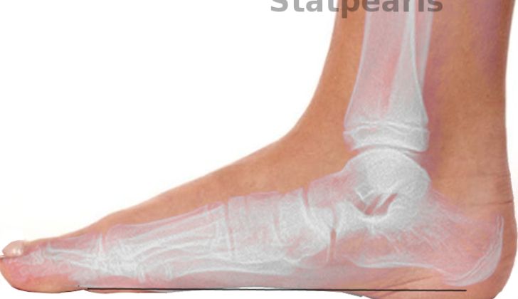
demonstrates PCFD with increased talar head exposure and forefoot abduction.
Preventing Progressive Collapsing Foot Deformity (adult-acquired flatfoot or posterior tibial tendon dysfunction)
A condition where the foot starts to collapse gradually, known as progressive collapsing foot deformity, is often seen in people with obesity, diabetes, high blood pressure, and other diseases that can be improved with healthy lifestyle choices and losing weight. Treating these health issues can also help the patient’s foot problem. In addition, if prescribed a device to support their foot, patients can slow down the worsening of their foot condition. Numerous individuals, who use this support device along with physical therapy or medication, won’t require surgery.


