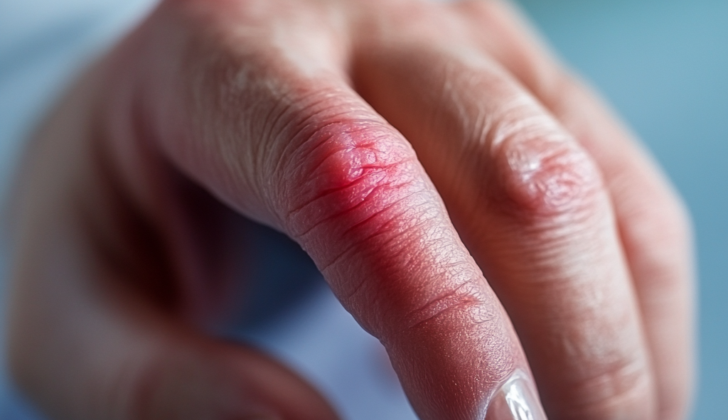
What is Pyogenic Flexor Tenosynovitis?
Pyogenic or suppurative flexor tenosynovitis (PFT) is a serious bacterial infection that develops in the closed area of the digital flexor tendon sheaths, which are coverings on the tendons in your fingers. PFT accounts for between 2.5% and 9.5% of all hand infections and can result in the death of tendons and finger tissues. This infection disrupts the smooth movement of the tendons inside the sheath and causes tissue adhesions that severely limit finger movement. PFT mainly occurs from a penetrating injury to the finger that involves the flexor tendon sheath, but it can also develop from a bloodstream infection.
Back in 1912, a physician by the name of Allen B. Kanavel came up with 3 main signs of PFT: tenderness in the flexor sheath, a bent/flexed position of the affected finger, and pain when the infected finger is extended. Later on, a fourth sign was added; swelling of the finger.
Understanding these 4 signs is critical for diagnosing PFT on physical examination, which has a high success rate of between 91.4% and 97.1%. As this condition can lead to serious complications, it is crucial to quickly diagnose and treat PFT.
Knowing the structure of the flexor tendon sheaths in the hand is key to understanding and managing PFT. In simple terms, it consists of two parts: the inner synovial layer and the outer fibrous layer. The flexor tendon sheath comprises 2 layers: an inner visceral layer that closely covers the flexor tendon, and an outer parietal layer that is connected to several tendon pulleys. The space between the layers contains synovial fluid that nourishes the tendons within the sheath, which receive their blood supply from the surrounding arteries. However, their blood supply can be unpredictable, creating an environment prone to infection.
As PFT develops, pus begins to accumulate in the synovial space within the flexor sheath, leading to pressures of up to 30 mm Hg in the enclosed area of the flexor sheath. This increased pressure then interferes with blood supply to the flexor tendon, which can lead to scarring and possible rupture.
Knowing the endpoints of the flexor tendon sheaths for the index, middle, ring, and little fingers also contributes to understanding and managing PFT. For example, they finish where the deep flexor tendon of the finger gets inserted into the end (or distal phalanx) of the finger bone. In about 80% of people, a small anatomic space within the distal forearm (known as the space of Parona) allows an infection in the little finger or thumb to create a horseshoe-shaped abscess.
So, as you can see, understanding the anatomy and function of the flexor tendon sheaths plays a pivotal role in understanding, managing, and preventing a serious condition like pyogenic flexor tenosynovitis.
What Causes Pyogenic Flexor Tenosynovitis?
Most people who experience a flexor tendon sheath infection, or an infection surrounding the tendons that helps bend the fingers, usually have it because of an injury where something sharp poked into their finger. However, other sources of these infections can come from neighboring infections, like an inflamed fingertip called a felon, a contaminated joint, or infections deeper in the hand or finger. A person can also get this infection from human or animal bites that break the skin.
It’s quite rare, but these infections can also be caused by bacteria getting into the blood, usually as a result of a widespread infection by a specific type of bacteria called gonococcus.
The main bacterial culprit behind these tendon sheath infections is a germ called Staphylococcus aureus, which is responsible for about 75% of these infections. So, about a third of these cases also involve a version of this bacteria that can resist treatment with methicillin, a common antibiotic. Other bacteria that might cause this infection includes Staphylococcus epidermidis, β-hemolytic streptococcus, and a kind of germ-negative bacteria called Pseudomonas aeruginosa.
Patients who have a weaker immune system might get these infections from multiple bacteria or germ-negative rods. The bacteria Eikenella corrodens can cause these infections through human bites. Cases that happen after animal bites might be due to infection by Pasteurella multocida bacteria.
Risk Factors and Frequency for Pyogenic Flexor Tenosynovitis
Pyogenic flexor tenosynovitis (PFT), a type of hand infection, is found in about 2.5% to 9.5% of all hand infections. This usually happens after the hand has been injured. The occurrence of this condition equally affects all age groups and both sexes. With the use of antibiotics delivered directly into the blood and quick surgical treatment, the impact of PFT has been lessened.
The severity of the infection and how the disease develops are closely connected to the patient’s ability to fight off infections. People who have conditions that reduce their ability to fight off infections, like malnutrition, chronic use of steroids, autoimmune diseases, peripheral vascular disease, and diabetes, are more likely to experience complications and worse outcomes after getting PFT.
- PFT is found in about 2.5% to 9.5% of all hand infections.
- It usually occurs after a hand injury.
- Incidence rates are equal among different age groups and sexes.
- Modern treatments include antibiotics delivered directly into the blood, as well as early surgery.
- The severity of PFT and how it progresses is directly tied to the patient’s ability to fight off infections.
- People with reduced immunity due to conditions like malnutrition, chronic steroid use, autoimmune diseases, peripheral vascular disease, and diabetes face a higher risk of complications and worse outcomes after PFT.
Signs and Symptoms of Pyogenic Flexor Tenosynovitis
Doctors should ask patients detailed questions to understand their physical condition better. This could include:
- Checking for any piercing wounds on the front side of the hand and fingers. This information can help identify the cause of an infection. Usually, a hand injury can occur 2 to 5 days before patients show signs of infection, but this could take longer for patients with a weaker immune system or diabetes. Sometimes, injuries may be minor and overlooked by patients, and many may not recall a previous injury.
- Establishing the exact time frame between the start of symptoms and when the patient sought medical help. The length and severity of symptoms can guide treatment decisions. Delayed treatment after the onset of symptoms can lead to poor outcomes and increased risk of finger amputation.
Understanding the patient’s social background, including their profession and whether they are right or left-handed, can provide useful information for planning post-surgery rehabilitation and support. Doctors should also identify and manage other existing health conditions before performing surgery. For instance, patients with diabetes, kidney failure, or peripheral vascular disease who develop infections are more at risk of poor health outcomes.
Testing for Pyogenic Flexor Tenosynovitis
A physical examination and clinical assessment are typically used to diagnose Pyogenic Flexor Tenosynovitis (PFT), an infection of the tendon sheath in your finger. The presence of four particular signs, known as the Kanavel signs, are generally strong indicators of PFT. These signs are:
* An evenly swollen finger.
* The finger being held in a bent position.
* Pain when trying to straighten the finger.
* Tenderness along the length of the tendon sheath from the tip to the base of the finger.
But in some cases, especially those with compromised immune systems, diabetes, or a history of intravenous drug use, these signs can be less clear.
Pain when trying to straighten the finger is usually the first thing patients notice, while tenderness of the tendon sheath is the last sign to appear. In fact, only about 17% of patients with PFT have a fever. During a physical examination, your doctor will also look at your hand for any wounds, and check for signs of infection spreading to other areas, such as redness, swelling, and tenderness in the padded areas at the base of your thumb or little finger.
In terms of the most important sign, there are varying opinions. Dr. Kanavel believed that tenderness over the tendon sheath was the most important, while others suggest that swelling of the fingers was more common. However, not every patient displays all four signs, raising questions about the effectiveness of these signs alone in diagnosing PFT.
In addition to a physical examination, imaging and lab tests can help diagnose PFT. X-rays can be used to look for any foreign objects and signs of chronic infection in the bone. A type of ultrasound scan, known as a hand ultrasound, can be an inexpensive and non-invasive way to confirm a PFT diagnosis. It helps by visualising the tendon and identifying any fluid buildup which is often associated with infection. However, it depends on the person doing the scan and can’t tell the difference between pus and blood.
An MRI scan can help to see the extent of the infection, but can’t tell the difference between PFT and other inflammatory conditions, so it is rarely used. A CT scan of the hand with contrast can also be used.
Your doctor may also ask for a blood test, which can show if there’s inflammation in your body through markers like white blood cell count, C-reactive protein level, and erythrocyte sedimentation rate level. While these tests can’t specifically diagnose PFT, they can offer a broad indication of infection and help track how well treatment is working. Another useful test is a culture of pus or necrotic (dead) tissue which can help identify the bacteria causing the infection and guide antibiotic treatment. A blood culture may also be run if there are signs of a widespread infection.
Treatment Options for Pyogenic Flexor Tenosynovitis
Pyogenic Flexor Tenosynovitis (PFT), a severe infection in the hand, can be managed without surgery if it’s detected within 48 hours of the hand being injured. Non-surgical treatment involves administering antibiotics through an IV, keeping the arm elevated, and immobilizing the hand with a splint. These measures are often most beneficial if the symptoms are less severe. The person will be monitored closely for changes in the infection through regular hand exams and lab tests. If there’s no improvement or if symptoms worsen, prompt surgical treatment may be needed. Past studies have shown that patients with less severe symptoms and a shorter timeframe from when symptoms began often do well with just antibiotics and don’t require surgery.
Despite this, many PFT cases will still require surgical procedures. There are several techniques a surgical team may choose, depending on the severity of the PFT. If the infection is severe and has been left untreated for a while, causing tissue death, then invasive surgery such as washout or debridement may be necessary.
One surgical option is closed flexor sheath catheter irrigation. This method involves making two minor cuts on the front of the hand, careful dissection to identify the infected area—taking care not to harm the nerves or blood vessels—and then draining the fluid for lab examination. From there, a catheter is passed through one of the incisions and sterile saline is injected to clean the infected area. Research suggests that this approach can be equally effective to more invasive procedures.
Another more invasive surgical approach is open flexor sheath irrigation and debridement. This involves a larger single cut on the front of the hand to expose the entire infected area for thorough cleaning. After nerve and blood vessels are identified and protected, the infected area is cleaned, then the opening is stitched shut. However, this treatment can come with a higher risk of post-surgery issues like finger stiffness and issues with the tendons.
After surgery, antibiotics are typically given according to lab results, but recent studies suggest that oral antibiotics over a 7 to 14-day period may be effective, which allows for outpatient care.
What else can Pyogenic Flexor Tenosynovitis be?
A felon is a type of infection that occurs in the soft padding at the end of your fingers, most commonly affecting the thumb or index finger. It’s usually caused by a bacteria known as Staphylococcus aureus, which can get into the skin through a small injury. However, different types of bacteria can also cause a felon, especially in people with weakened immune systems. This infection causes severe pain and often requires surgical drainage.
Septic arthritis is another condition that affects the fingers and can cause signs of infection in the joints. The fingers may have limited movement and cause pain, due to swelling and tension in the joint capsule. This condition is different from a felon as it typically does not cause swelling or tenderness along the finger.
Herpetic whitlow is a rare infection caused by the herpes simplex virus that affects the fingertips. It causes painful blisters filled with clear fluid that can merge to form larger blisters. This condition does not require surgery but is typically treated with antiviral medicines like acyclovir.
Hand cellulitis is an inflammation of the hand that does not involve pus. It usually causes widespread swelling and redness without any abscess (a swollen area filled with pus). This condition is usually treated by resting the hand in an elevated position and taking antibiotics to treat the infection.
What to expect with Pyogenic Flexor Tenosynovitis
PFT, or Pyogenic Flexor Tenosynovitis, is a condition that can lead to severe consequences like finger amputation and spreading infection if not treated early. It affects the ability to fully move the finger in 10% to 25% of affected patients. Those with conditions such as diabetes, peripheral vascular disease (a circulation disorder affecting blood vessels outside the heart), and renal failure (when the kidneys stop working) face a higher risk of finger amputation after PFT.
Possible Complications When Diagnosed with Pyogenic Flexor Tenosynovitis
Finger stiffness and limited movement
Stiffness after PFT – that is, Pyogenic Flexor Tenosynovitis, an infection of the spaces around the tendons of the fingers – may be due to either the infection itself or the surgery needed to clear it out. This infection can cause the finger’s moving tendons to stick together, make the joint capsules thicker, and damage the pulleys which help the fingers to bend. To avoid stiffness, it’s recommended to start exercises for the affected finger early. Once the infection is entirely gone, a procedure known as flexor tenolysis might be required to restore movement to the affected finger.
Flexor tendon scarring and breaking
High pressure inside the flexor sheath can interfere with the blood flow and nutrition to the flexor tendons in the fingers. These unhealthy, scarred tendons can lose their natural springiness and might break with further activity.
Tissue Death and Finger Ischemia
A severe lack of blood flow to the finger (critical ischemia) can happen if the blood flow is interrupted due to high pressure in the finger or a blood clot caused by the infection.
Formation of hand horseshoe abscess
A horseshoe abscess is a u-shaped collection of pus. This can result from the spread of the infection from PFT in the thumb or little finger, especially in patients whose radial and ulnar bursae (sac-like structures around the tendons) can communicate or connect.
Amputation of the finger
In some cases, it might be necessary to amputate the finger at various levels. This could be due to a deep-seated infection, significant damage to the soft tissue, or if the finger loses all function and becomes stiff.
Summarized Complications:
- Finger stiffness and limited movement
- Scarring and breaking of flexor tendons in the finger
- Death of finger tissue due to lack of blood flow
- Formation of hand horseshoe abscess
- Amputation of the finger
Preventing Pyogenic Flexor Tenosynovitis
As a part of the treatment process, patients should be given detailed explanations and guidance about their condition, which is sometimes referred to as PFT. It’s crucial for doctors to break down medical jargon and use easy-to-understand language to explain the importance of treatment and the possible risks as part of giving their consent to the treatment. They should know that their condition is a kind of emergency, requiring prompt treatment to maintain the health and function of the affected finger and hand.
The doctor must alert the patient about possible complications caused by the infection. The main potential issues are limited movement and, in severe cases, the need for amputation. If the infection is in the hand you use the most, typically called the dominant hand, you may need extra support during your recovery period, which might take longer than expected.


