What is Septic Bursitis?
Bursae are like little sacs filled with fluid. They are located between moving parts of your body’s muscular-skeletal system, like between skin and bone, or joints. Think of them as tiny shock absorbers. Your body has over 150 of these bursae, both deep within your body and near the surface, cushioning areas between bone, muscle, and skin. They contain a small amount of synovial fluid that acts like oil in a car, reducing friction by making things slippery.
Sometimes, these bursae can become inflamed, leading to a condition known as bursitis. This happens when more fluid than usual is produced, causing the bursa to swell and become irritated. This inflammation can occur for various reasons such as sitting or leaning too long on a hard surface, doing the same motion too many times, some forms of arthritis, and direct injuries like a blow or puncture. Common areas where bursitis occurs include the knee, elbow, and hip.
It’s possible for the bursa to also get infected, a condition called septic or infectious bursitis. This happens when bacteria get into the bursa, either directly through the skin (usually for the bursa near the skin) or from somewhere else in the body (for the deep bursa). Septic bursitis can be sudden, slowly developing, or even recurring. It can sometimes be hard to tell the difference between infected and non-infected bursitis, so doctors may need to remove and test some of the fluid from the bursa to make a correct diagnosis.
What Causes Septic Bursitis?
Septic bursitis happens when bacteria infect the bursa, a small, fluid-filled cushion that helps reduce friction in your joints. This can often happen due to small injuries, or when the skin above the bursa is punctured, leading to an infection. Another common cause is the spread of skin cellulitis, which is a skin infection, to the bursa.
In 80-90% of cases, a type of bacteria called Staphylococcus aureus causes septic bursitis, followed by Streptococcus species. Other bacteria like Escherichia coli, Enterococcus, Pseudomonas aeruginosa, and coagulase-negative staphylococci can also cause this condition.
When bursitis becomes chronic and lasts for a long time, it is likely due to unusual bacteria and fungi. If this happens, it is important to get evaluated for a full-body infection promptly.
Risk Factors and Frequency for Septic Bursitis
Septic bursitis, an inflammation of the bursa due to infection, is more common in males around the age of 50. Although it can occur in those with certain existing health conditions, it mostly happens due to continuous strain or injury from work activities. Professions like plumbers, carpenters, roofers, clergy, and athletes are often affected. Surprisingly, sometimes a treatment to ease non-infectious bursitis, like joint steroid injections, can cause septic bursitis. Also, people with conditions that cause more fluid in the bursa, such as Gout, or inflammatory conditions like rheumatoid arthritis, have a higher risk.
- Septic bursitis is more frequent in males, and it typically starts around 50 years of age.
- It can happen more often in people with certain health conditions, but most of the time it’s caused by repetitive injuries from work.
- People in professions like plumbing, carpentry, roofing, clergy, and athletics are more likely to get it.
- Sometimes it can be caused by injections meant to treat non-infectious bursitis.
- People with conditions like Gout that cause more fluid in the bursa are at higher risk.
- Those with inflammatory conditions, like rheumatoid arthritis, are also more susceptible.
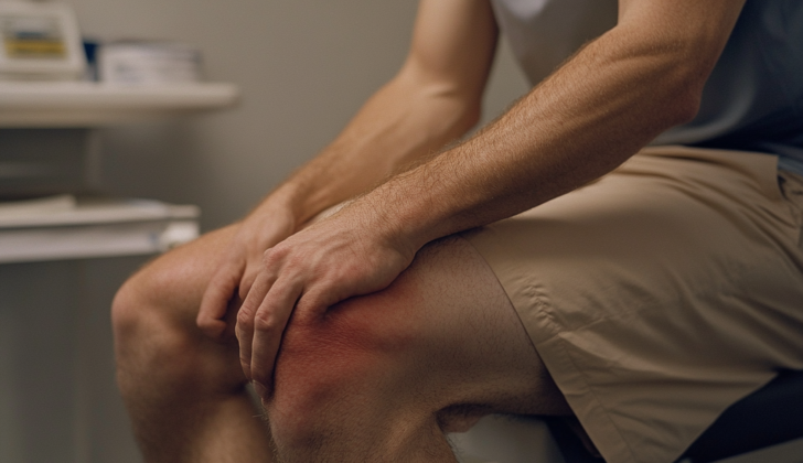
Signs and Symptoms of Septic Bursitis
Septic bursitis refers to the infection of the bursa, a small fluid-filled sac that reduces friction in your joints. Determining if bursitis is septic or non-septic can be challenging as the symptoms are often similar. Both types might cause pain and swelling over the affected area. However, in septic bursitis, it’s more common to also see redness and warmth, and there might be signs of skin injury or infection too. A fever may accompany these symptoms but isn’t always present. Importantly, joint movement is usually normal in septic bursitis, unlike in septic arthritis where it’s often limited. These symptoms aren’t foolproof though, so further tests are usually necessary to confirm the diagnosis. When examining a patient suspected to have bursitis, healthcare professionals focus on a few specific factors.
- The size of the swelling
- The consistency of the swelling (is it soft, firm, or hard?)
- Presence of fluctuance (a wavy feeling when the skin over the swelling is pressed)
- Signs of cellutitis (a skin infection)
- Any swollen lymph nodes (lymphadenopathy)
- The range of motion and pain in the nearby joint
Testing for Septic Bursitis
Before starting antibiotic treatment for bursitis, it’s helpful to perform certain tests. These tests look for signs of inflammation and check for bacteria in your blood. Other routine blood work may not be as helpful in figuring out whether bursitis is caused by an infection or not. For example, the white blood cell count, which usually goes up during infections, might not be different or even high with infectious bursitis.
However, tests for two substances, called C-reactive protein and erythrocyte sedimentation rate, often provide useful clues. These levels usually go up in cases of infectious bursitis. Your doctor might also want to check your blood levels of uric acid. This is especially true if there is a possibility that the bursitis is caused by tiny crystals forming inside your joints – a condition known as crystal arthropathy. Tests for antinuclear antibody and rheumatoid factor, which check for autoimmune diseases, might also be needed if the bursitis is chronic or if an underlying autoimmune disease is suspected.
Plain X-rays are often done initially, but they don’t usually help much in diagnosing infectious bursitis. X-rays can sometimes show bone spurs, which are bony growths that can occur alongside chronic bursitis, but swelling of the joints isn’t usually present. More advanced imaging techniques like computed tomography (CT) and magnetic resonance imaging (MRI) aren’t usually needed unless your doctor suspects a bone infection, an infected joint, or is considering surgery for severe bursitis. Importantly, if an MRI doesn’t show any signs of inflammation around the bursa, then infectious bursitis can be ruled out.
The most reliable way to figure out if bursitis is infectious or caused by crystals is to draw out a small amount of fluid from the bursa and test it. This fluid is tested for its white blood cell count, bacteria (gram stain and culture), and crystals. The white blood cell count inside the bursa usually ranges all the way up to 63,000/mm, with more than 2,000/mm suggesting a high likelihood of infectious bursitis. Infectious bursitis usually causes an increase in a specific type of white blood cells called polymorphonuclear leukocytes, while non-infectious bursitis causes an increase in another type called mononuclear cells. Bacterial testing of the fluid can vary widely in terms of its sensitivity and may only be positive in half of the cases. However, even if the bacterial test (gram stain) is negative, a high white blood cell count and clinical suspicion of infectious bursitis should be treated accordingly. Testing for bacterial growth in the bursal fluid (culture) needs to be done, since it can help guide the treatment.
Treatment Options for Septic Bursitis
Bursitis is an inflammation in the bursa which are small, fluid-filled sacs that cushion the bones, tendons and muscles near your joints. There are two types of bursitis, infectious and non-infectious. While non-infectious bursitis can be managed with treatments aimed to reduce inflammation, the treatment for septic or infectious bursitis always includes antibiotics.
Patients with bursitis may be offered injections of non-steroidal anti-inflammatory drugs (NSAIDs), which help control pain. In cases where the pain is not controlled by NSAIDs, corticosteroids, a type of medicine that reduces inflammation, may be injected. Treatment can usually be done on an outpatient basis, meaning you can go home the same day. However, some might need to stay in the hospital for treatment with intravenous (directly into a vein) antibiotics. This is especially common in patients who have a weaker immune system, show body-wide symptoms, or have inflammation in a joint.
Initial antibiotic treatment is chosen based on what bacteria are most likely to be causing the infection, and then this can be adjusted based on the results from a laboratory test of a fluid sample from the bursa (the gram-stain and culture results). Clindamycin, doxycycline, or trimethoprim-sulfamethoxazole are often recommended at the beginning of treatment until these results are available. If the infection is severe or if the patient has a weakened immune system, they may need to be admitted to the hospital for treatment with a strong antibiotic called vancomycin, given directly into a vein. For patients with a penicillin allergy, two other antibiotics, ciprofloxacin and rifampin, are recommended.
There is debate over how long and what type of treatment should be used when treating bursitis. Some suggest a minimum of 10 days of treatment in mild cases, while others recommend treatment until the fluid in the bursa no longer shows signs of infection in more severe cases. The course of treatment will be guided by how well the patient responds and what the cultured bacteria show.
In some cases, doctors may decide to perform a procedure. This is usually done in case of large swellings or severe or repeated cases of infection that don’t get better with just antibiotic treatment. There are several procedures available. The simplest one is a needle aspiration, where a needle is used to remove fluid from the bursa along with drug treatment. If this does not work due to the presence of small pockets of pus or bacteria (loculations), an incision might be made to drain the pus or other fluid. Another method used is suction irrigation. In some cases, a surgery might be needed to remove the bursa. This can be done through an open surgery or an arthroscopic procedure (looking inside the joint with a small camera and performing the surgery with miniaturized instruments). It has been reported that performing this surgery in single stage has more chances of recurrence as compared to a two-stage surgery with a gap in between the two stages. Arthroscopic procedures also tend to have fewer wound complications in comparison with open procedures.
What else can Septic Bursitis be?
When a doctor is trying to figure out what’s causing knee pain, they consider several possibilities. These could be:
- Cellulitis, a bacterial skin infection
- Gout or Pseudogout, which are forms of arthritis caused by crystal deposits in the joints
- Rheumatoid Arthritis, a disorder that causes the body’s immune system to attack its own tissues
- Soft Tissue Knee Injury, which could be a sprain or strain
- Tendonitis, inflammation of a tendon
These conditions can create similar symptoms, so careful investigation is needed to make an accurate diagnosis.
Possible Complications When Diagnosed with Septic Bursitis
Complications of surgical procedures may be many and varied. One possible issue is the rupture of a bursa, which is a small fluid-filled sac that acts as a cushion between bones and other body parts. Another is Osteomyelitis, a severe infection affecting the bone. Wound healing complications can also occur, such as a wound reopening (dehiscence), excessive fluid drainage (exudate), chronic sinus problems, and death of skin tissue (necrosis).
The risk of the disease returning is another significant aspect to consider. It is crucial to understand that failing to take operative action when needed significantly increases the chances of the disease coming back. The rate of recurrence is 14.6% when operation is taken as against a huge 80% when it’s neglected. Along similar lines, for those who have undergone surgery, having a weakened immune system contributes to the likelihood of disease recurrence.
Key Complications:
- Bursa rupture
- Osteomyelitis
- Complications in wound healing, such as dehiscence, excessive exudate, chronic sinus, and skin necrosis
- Recurrence of the disease, particularly if operative action isn’t taken when indicated or in the case of a weakened immune system


