What is Spondylolysis?
Spondylolysis is a defect that can either appear on one or both sides of a specific part of your spine, known as the pars interarticularis. This can occur with or without the slipping of a vertebral body. This condition is commonly seen as a result of repeated injury to a growing, immature skeleton among individuals who are genetically more likely to develop it.
The pars interarticularis is a bridge-like section of bone that connects the lower and upper joint surfaces of an individual vertebra, which is one of the small bones that make up the spine.
What Causes Spondylolysis?
Spondylolysis, a condition affecting the spine, can either be present from birth or develop over time. While the exact causes are still not fully understood, it’s often linked to a fracture in a specific part of the spine (the pars interarticularis), which fails to heal properly. This condition typically develops in children and teenagers who are genetically predisposed, have abnormal spine mechanics, and frequently undergo repeated minor injuries to the spine due to activities like bending backwards excessively with twisting movements.
Risk Factors and Frequency for Spondylolysis
Spondylolysis is a condition that starts appearing in childhood, with 4% of kids showing signs by age 6 and 6% by age 14. After that, the rate stays the same into adulthood. Certain groups are more likely to experience this condition, including males, who are twice as likely as females to have it, people of Alaskan Eskimo descent, children of people with the condition, and those with certain other conditions like spina bifida occulta, Marfan syndrome, osteogenesis imperfecta, and osteopetrosis.
Teenagers who play sports have a higher rate of spondylolysis compared to those who don’t participate in sports. The average age when this condition is diagnosed is 15. Certain high-risk sports contribute to an increased incidence rate. These are activities that cause repeated stress and extensive movement of the lumbar spine, such as gymnastics, dance, and many types of athletics.
- Spondylolysis is more common in:
- Males (they are twice as likely to get it).
- People of Alaskan Eskimo heritage
- People whose parents have the condition
- People with spina bifida occulta, Marfan syndrome, osteogenesis imperfecta, or osteopetrosis.
- Teenagers who play sports are affected more than non-players.
- The usual age of diagnosis is around 15.
- Activities that stress the lower back increase the risk, including gymnastics, dance, football (especially for linemen), rugby, wrestling, martial arts, soccer, basketball, cheerleading, pitching, golf, tennis, serving in volleyball, weightlifting, and the butterfly and breaststroke in swimming.
Signs and Symptoms of Spondylolysis
Spondylolysis is a condition that often does not show symptoms. However, about 10% of those affected may experience recurring lower back pain which becomes more intense with activity. This pain can be intensified with an arched back posture and may or may not be accompanied by pain originating from the nerves. The severity can range from a mild ache to severe pain in the lower back, buttocks, and thighs. If there are neurological signs or symptoms, these often occur due to the narrowing of nerve passages, which can pinch the nerves.
Furthermore, it’s also important to know that if spondylolysis has turned into spondylolisthesis (slipped vertebrae) and is causing pain, there’s not a direct relationship between the amount of pain felt and the degree of vertebrae slippage. This can make diagnosing the condition a challenge, often leading to a delayed diagnosis.
During a neurological examination, the doctor might notice an increased inward curve of the lower back, tight hamstrings, and reduced trunk motion, especially when trying to lean back. There could be tenderness at the stress fracture site, and there might be an increase in discomfort when standing on one foot and twisting the spine. In such cases, the existence of spondylolysis is often presumed until proven otherwise. This test is called the stork test. The presence of tingling or numbness radiating from the spinal nerve is less common, but it can occur.
- Recurring lower back pain
- Mild to severe pain in the lower back, buttocks, and thighs
- Pain or discomfort might increase with arched back posture or activity
- Numbness or tingling sensation in extremities (less common)
- Tight hamstrings
- Increased inward curve of the lower back
- Reduced trunk motion, especially when leaning back
- Tenderness at the stress fracture site
- Pain might increase when standing on one foot and twisting the spine
Testing for Spondylolysis
If your doctor suspects a certain type of bone injury, they will likely check your nerve function and run some blood tests, including tests for signs of inflammation. In most cases, these test results will be largely normal.
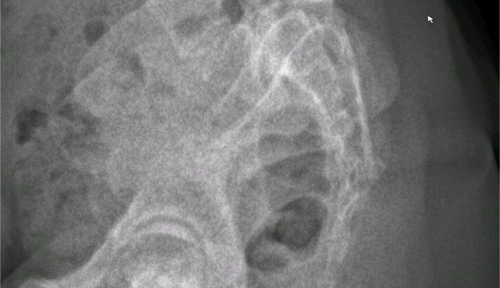
There aren’t any universally agreed-upon guidelines for imaging in this situation, but one common initial step is taking plain x-rays of the lower back region from three different angles: from the back, from the side, and from an angle. If an injury (often referred to as a “stress fracture”) is present, it usually shows up best in the angled view, where it looks like a collar on a dog-shaped part of the bone. However, it’s important to note that an x-ray might not be able to detect this injury within the first two weeks after it happens. What’s more, x-rays only spot this issue about 33% of the time.
Some experts champion CT scans as the go-to test for this condition. However, these scans do involve some radiation exposure, so doctors are often mindful about using them too frequently, especially in children, who are more likely to have this injury. If an x-ray doesn’t provide a clear diagnosis, doctors might order an MRI next. This type of scan is really good at detecting changes in bone marrow that can signal this kind of stress injury, as well as showing details about nerves and soft tissues. As an alternative to an MRI, a SPECT scan can also be effective in identifying acute stress fractures and determining how fresh the fracture is. However, like CT scans, SPECT scans come with some radiation risks, so they are preferably avoided in children.
Treatment Options for Spondylolysis
Most individuals diagnosed with spondylolysis, a type of back issue, including athletes, can be treated without surgery. In fact, those who don’t have symptoms but are diagnosed based on imaging scans can continue their normal physical activities without restrictions. However, if a patient is experiencing symptoms caused by spondylolysis, as confirmed by a specialized imaging scan called SPECT or MRI, treatments that don’t involve surgery are recommended. Here’s what they usually include:
One method is to wear a special back brace, aimed at limiting back movements and reducing stress on a specific part of the spine. However, recent medical data shows that regardless of whether or not they wear a brace, 83% of patients not opting for surgery experience improvement in their conditions.
It’s also very important to change one’s activities, especially stopping activities that involve overstretching the spine. As the pain subsides, athletic activities can be slowly started again.
Physiotherapy that focuses on enhancing stability of the spine is often advised. This usually starts with stretching exercises and includes techniques to strengthen abdominal and back muscles. The therapy takes a step-by-step approach to increase resistance over time for complex muscle training.
Additional treatments, including heat and cold therapy, anti-inflammatory drugs, epidural injections (an injection to relieve pain around the spinal area), massage, osteopathy or chiropractic adjustments and cognitive-behavioral therapy (CBT) are generally safe, beneficial and should be considered as part of the overall treatment plan.
With such non-surgical treatment practices, 75% adolescents improve, and the bone defects eventually heal. Bone defects occurring on one side of the vertebra are more likely to heal than defects on both sides. However, if a physical issue called spondylolisthesis is present along with spondylolysis, the bone defects are unlikely to heal even with conservative treatment. But these treatments often help the patients get relief from their symptoms and return to their previous level of activity. A recent study found that outcomes were similar for patients with spondylolisthesis whether they chose non-surgical or surgical treatments. As surgical treatments are expensive and could have potential side-effects, this is significant information. Moreover, there is currently no clinical evidence supporting the use of back bracing in preventing the spinal displacement among patients with spondylolysis and spondylolisthesis.
Surgery is typically used as a last resort. It’s only recommended if non-surgical treatments have been trialed for at least 6 months with no success, or if the patient has very serious symptoms like loss of sensation, problems with bowel or bladder function, unbearable pain, or develops a severe case of spondylolisthesis. The most common surgical procedures performed for spondylolysis involve direct repair of the defect in the spine or a spinal fusion surgery.
What else can Spondylolysis be?
Possible causes for back pain can be vast. They can range from muscle injuries to more serious conditions:
- Muscle strains and sprains
- Issues with the lumbar region due to degenerative disc disease, disc bulge, or herniation
- Narrowing of the spinal canal, also known as spinal canal stenosis
- Abscess or a collection of pus in the area surrounding the spinal cord
- Fractures of certain parts of the vertebrae
- Bone tumors such as osteoid sarcoma
- Fractures caused by weakened bones due to osteoporosis, cancer, infections, or other conditions
- A condition popularly known as “slipped vertebrae” in adults, also termed as degenerative spondylolisthesis
- Ankylosing spondylitis, a type of arthritis affecting the spine
What to expect with Spondylolysis
The outlook for individuals with a condition called spondylolysis is usually very good. This condition doesn’t require any specific treatments or lifestyle changes for people who don’t experience any symptoms. Even those who do have symptoms typically have a very positive outcome. A recent thorough review of multiple studies found that 92% of young athletes could go back to their sports after non-surgical treatment, and 90% were able to return after surgical intervention.
Possible Complications When Diagnosed with Spondylolysis
Most people with spondylolysis, a condition related to the spine, do not experience any symptoms throughout their lives. However, aging can lead to a related condition called degenerative disc disease, which might be more common and accelerate in those patients with spondylolysis. As a result, they could develop spinal stenosis, a condition where the spaces within your spine become narrow and cause issues with the nerves, and lumbar radiculopathies, which refers to the problems caused by the pinched nerves in the lower spine. These problems can occur in 50% to 75% of patients with spondylolysis, particularly when their vertebral body, a part of the spine, slips out of place.
If a surgical intervention is necessary, potential complications can occur, including an unsuccessful fusion of the spine bones, infections, constant pain, deteriorations of the nervous system, and failed back surgery syndrome, a chronic pain condition.
Potential Risk and Complications:
- Acceleration of degenerative disc disease due to aging
- Development of spinal stenosis
- Lumbar radiculopathies or problems with lower spine nerves
- Slippage of the vertebral body
- Unsuccessful fusion during surgery
- Infections associated with surgery
- Constant chronic pain
- Deterioration of the nervous system
- Failed back surgery syndrome
Recovery from Spondylolysis
It’s quite rare that someone would need surgery to fix spondylolysis, a condition where there’s a defect or fracture of one or both of the wing-shaped parts of a vertebra. If it does happen, there are several steps they should follow after surgery to aid in their recovery. These include:
1. Protecting the area where the surgery was performed until the wound has fully closed. The use of a brace to do this will depend on the surgeon’s preferences.
2. Proper pain relief. This could be in the form of medication, ice packs, or other methods to help keep the patient comfortable as they heal.
3. Gradual physical therapy. This means slowly increasing the amount and intensity of physical activity over time, to help regain strength and flexibility without causing injury.
4. Learning about proper way to move and hold their body, with a focus on maintaining good posture and how to properly position themselves when sleeping. This can help prevent reinjury and speed up recovery.

dysplasia of upper endplate of S1, Slip angle (SA = angle between inferior
endplate of L5 and line perpendicular to the S1 posterior wall) and lumbo sacral
angle (LSA = angle between the superior endplate of L5 and posterior wall of the
S1).
Preventing Spondylolysis
People who do not show symptoms, and whose disease is discovered by chance through medical scans, shouldn’t worry. This disease often doesn’t cause harm throughout a person’s life and does not require any changes to routine activities. However, those who are experiencing symptoms and are actively receiving treatment for a “pars interarticularis defect,” a kind of crack or fracture in a specific part of your spine, need to learn and understand specific movements to help with their comfort, reduce strain and facilitate the healing process. Examples of these techniques are:
* ‘Log-rolling’ when getting into and out of bed and moving the body in only one direction at a time during walking and shifting positions. This movement helps to avoid twisting and turning your body which could further hurt your spine.
* Using a lumbar roll while sitting, putting a pillow under the knees when lying flat on your back, and placing a pillow between the knees when lying on your side can help maintain a healthy position for your spine. This posture reduces strain on the affected spinal bone, supports healing, and maximizes the patient’s comfort.


