What is Stress Reaction and Fractures?
Stress injuries are a type of injury that range from inflammation of the membrane surrounding the bone to a complete break in the outer layer of a bone. These injuries are often seen in athletes due to repetitive strain on a bone, especially in those sports that involve running and jumping. They typically occur after a heightened volume or intensity of training, and the lower parts of the body, specifically, the legs, are the most common sites. However, despite being less frequently observed, they can also occur in the upper parts of the body, particularly the ulna, the larger bone in the forearm.
Certain types of athletes are more prone to fractures in particular regions. For instance, throwing and jumping athletes, as well as dancers, tend to be more prone to fractures in the first rib, whereas rowers usually experience stress fractures in ribs 4 through 9. Golfers, on the other hand, can end up with stress fractures in the back part of their ribs.
Pelvic stress fractures can mimic other causes of thigh and hip joint pain. These are usually seen in the two flat bones (ischiopubic ramus) and sacrum (triangle-shaped bone at the base of the spine). Runners are more susceptible to these injuries.
Hip joint fractures account for roughly 11% of stress injuries in athletes. These can be classified into tension-type fractures, including the top-outside of the joint and are at the highest risk for a complete fracture, and compression-type fractures, typically seen in younger athletes and involving the bottom-inside hip joint. For patients who do not show a visible fracture line on X-ray in the compression type injuries, it is possible to avoid surgery. This injury is prevalent in runners.
Stress fractures of the thighbone are represented 22.5% of all stress fractures in one military study. These individuals typically experience poorly localized, long-lasting pain in the leg, often mistaken for muscle injury. In these cases, if there’s no evidence of a bone break on imaging, a non-surgical approach can be attempted.
The kneecap is a rare location for a stress fracture. These types of injuries are either transverse or vertical, with the former having a higher risk of displacement, requiring immobilization.
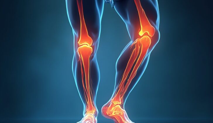
The shin bone is the most common site for stress reactions and fractures. It is sometimes challenging to differentiate between shin splints and shin fractures, as the pain associated with them can be similar. However, shin injuries cause pain during everyday activities, whereas shin splint pain is generally limited to physical activity. If the front part of the shin bone is affected, as seen in jumping athletes, it could lead to a nonunion (failure of bones to heal) and need aggressive therapy or potential surgery if that fails. Stress fractures of the top part of the shin bone are rare but can be confused for knee cartilage injury or inflammation of a little sac of fluid located in the shinbone, meaning doctors need to be suspicious.
Fractures in the smaller leg bone (fibula) are common and are often situated in its lower third. Patients will generally feel pain when the affected part is touched.
Fractures in the bump on the inside of your ankle (medial malleolus) are less frequent and can develop in athletes participating in running and jumping activities. If a full break is identified, surgical fixation is usually required.
Heel bone fractures are identified as localized tenderness over the heel of the foot posterior to the ankle bone. Patients will have a positive squeeze test.
Stress fractures can occur in the navicular, medial cuneiform (bones in the middle of the foot), and lateral process of the ankle bone. Navicular stress fractures are hard to diagnose early and are at a high risk of non-union due to poor blood flow, particularly in its middle third. These often occur in basketball players and runners.
Toe bone fractures in athletes represent 9% of all stress fractures, especially affecting the second and third toes. They tend to be tender with localized swelling over the affected bone. A particular type of toe bone fracture called a “dancer’s fracture” occurs at the base of the second toe. It’s important to distinguish these fractures from acute ‘Jones fractures’ which occur in the part of the toe bone near the small toe.
Injuries to the pair of tiny bones embedded within the tendons of the big toe often show up as gradual pain on the underside of the foot, with usually the inside bone being the most frequently affected. Direct tenderness or pain with passive extension of the toe aids in diagnosis.
What Causes Stress Reaction and Fractures?
Stress fractures, which are partial or total breaks in a bone, can happen due to repeated physical stress. This is kind of like how buildings or bridges can develop cracks from constant use. Normally, such stress doesn’t cause a fracture, but if the bone doesn’t get enough rest time to heal, stress fractures can occur. This could be due to the muscle forces acting on the bone or because the structures supporting the bone get tired. Likely, both of these play a part.
Stress fractures can be classified as high or low risk. High risk means the fracture is likely to get worse or not heal on its own, which may require surgery. High-risk areas include the heel, ankle, foot bones, shin, knee, hip, and lower spine. Low-risk injuries can occur in the pelvic bone, backbone, ribs, upper arm, shin, lower leg, and foot bones
If you’re not getting enough energy from your food or you’re low on vitamin D, this can increase your risk of stress fractures. The risk is also higher for female athletes who have eating disorders, irregular periods, and weak bones. Other factors that increase risk include starting your periods late, having bone disorders like osteomalacia and Paget’s disease, and high cortisol levels in your blood. Stress fractures can also occur if you suddenly increase your exercise intensity or switch to a different form of exercise without giving your body enough time to adjust. Not taking enough rest days in between intense workouts can also lead to injury.
Stress fractures often happen when someone suddenly changes their training routine. They’re common in runners and military recruits. For runners, this could mean increasing their training intensity, changing their shoes, or running on a different surface. In the military, these injuries often occur when recruits start basic training or increase the distance covered during their runs or marches.
There are also certain physical characteristics that can increase someone’s risk of stress fractures, but these are harder to study. For example, military recruits with narrow shinbones and greater external rotation were found to be at greater risk of stress fractures. Similarly, female runners with stress fractures were found to have thinner calves and less muscle mass in their lower limbs. Also, runners with stress fractures tend to have smaller cross-section of their shinbones than runners without stress fractures, suggesting that the shape of the bone plays a role in the development of stress fractures.
Risk Factors and Frequency for Stress Reaction and Fractures
Stress injuries account for up to 20% of all injuries seen in sports medicine clinics. They are so prevalent among military recruits that they are the primary reason for lost training days. In high school athletes, around 0.8% of all injuries are stress fractures, or cracks in the bone due to repeated stress. These fractures are most common in cross-country athletes, with a rate of 1.54 per 100,000 experiences. The majority of these injuries occur in the lower leg (40.3%) and foot (34.9%). Also, elite soccer players experience an incidence rate of 0.04 injuries per 1000 hours, most of which impact the lower part of the body, particularly the fifth metatarsal or a long bone in the foot. In US Army recruits, the rate of stress fractures is 19.3, with 79.9 cases per 1000 recruits.
Weight-bearing limbs, or those that support the body’s weight, suffer from stress fractures more frequently than non-weight-bearing limbs. This type of fracture often affects the tibia (shin bone), metatarsals (foot bones), and fibula (smaller lower leg bone). One of the most common forms of early stress injury is medial tibial stress syndrome, commonly known as shin splints, which is pain along the inner edge of the shin bone.
The sport an individual participates in can influence the location of stress injuries. For example, track athletes often get fractures in the navicular bone (in the foot), tibia, and metatarsals. Distance runners often fracture their tibia and fibula. Dancers frequently experience stress injuries in their metatarsals, while military recruits often fracture their calcaneus (heel bone) and metatarsals. When it comes to the upper extremity, arms and hands, the ulna (inner forearm bone) is most often affected.
Signs and Symptoms of Stress Reaction and Fractures
When someone has a stress injury from exercise, it can start with a vague pain that doesn’t seem connected to any particular injury or accident. They might have noticed it after doing a lot of one kind of exercise, like running, or after they increased how hard or how often they were training. A change in the surface they were training on, like switching from a treadmill to the road, might also set it off. At first, the pain might only come on, or get worse, when they’re exercising. But if it gets more severe, they might start to feel it during their everyday activities, too. If they stop exercising, the pain often gets better.
During a medical exam, the doctor will check the area where the patient feels pain. They might find that it’s tender or swollen. When the tender area is in the soft tissues (the muscles, tendons, and ligaments), it might mean there’s a muscle injury or a stress reaction, which is the earliest stage of a stress fracture. If the bone is tender, it might point to a stress fracture. Sometimes, the area of the stress injury is hard to check over, like the pelvis and the sacrum (the lowest part of the back, located between the hips). In these cases, the doctor might rely more on the patient’s account of their symptoms.
Here are a couple of tests doctors might use to help diagnose stress injuries:
- One Leg Hop Test: This test is used to tell the difference between shin splints (medial tibial stress syndrome) and a stress fracture in the shin bone (tibia). Those with a stress injury can hop repeatedly without pain, whereas those with a stress fracture in the shin bone will feel pain during the hop test. The pain is often felt when landing.
- Fulcrum Test: This test can help diagnose a stress fracture in the femur (the thigh bone). The doctor’s arm acts as a lever under the patient’s thigh, while pressure is applied to the knee. If the test is positive, the patient will feel pain or apprehension where the fulcrum (the lever) is located.
Testing for Stress Reaction and Fractures
If a doctor suspects you may have a stress injury, such as a fracture, they’ll first take an X-ray of the affected area. However, X-rays taken in the first few weeks after the injury are likely to appear normal and therefore, aren’t very sensitive in detecting stress injuries. Changes your doctor would be looking for to confirm a stress injury include an elevated bone surface, hardened areas of bone tissue, increased thickness of the bone’s outer layer, and possible fracture lines. A decrease in bone density could also indicate an early stress injury. Specific warning signs could also be seen, such as a ‘dreaded black line’ which can be an indicator of a stressful fracture in the shin or thighbone.
If the X-ray images do not reveal any stress reactions or fractures, then your doctor might recommend repeating the x-rays, as well as getting Computed Tomography (CT) scans, Magnetic Resonance Imaging (MRI), or bone scans.
A CT scan can find similar signs to an X-ray, such as hardened areas of bone tissue, new bone formation, an elevated bone surface, fracture lines, especially in long bones. CT scans are particularly useful for ruling out other possible causes of your pain if the diagnosis is unclear from the other imaging methods.
Bone scans use radioactive substances to visualize bones on a scan. While this method can successfully detect stress injuries 74% of the time, they do have limitations. These scans can also light up for reasons other than stress fractures (like bone death, bone infections, cancer, etc.), making it less specific in identifying stress fractures.
The most effective way to diagnose stress injuries is through MRI scans. They have an approximately 88% success rate in identifying stress injuries and are becoming the gold standard for diagnosing these kinds of injuries. In addition to finding the fracture line usually extending through the bone’s outer layer into the bone marrow, the MRI can detect swelling in the surrounding bone. Importantly, MRI scans can also help check for any other possible injuries including damage to muscles, ligaments, and cartilage.
Treatment Options for Stress Reaction and Fractures
The treatment for stress injuries such as stress reactions or fractures depends on the severity and location of the injury, and the person’s needs for recovery. Identifying these injuries early is beneficial as it can speed up the healing process. High-risk stress fractures may be treated conservatively or with surgery, based on the individual’s work or sports involvement.
Generally, stress injuries at low-risk sites are treated conservatively in two phases. The first phase includes pain relief, changing weight bearing and activity adjustments, like stopping any activities that may worsen the injury. If the patient has pain when walking, temporary immobilization may be recommended. Ways to stay fit and strong during this phase can include water fitness, cycling, or using an elliptical machine. The second phase begins after a period of rest with no pain and involves a gradual return to activity over a few weeks, with continued therapy. For example, a runner might start by running slower and shorter distances every second day, and then gradually increase distance, frequency, and speed over time, aiming to return to their normal levels. The length of each phase can vary.
For athletes who have feet that roll inward or outward excessively, orthotics could be beneficial. Changing or addressing footwear can also help those with inadequate shock absorption. Running shoes, for example, should be replaced every 300 to 350 miles. Medical professionals who specialize in running may be able to conduct gait analyses and suggest changes to the form that can reduce the risk of injury again.
Female runners who started their menstrual cycles late, have fewer periods, or have lower bone mineral density, are at a higher risk of stress fracture. If these risk factors are present, a bone mineral density test and hormone check-up may be suggested. Vitamin D and calcium supplementation is generally not needed unless the diet is inadequate. Athletes with repeated stress fractures may need their vitamin D and calcium levels checked, supplements may be recommended if these are low. Athletes with eating disorders should get evaluated, and if needed, undergo mental health testing and nutritional counseling. Certain medicines that help improve bone mineral density are showing early promise for treating stress fractures, but more research is needed.
Specific types of stress fractures may need different management approaches. For instance, rest is often the treatment for rib stress injuries, with changes in training or techniques that could be contributing to the injury. Pelvic stress fractures typically require rest, crutches if necessary, and a slow return to the sport. Stress fractures in the femoral neck, the part of the thigh bone that joins with the hip, may need non-weight bearing and activity restrictions, or surgical intervention depending on severity and location. Most stress fractures in the femoral shaft, the long part of the thigh bone, can be managed conservatively, while some may need surgical correction, especially in older adults, individuals with low bone mineral density or more severe fractures. Patellar stress injuries typically require immobilization and a slow return to activity.
Different types of tibial shaft stress injuries may also be managed conservatively, but some may require surgical correction if the injury is severe, persistent, or associated with particular features. Fibular stress injuries can often be treated with rest, immobilization, activity changes, and a gradual return to play. Calcaneal stress injuries usually respond well to conservative management with quick healing and return to activity. Other bones in the foot such as the navicular, medial cuneiform, and some parts of the talus can also be managed conservatively. On the other hand, fractures in certain metatarsals and sesamoid bones may require additional measures such as padding, non-weight bearing, immobilization, and possible surgery.
What else can Stress Reaction and Fractures be?
Doctors diagnosing stress fractures or reactions must consider a range of possible causes depending on the area affected. The list of considerations can be wide and diverse. This can include different types of bone and joint issues, including inflammation of the bursa (bursitis), ligaments (tendonitis), muscle strains, or deteriorating joints. Moreover, bone bruising, a lack of blood supply to the bone, bone growths (osteosarcoma), or infections in the bone (osteomyelitis) can also be considered.
Other conditions that aren’t related to the bone or muscles must also be thought of, depending on the location of the injury. These could include skin, blood vessel or nerve issues, or even issues related to reproductive organs or the digestive system. For instance, stress injuries in the pelvic area or the upper thigh bone could cause pain in the pelvic region, the hip, thigh, or groin. In this case, the doctor must consider all the medical possibilities related to these areas. Similarly, stress fractures in the fibula, a leg bone, will cause leg pain and physician needs to think about all the medical conditions related to the leg.
In the case of tibial stress injuries, which are injuries to the shin bone, doctors may consider inflammation of the outer layer of the bone, more serious stress fractures or conditions related to overexertion or arteries trapped in the knee area.
What to expect with Stress Reaction and Fractures
With the right amount of rest and rehabilitation, most athletes can get back to their sports with little pain and normal functioning. However, if they rush back into activity or don’t get enough rehabilitation, the pain could become chronic and lead to long-term injury. Taking enough rest, immobilizing the injury and not putting weight on it when necessary, and slowly getting back into activity should help athletes get back to their original playing level. The main problem with stress injuries is the amount of playing time that gets missed.
Possible Complications When Diagnosed with Stress Reaction and Fractures
Complications from stress injuries can be immediate or long-term. Immediate complications include things like pain, swelling, and the inability to continue playing. If the stress injury is severe enough to cause a full break in the cortical bone, surgery may be needed, which carries its own risks. On the other hand, long-term complications can include chronic pain, the inability to return to the original level of play, and the recurrence or repetition of stress fractures.
These complications can be summarized as:
- Pain
- Swelling
- Missed playing time
- Potential need for surgery with all associated risks
- Chronic pain
- Inability to return to the original level of play
- Repeat or recurrent stress fractures
Preventing Stress Reaction and Fractures
Almost all injuries caused by stress are due to excessive use or intense training without enough time to recover. So, to prevent these types of injuries, it’s important to focus on proper body movements, wearing the right shoes, training on suitable surfaces, and increasing the intensity of training in a gradual manner. Mixing in different types of exercises, also known as cross-training, can also reduce the chances of getting stress injuries. It’s also crucial to maintain a healthy diet and ensure enough time for recovery for prevention.


