What is Swan-Neck Deformity?
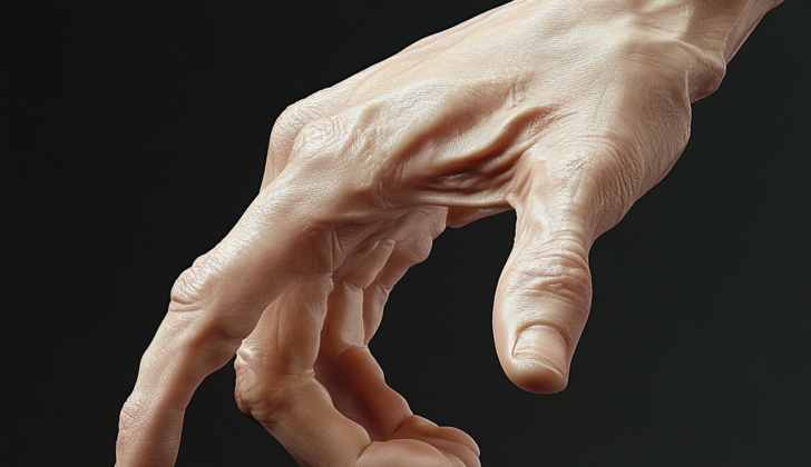
Swan neck deformity is a condition where your finger abnormally bends – it’s hyperextended at the first knuckle from the tip (proximal interphalangeal, or PIP joint) but flexes or bend at the tip knuckle (distal interphalangeal, or DIP joint). There’s also notice of the same bending at the base of the finger (the metacarpophalangeal, or MCP joint). This happens due to an imbalance in the finger’s extensor mechanism – the parts of your finger that help it straighten.
The cause of this imbalance could be a major cut or stretching of the outside tendons (extensive tendon forces) that support the end phalanx, which is the tip of your finger, or it could be due to tightness and pulling of the extensor mechanism both within and outside the finger at the first knuckle from the tip (PIP joint).
What Causes Swan-Neck Deformity?
Swan neck deformity is a condition that results when the extension mechanism of a finger joint becomes impaired. This could be because it’s too loose or too tight at either the distal or proximal phalanx, which are the different segments of your finger.
Initially, swan neck deformity might happen because of a loss of the extensor tendon, the structure that helps extend or straighten your finger, at the distal phalanx. This usually happens over time and develops into the classic form of the condition. This kind of injury can happen through:
* A severe cut to the extensor mechanism
* Closure of the avulsion (where a piece of bone is torn away) after a direct hit on the distal phalanx of a straightened finger
* Gradual weakening due to long-term inflammation at the DIP joint (the last joint of a finger), along with dislocation of the extensor tendon due to rheumatoid arthritis or any other inflammation-related condition.
The PIP joint (the middle joint of a finger) can stick out too much due to the tightness of the muscles inside the finger and increased pull at the central slip, which is a part of the extensor mechanism. This could be a result of rheumatoid arthritis, extreme reflex reactions from traumatic brain injury or stroke, or just tightness of these muscles alone.
Lastly, looseness of the volar plate, a thick ligament that stops the PIP joint from extending too much, can also cause swan neck deformity and the PIP joint to extend too much.
Risk Factors and Frequency for Swan-Neck Deformity
Swan neck deformity, which can be caused by a traumatic event, does not occur more frequently in any particular age group or gender. However, if swan neck deformity is due to rheumatoid arthritis, it is seen more commonly in women than in men.
Signs and Symptoms of Swan-Neck Deformity
Swan neck deformity, often associated with conditions like rheumatoid arthritis or caused by a certain injury, can be identified during an examination. Visually, the finger will show evident bend at the base and tip, but will be straight in the middle joint. In the early stages, this middle joint, referred to as the Proximal Interphalangeal (PIP) joint, can still move quite a bit. But as the condition progresses, it may stiffen and become unmovable. In testing the joints, a doctor will look at both active and passive motion, and may use a specific test to ascertain if the problem is due to a tight joint lining or tightened muscles.
During a physical exam:
- Doctors will evaluate your medical history, specifically instances of inflammatory diseases or injuries to the extensor mechanism.
- The affected digit will display a noticeable swan neck deformity, characterized by hyperextension at the PIP joint and flexion at the Distal Interphalangeal (DIP) joint.
- The movement of the finger’s three major joints (MCP, PIP, and DIP) will be assessed.
- In the early stages, you may still have a good deal of active or passive movement in the PIP joint – this can change as the deformity progresses.
- You may use your other hand to flex the PIP joint out of hyperextension – once it’s no longer hyperextended, you can still actively flex the joint in the early stages.
- In later stages the PIP may stiffen and become unmovably hyperextended; this can result in further joint pain and loss of motion.
- Your doctor may perform the Finochietto-Bunnell test to help pinpoint whether the issue is due to a capsular restriction or intrinsic muscle tightness.
Testing for Swan-Neck Deformity
Along with tests to check for rheumatoid arthritis, doctors also use X-rays. These help them see if there’s any severe damage to your joints, like a serious case of arthritis, or if the small bones in your wrist (the carpal bones) have collapsed. This is especially important in the later stages of the disease.
Treatment Options for Swan-Neck Deformity
Conservative treatment, which means non-surgical treatment, may involve hand therapy for gentle stretching and the use of corrective splints. These methods can be beneficial even for chronic conditions, potentially increasing mobility and flexibility in the joints of the fingers. One type of splint, called an extension block splint, can help address excessive straightening of the finger joint. Another type, known as progressive extension splinting, can assist with bending deformities of the fingertip joint. However, it’s important to note that while these methods can improve movement, severe or long-term deformities are unlikely to be corrected fully without surgery.
The more advanced stages of swan neck deformity, a condition where a finger develops a distinctive curved shape, may require surgical treatment. In these cases, if the patient can still move their finger joint without assistance, a structure called the volar plate needs to be recreated during surgery. This procedure is done to prevent excessive straightening of the finger joint. Sometimes, a technique called FDS tenodesis is used for this purpose. However, if the side structures of the finger joint have become lax, a different surgical technique known as conjoint lateral band tenodesis may be needed.
If the finger joint is excessively straightened, it can be corrected by lengthening a structure called the central slip. This can also be achieved by using an FDS tenodesis or performing a dermodesis, which involves removing a small oval shaped area of skin on the side of the joint facing the palm. If there’s also excessive tightness in the muscles contributing to the finger deformity, a surgical release of these muscles may be done.
In later stages, if there’s evidence of joint decay, joint fusion or replacement may be necessary. These are additional to the soft tissue procedures that would still be needed. If the joints at the base of the fingers are also slipping out of place due to severe degenerative arthritis, these joints may also need to be addressed surgically, possibly involving joint replacement.
What else can Swan-Neck Deformity be?
It’s easy to confuse one hand condition called Boutonniere deformity with another called Swan Neck deformity. In Boutonniere deformity, the end joint of a finger (known medically as the DIP) is overly stretched, while the middle joint (the PIP) is bent. On the other hand, Swan Neck deformity is just the opposite. Recognizing these differences is important.
Possible Complications When Diagnosed with Swan-Neck Deformity
Complications from the surgery might involve not fully fixing the deformity, the deformity coming back, stiff PIP joints, and not being able to fully extend the DIP joint because of too much correction.
Potential Complications:
- Not fully fixing the deformity
- Deformity coming back
- Stiff PIP joints
- Not being able to fully extend the DIP joint due to too much correction
Recovery from Swan-Neck Deformity
After a medical procedure, the patient’s hand is typically secured and kept still by a hard plaster splint. The MCP (metacarpophalangeal) joints, which are the large knuckles where the fingers meet the hand, should be fully straightened, while the PIP (proximal interphalangeal) joints, which are the middle knuckles on the fingers, should be bent.
If the patient has undergone a flexor tenosynovectomy, a surgery to remove inflamed tissue around the tendons of the hand, they should start flexing exercises within 4 to 5 days. These gentle movements help the hand regain strength and flexibility.
A dorsal extension block, a device to limit the extension (straightening) of the fingers, should be used for 4 to 6 weeks after surgery. This helps to protect the operated area, while the healing process takes place.


