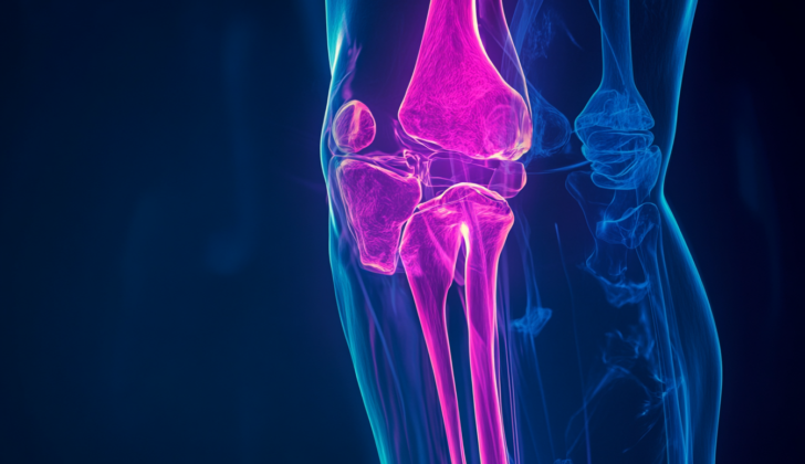
What is Tibia Nonunion?
The healing of broken bones needs a careful balance of natural processes and stabilization during recovery. There are four key components for good bone healing: mechanics (the physical structure and support), osteogenic cells (cells that create new bone), scaffolds (structures that support the growth of new tissues), and growth factors (substances that aid in cell growth).
Sometimes, things do not go as planned, and the bone doesn’t heal correctly, requiring additional medical help. Though different studies may have varying definitions, generally, if there’s no sign of the healing process progressing for 3 months, or if no healing has occurred after 9 months from the injury, we can say that the healing is not happening as expected.
This condition can seriously impact a patient’s quality of life, making life as difficult as living with advanced hip arthritis and even worse than living with congestive heart failure. Besides, it also significantly increases the overall cost of care, often doubling the costs associated with fractures that heal properly.
Despite having a variety of treatment options available, treating non-healing fractures in the shinbone (tibial nonunions) remains a difficult task for doctors.
What Causes Tibia Nonunion?
The “diamond concept” is a term coined by a group of scientists led by Giannoudis. It was created to explain the elements necessary for a bone fracture to heal properly. The idea places emphasis on three biological elements: cells that create bone (osteogenic cells), structures that support these cells (osteoconductive scaffolds), and substances that stimulate growth (growth factors). In addition to these biological elements, a fourth factor, mechanical stabilization (keeping the bone steady), is also important. If any of these factors are influenced negatively, the healing of the bone fracture could be at risk.
Risk Factors and Frequency for Tibia Nonunion
Each year in the United States, about 6 million people suffer from fractures. Among these, between 1.9% to 15% don’t properly heal, a condition known as nonunion. The likelihood of this happening varies based on the specific bone that’s broken and certain factors about the fracture itself, such as whether it’s an open or closed fracture.
- About 6 million fractures occur annually in the United States.
- Nonunion, when the bone doesn’t heal properly, occurs in 1.9% to 15% of these cases.
- The risk of nonunion varies depending on the specific bone and features of the fracture.
- For example, tibial (shin bone) fractures have a nonunion rate of about 1.1% for non-surgical treatment and almost 5% for surgical treatment.
- If the fracture is open (where the bone has pierced the skin), the risk of nonunion significantly increases.
Signs and Symptoms of Tibia Nonunion
If there’s a suspicion that a person might have a non-healing bone fracture, called a ‘nonunion,’ doctors will start by getting to know their full medical history. They’ll identify any potential risk factors, and will also gather information about the type of fracture they had, how it was treated, and any past treatments they’ve undergone. People with nonunion fractures often continue to feel pain at the site of the fracture and may also have difficulty moving.
- Full past medical history
- Type of fracture
- Fixation method and past treatments
- Continuous pain at the fracture site
- Functional limitations
Another helpful indicator that a nonunion fracture is not present is if the patient doesn’t feel pain when walking or using the affected limb. The doctor will also carry out a physical exam to check the nervous system and blood supply status and range of motion. They’ll pay special attention to any signs of infection like changes in skin or soft tissue, and to any movement at the fracture site, as this could signal that the bone healing process, known as ‘callus formation,’ isn’t adequate.
- No pain when walking or using the affected limb
- Neurovascular status and range of motion
- Signs of infection
- Mobility at the fracture site
Testing for Tibia Nonunion
Diagnosing septic (infected) nonunion versus aseptic (non-infected) nonunion can be tough. It’s crucial though, because the treatment plan changes a lot depending on if there’s an infection or not. This is something important your doctor has to consider when evaluating nonunion cases – where a broken bone fails to heal.
For patients suspected with nonunion, there are usually some initial steps of evaluation:
As part of the examination, your doctor will look at x-ray pictures taken from different angles, including you standing up. They’ll be checking for signs of healing or the lack of it. They’re also going to keep an eye out for any deformities, the quality of your bone and any signs of infection. Furthermore, they’ll pay attention to how well the broken ends of the bone are lining up and how stable they are. Comparing current and past x-rays can help them understand how the nonunion has progressed over time.
If the x-rays aren’t clear because of medical devices obstructing the view or the bone pattern, your doctor may advise a CT scan. This provides a more detailed look at the fracture and how it’s positioned. Another test – known as a bone scan could be used to study bone metabolism, and give clues about how the bone is healing.
Some blood tests might be helpful too. By looking at a complete blood count, an inflammation marker called C-reactive protein, and your erythrocyte sedimentation rate – which is another inflammation gauge – your doctor may be able to rule out an infection. But elevated inflammation markers on their own are not always definitive as they can rise for many reasons. CRP can be a good signal of infection in cases of suspected nonunion. It usually increases within 6 hours of an infection starting or when there’s a tissue injury, and reaches the peak between 48 and 72 hours, finally returning to normal around 5 to 21 days after the infection is treated. Checks for certain metabolic and hormonal diseases can also help show why the nonunion may have occurred.
To positively identify a septic nonunion, a tissue sample, usually taken from an open biopsy, needs to be evaluated in the lab for any presence of microbes. Swabs are generally not reliable, as they can often accidentally pick up other bacteria in the process.
Nonunion can be classified based on its cause into various types:
– Septic (infected) or aseptic: Septic non-union in the lower leg (tibia) bone, for instance, can be usually diagnosed if there’s an open wound, exposed bone, or loosened implants.
– Pseudarthrosis, a type of nonunion where the bone attempts to heal but fails.
– Hypertrophic type involves a well-supplied blood flow, but inadequate immobilization.
– Atrophic characterizes where there is both inadequate immobilization and blood supply early in the healing process.
– Oligotrophic category involves cases where the broken bone ends have not been aligned well resulting in lack of bone healing or callus formation.
Treatment Options for Tibia Nonunion
The goal of treating a non-healing bone fracture, known as a nonunion, is to allow the bone to heal while keeping it functional. There are several treatment options, and the choice depends on the individual patient’s situation. Here are some of these options:
Non-surgical Treatments:
– Conservative treatment/weight-bearing: Some people, usually older people who can’t have surgery, may be treated just by weight-bearing and watchful waiting. This can also be combined with other treatments such as bone excision.
– Electrical stimulator/electromagnetic fields: Small electric and electromagnetic fields can stimulate the production of growth factors. These growth factors can help the bone heal.
– Ultrasound (low-intensity pulsed ultrasound (LIPUS)): Low wave ultrasounds can promote bone healing by increasing the activity of bone-forming cells (osteoblasts).
– Injection of bone marrow: This involves injecting bone marrow fluid into the patient.
– Extracorporeal shock wave therapy: High-energy sound waves can be as effective as surgery for stable nonunions that aren’t healing.
Surgical Treatments:
Surgical strategy for non-healing tibia (shin bone) depends on the absence or presence of infection. The patient’s condition, the exact location of non-union, imaging results, and the presence of bone deformation also matters. The following are types of surgical procedures:
– Single stage approach: Used to treat a non-infected nonunion, it involves cleansing the fracture of dead tissue, then using surgical methods like open reduction and internal fixation (putting the bones in their original position and using screws and plates), exchange nailing (replacement of bone-stabilizing rod), etc.
– Staged approach: Used in infected tibial nonunion, it is a two-step process comprising firstly infection control and secondly definitive surgery.
Additional surgical techniques mentioned in medical literature include:
– Nail dynamization: Helps in healing after the bone is stabilized by removing the interlocking bolts from distances of the fracture site.
– Nail exchange: Involves removal of the prior nail used to stabilize the bone, reaming the bone to stimulate healing and using a larger nail.
– Fibular Osteotomy: Procedure used to correct alignment of the leg due to nonunion.
– Open Reduction and Internal Fixing with plate and screws: Used when intramedullary nailing (stabilizing bone with a rod) is not possible or not recommended.
– Locking compression plates: Helpful when the breakpoint is near the joint or part of bone closer to the middle.
– External fixation: Helpful in complex nonunions where rod insertion isn’t possible.
– Bone grafting: Bone from another part of the body is used to encourage healing.
Sometimes, more novel approaches, like cell therapy can be used. This therapy uses special cells, called mesenchymal cells, to create a healing environment in the bone. Very rarely, amputation might be considered if a functioning leg cannot be achieved.
What else can Tibia Nonunion be?
A ‘delayed union’ is a term used to describe a broken bone that is healing slower than usual and generally needs some kind of treatment to help it heal.
What to expect with Tibia Nonunion
The chances of recovery from tibial (shin bone) fractures can be evaluated using something called the RUST score, which involves examining the entire fracture to check for signs of healing, like disappearance of the fracture line or the presence of new bone growth (known as a bone callus).
The Nonunion Risk Determination (NURD) score is another method that doctors use. This considers the RUST scores alongside some other factors. These can include whether the fracture was open or closed, if there’s a condition called compartment syndrome, how severe the fracture is (like a IIIB tibia), the presence of ongoing illnesses, whether the patient smokes, if there’s an infection, the patient’s gender, and their general health status as measured by the American Society of Anesthesiologists (ASA) score. This model helps doctors identify those patients who might have a longer period of recovery (nonunion) within three months of injury.
According to studies, the average time it takes for healing of aseptic tibial nonunion (where the bone isn’t healing as expected, but there’s no infection) when treated with reaming and exchange nailing (kinds of surgery) ranges from 5 to 9 months./p>
The percentage of patients who experienced healing, or “union,” with this treatment varies and is reported to be between 72% and 100%.


