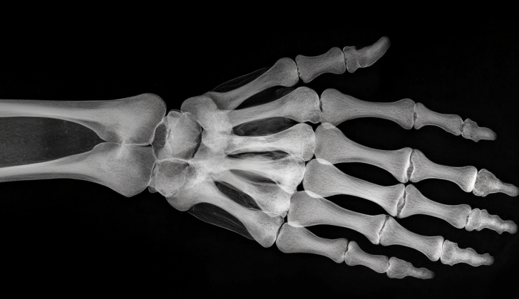
What is Triangular Fibrocartilage Complex?
The triangular fibrocartilage complex (TFCC) is a part of your wrist that helps to stabilize it. It’s located between three small bones in your wrist: the lunate, triquetrum, and ulnar head. Sometimes, the TFCC can get injured due to sudden trauma or wear and tear over time. Certain movements, like moving your wrist in the direction of your little finger with a lot of force, can increase the risk of getting these injuries.
If your TFCC is injured, you might feel pain on the side of your wrist closer to your little finger. This pain might come with a clicking sound or feeling, or you might be able to pinpoint a specific area of tenderness between the ‘pisiform’ (a small wrist bone) and the bottom of your ulna. A doctor often uses an MRI scan, a special kind of imaging technique, as an initial method to check for TFCC injuries. However, arthroscopy, a procedure where a tiny camera is inserted into your wrist joint, is the most accurate way to diagnose these injuries.
Treatment for TFCC injuries can range from non-invasive options to surgery. Non-invasive treatments could involve resting your wrist, over-the-counter anti-inflammatory medications like NSAIDs, and injections of corticosteroids (medicine to reduce inflammation). If these don’t work or the injury is severe, your doctor may recommend a surgical procedure.
What Causes Triangular Fibrocartilage Complex?
An injury to the TFCC (Triangular fibrocartilage complex), a part of your wrist, often happens when pressure is put on it while your wrist is bending towards your little finger – a movement known as ulnar deviation. You can experience this kind of movement when you swing a racket or a bat, for example.
TFCC injury can also be related to something called positive ulnar variance, which means that the side of your ulna bone (one of two bones in the forearm) furthest from the body is lower down than the corresponding side of the radius bone (the other bone in your forearm). This condition usually happens after surgery or after breaking a bone.
Risk Factors and Frequency for Triangular Fibrocartilage Complex
Research has shown that injuries to the TFCC (triangular fibrocartilage complex), a part of the wrist, are more common in older age groups. One study found these injuries in 49% of people aged 70 and above, while only 27% of people 30 years old or younger were affected. Another study found similar rates among the under-30s, but also showed that TFCC injuries were just as common among people who had wrist scanning for reasons other than pain on the ulnar side (the side of the wrist closest to the pinky finger). This indicates that not all TFCC injuries result in ulnar-sided pain.
- TFCC injuries are more frequent in older age groups.
- One study showed a 49% prevalence in those aged 70 or older, and a 27% prevalence in those 30 or younger.
- Another study confirmed these rates in under-30s, but also found TFCC injuries among people getting wrist scans for other reasons, not just ulnar-sided pain.
- This suggests that not every TFCC injury results in pain on the ulnar side of the wrist.
Signs and Symptoms of Triangular Fibrocartilage Complex
Patients with TFCC (Triangular Fibrocartilage Complex) injury often experience wrist pain on the side of their little finger, especially during physical activity. Other symptoms may include a weakened grip, instability in the wrist joint, or a clicking sensation. Certain sports activities can lead to specific injuries. For example, in baseball, a sudden wrist injury might arise from forcefully stretching the wrist during a head-first slide or when a batter tries to hit a close-range pitch and gets jammed. Chronic injuries can happen over time due to the high stress placed on a baseball player’s wrist during batting. Even players with perfect wrist structures can suffer from TFCC injuries.
Patients with instability in their distal radioulnar joint (the joint between the two bones of the forearm) may have weakness when turning their wrist up and down – this weakness can also be a sign of a TFCC injury.
The TFCC can best be felt with the wrist turned towards the little finger side. It’s located between the flexor carpi ulnaris (one of the muscles of the forearm), the ulnar styloid (a prominent bone bump on the little finger side of the wrist), and the os pisiform (a small wrist bone). Various physical tests can suggest a TFCC injury:
- TFCC compression test: Patient’s symptoms get worse when the forearm is in a neutral position and the wrist flexes towards their little finger side.
- TFCC stress test: Symptoms occur when a force is applied across the length of the forearm while the wrist is flexed towards the little finger side.
- Press test: Patient shows pain when lifting themselves from a seated position using their wrists in an extended position.
- Supination test: When patient holds the underside of a table with their palms up, which loads the TFCC and causes a tearing sensation on the back of the wrist if there’s an injury.
- Piano key test: With palms pressed on the table, if the bone on the little finger side of the wrist is more visible on the affected hand, it indicates possible distal radioulnar joint instability, often associated with a TFCC injury. If relaxing the palms causes this bone to return to normal position, this confirms the test.
- Grind test: Pain experienced when the two forearm bones are pressed together and the forearm is rotated could signify a degenerative process.
Testing for Triangular Fibrocartilage Complex
If you’re suspected of having a Triangular Fibrocartilage Complex (TFCC) injury, which are injuries to the cartilage and ligaments located on the outside, or ulnar side, of your wrist, your doctor will first order an X-ray to check for any fractures and assess your specific wrist anatomy. If further tests are needed, the next step is usually to get an MRI.
An MRI is a scanning technique that uses magnetic fields and radio waves to create detailed pictures of the inside of the body. Sometimes a special dye (arthrogram) is used to further enhance the images, but it might not be necessary since it can cause more discomfort and adds to the cost of the test. If an MRI isn’t an option, a CT scan can be used, although it may not be as sensitive.
The most accurate method to diagnose a TFCC injury is through a procedure called arthroscopy, which is a minimally invasive surgical procedure that allows the doctor to see inside your joints using a small camera.
It’s important to figure out if the lunotriquetral ligament, which is one of the ligaments in your wrist, is intact or torn to guide the proper treatment. This can be determined by looking at X-ray images for specific signs or by direct visualization during arthroscopy.
Studies have shown that over half of the people who needed surgery for fractures on the radius bone, one of the two bones in your forearm, also had a TFCC injury confirmed by arthroscopy. However, initial X-ray images of a radius fracture can’t predict the presence of a TFCC injury, so additional tests are typically needed.
Treatment Options for Triangular Fibrocartilage Complex
If you have a ligament injury in your wrist known as a TFCC tear, the first steps for treatment typically include rest, physical therapy, and injections to reduce inflammation – what doctors often refer to as “conservative treatment.” If there’s no instability in the wrist joint, trying these methods for about six months before considering surgery may be a good idea.
There’s limited evidence that wearing a brace can help treat TFCC tears. In one case, a patient who had not improved with conservative management wore a brace for 12 weeks instead of having surgery. This patient was able to use their upper arm more immediately after taking off the brace, and improvements remained after a year. More research needs to be done on the effectiveness of using braces to treat TFCC.
If conservative treatments aren’t working or if the wrist joint is unstable, surgery could be considered. Various surgical procedures are available, including using a tiny camera to make small repairs (arthroscopic repair), removing damaged tissue to stimulate healing (debridement), shortening the ulna bone in the forearm (ulnar shortening), and a type of surgery known as the Wafer procedure.
Which treatment option is best will depend on the classification of the injury, for example:
- 1A: This type of injury is in a region with little blood supply, which doesn’t heal well. In such cases, debridement is usually the best choice.
- 1B: This area has adequate blood supply, so direct repair is an option. If the TFCC is fully detached from the bone, a tendon graft might be needed as part of the repair.
- 1C: Both arthroscopy and debridement are options. If the ligaments are damaged beyond repair, debridement is an option.
- 1D: If the ligaments that connect the wrist bones are damaged, surgery to reattach them is the best option. If these ligaments are not damaged, removing part of the TFCC through arthroscopy is a possibility.
For Type 2 injuries, the treatment is decided based on whether a particular ligament is torn or intact. If conservative treatment fails, the Wafer procedure (removing the end of the ulna bone) is a possible next step.
Patients with chronic TFCC tears who have surgery generally report high satisfaction rates. On average, it takes about nine weeks for them to return to their jobs.
Treatment for athletes, especially those wanting to continue their sports careers, may vary. Non-surgical treatment options could include resting and wearing a plaster cast for a number of weeks, followed by a gradual return to the sport. If there’s a chronic tear, athletes may either decide to play through the injury until the end of the season or opt for immediate surgery. Corticosteroid injections, which help to reduce inflammation and pain, are also an option, particularly for professional athletes who are looking to postpone surgery and finish the season.
What else can Triangular Fibrocartilage Complex be?
To diagnose conditions affecting the muscles and bones of the hand and forearm, doctors might consider the following possibilities:
- Hypothenar hammer syndrome: This one can be identified when there’s discoloration, fingertip ulcers, or splinter hemorrhages on the fourth or fifth digits. An angiogram, a type of X-ray, could be used to diagnose this condition.
- Ulnar carpal impingement: This is often due to a shortening of the ulna bone, usually as a result of surgical resection from a prior injury.
- Ulnar extensor or flexor muscle tendonitis: Sensations of pain are provoked by movements that cause the muscle to contract, and the pain may spread along the muscle belly depending on the level of inflammation.
- DRUJ chondral lesions or osteoarthritis: These are identified through X-ray images that suggest a chondral lesion or osteoarthritis, which are both types of joint damage.
- Ulnar styloid impingement syndrome: This condition presents with symptoms similar to a TFCC injury, but the TFCC – a cartilage structure in the wrist – is actually intact.
The doctor would need to conduct specific tests to recognize each condition and make an accurate diagnosis.
What to expect with Triangular Fibrocartilage Complex
The outlook for a TFCC (triangular fibrocartilage complex) injury is usually positive. Common surgical treatments for this injury, such as a procedure where the surgeon clears out damaged tissues (arthroscopic debridement), or surgery to repair the injury (arthroscopic repair), have proven to be successful. These procedures are often performed alongside a surgery that shortens the ulna bone (ulnar shortening osteotomy).
Surgical treatments are also beneficial for children and are found to have positive outcomes especially for young athletes who want to get back to their sports. In a study of 71 patients under age 45 who had a central TFCC tear, 70% were satisfied after surgery removing damaged tissue.
However, it was also discovered that those having degenerative tears or a greater positive ulnar variance, meaning the ulnar bone of the forearm is longer than usual, generally saw poorer results. Likewise, other factors indicating a poorer prognosis include a negative DRUJ (distal radioulnar joint) stress test result, being female, and longer duration of symptoms.
In the long term, patients who strictly follow the post-surgery instructions tend to have the best outcomes.
Possible Complications When Diagnosed with Triangular Fibrocartilage Complex
Complications mainly occur due to the surgical process. After surgery, patients may face issues like infections, overgrown scars, tendon injuries, nerve injuries, reflex sympathetic dystrophy, and stiff joints with a limited range of movement.
Possible Complications:
- Infections after surgery
- Overgrown scars
- Tendon injuries
- Nerve injuries
- Reflex sympathetic dystrophy
- Stiff joints with limited movement
Recovery from Triangular Fibrocartilage Complex
Recovery time after undergoing surgery differs in individuals, but on average, patients can expect a healing period of around four to six weeks for arthroscopy (a minimally invasive surgical procedure on the joint) and roughly three months for surgeries that require an open approach. Patients will need to participate in physical therapy after their procedure. The start date and duration of physical therapy will depend on the type of surgery carried out and what the surgeon thinks is best.
If the surgery involved shortening the ulna bone in your arm (a procedure known as an osteotomy), your arm will need to be immobilized for approximately 4 weeks before you can start exercises to help regain your range of motion. Figuring out the right time to begin strength exercises can be done by measuring grip strength. It’s estimated that your dominant hand has a grip strength that is 10% higher than your non-dominant hand. Once your grip strength is around 80% of what it should be, you can start strength-based exercises and slowly begin to reintroduce physical activity.
If the surgery was done on the arm typically used for throwing in sports, high-performing athletes might be able to resume playing as quickly as 8 to 12 weeks later. But if the surgery was performed on the non-dominant, non-throwing arm, it might be possible to begin playing again in as little as 6 to 8 weeks.
Preventing Triangular Fibrocartilage Complex
Patients should be informed to avoid any activities that cause them pain as soon as they first notice the pain. If they start resting and doing strengthening exercises early, it helps increase the chances of stopping their condition from getting worse.


