What is Horseshoe Kidney?
Horseshoe kidneys, a congenital disorder where the kidneys fuse together at the base forming a “U” or “horseshoe” shape, are quite rare, occurring in about 0.25% of people. This condition was first observed by da Carpi in autopsies conducted in 1522. In this condition, the affected kidneys are positioned abnormally and may also have incorrect rotation and blood supply.
People with horseshoe kidneys have kidney tissue present on both sides of their spine which is connected in the middle. The tubes carrying urine (ureters) from each kidney to the bladder do not cross each other, running a straight path from the kidney (renal hilum) to the bladder. In most cases, the bridge-like structure (isthmus) between the two kidney parts is at the center while in others it is on one side, leading to a tilted horseshoe shape – about 70% of these are left-leaning.
The isthmus is usually made up of kidney tissue in 80% of cases, while in the others it is composed of a fibrous band. The fusion typically occurs at the lower end of the kidneys in over 90% of cases, although it can sometimes occur at the top end in a few instances.
What Causes Horseshoe Kidney?
Horseshoe kidneys, which occur when both kidneys are connected, don’t have a clear genetic cause, despite some families having multiple cases. However, several factors can play a role in the development of horseshoe kidneys. These include things such as abnormal movement of the cells that form the kidneys, changes in the womb’s environment (such as using certain drugs like thalidomide, drinking alcohol, and controlling blood sugar levels can increase its occurrence), and physical factors like a bend or twist in the lower spine and narrow artery forks.
Generally, medical textbooks state that the kidney fusion occurs between the fourth and sixth week of development. However, there’s some evidence that it could happen later, especially when the connecting tissue (isthmus) is fibrous instead of renal parenchyma. (Renal parenchyma is the functional part of the kidney).
Risk Factors and Frequency for Horseshoe Kidney
The horseshoe kidney condition affects about 1 in 500 people in the general population. It is more commonly seen in males, with a ratio of 2 males for every 1 female. If you look at people visiting urology clinics, the numbers are slightly higher with 1 in 304 having the condition. Certain genetic disorders are also associated with a higher chance of having a horseshoe kidney. The following genetic disorders have different prevalences as:
- Edward syndrome – about 67% of people with this condition have a horseshoe kidney,
- Turner syndrome – between 14% to 20% of people with this condition have a horseshoe kidney, and
- Down syndrome – about 1% of people with this condition have a horseshoe kidney.
Signs and Symptoms of Horseshoe Kidney
Horseshoe kidneys, named so because of their distinct shape, often don’t have any symptoms which is why they’re usually discovered by accident during an unrelated check-up. If symptoms do occur, they’re generally vague, and can include abdominal pain or issues associated with a urinary tract infection, which are the most common symptoms in children with this condition.
The normal position of kidneys is in the back of your abdomen, with one kidney on either side of your spine and generally between the levels of your lower chest and lower back, specifically, between the T12 and L3 vertebrae. The left kidney is usually a little higher than the right one. These organs are slightly tilted such that their upper parts are positioned a bit toward the middle and backward relative to their lower parts.
However, horseshoe kidneys are a little bit different. They are distinguished based on their:
- Location: These kidneys often stay lower in the abdomen or pelvis as they don’t move to the normal position during the development process. The reason is said to be because they’re restricted by a specific artery – the inferior mesenteric artery.
- Orientation: Unlike regular kidneys, which rotate 90 degrees toward the middle during development, horseshoe kidneys can’t rotate normally because they are joined together, leading to a condition called malrotation. This affects the path of the tubes (ureters) which transport urine, causing potential issues with urine flow.
- Vasculature: Horseshoe kidneys have more varied and numerous blood vessels (arteries and veins). For instance, in a study of 90 horseshoe kidneys, a total of 387 arteries were found. However, even with this variation, the basic pattern of blood vessels inside the kidney remains the same. Ligating (tying off) or dividing these arteries can cause a section of the kidney to be starved of blood, resulting in harm to that part of the kidney because these kidneys have a poor secondary supply of blood. The rate of abnormal renal veins is also high in horseshoe kidneys, affecting as many as 23% of cases.
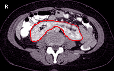
Testing for Horseshoe Kidney
A horseshoe kidney is a condition where your two kidneys are joined together and form a shape like a horseshoe. This condition can be identified through various tests that scan the abdomen. This condition is usually recognized during an ultrasound or CT scan, which are types of imaging tests.
CT scans and MRI scans are helpful in clarifying the structure of the kidney. They can show any additional blood vessels and related structures in the area. The best method is usually a CT urogram. This is a type of CT scan that allows your doctor to see the abdomen and pelvis, both with and without a special dye injected into your veins. This helps to spot kidney stones, blockages in urine flow, and potential obstructions at the point where the urine leaves the kidney (known as the ureteropelvic junction or UPJ).
An MRI can be used if there is a need to avoid procedures that involve radiation. It’s also used if you cannot tolerate the special dye used for contrast in regular CT scans. Sometimes, horseshoe kidneys can be spotted in X-rays. In these images, the kidney is positioned more inward and sits lower in the abdomen than normal kidneys.
Moreover, nuclear medicine scans that use radioactive materials can help to tell the difference between a real blockage and a kidney system that is simply wider than normal. This test can also help in diagnosing UPJ obstructions. Another test, known as a ‘voiding cystourethrogram’ can be used to identify a backflow of urine into the kidneys from the bladder, which is more common in people with horseshoe kidneys than in the general population.
Treatment Options for Horseshoe Kidney
Shockwave therapy that breaks kidney stones, also known as lithotripsy, may not work as well in people with horseshoe kidneys. This is a condition where the two kidneys are fused together at the base, forming a horseshoe-like shape. The reason for this decreased efficiency is that it can be difficult to accurately target the energy for kidney stones located in the pelvis and remove the broken pieces due to poor kidney drainage.
In some cases, kidney stones that are larger than 2.5 cm or cannot be removed by standard methods may require a less invasive percutaneous or mini-percutaneous surgery. This involves a surgeon making a small incision and inserting a special instrument to remove the stones. Before any surgical procedure, it’s crucial to conduct imaging tests like CT scans. These scans offer clear pictures of the body’s internal structures, helping doctors plan the surgery and understand the patient’s unique anatomy. This is especially important for people with horseshoe kidneys due to the highly irregular blood supply and the presence of a segment of colon at the back, increasing the risk of accidental damage during surgery.
A method called alternate biplanar fluoroscopy could be especially useful during surgery on patients with horseshoe kidneys. This involves using X-rays from two different directions to create better images of the stones and the complex kidney structures. It has several advantages, including less radiation exposure for the patient, shorter operation time, and no need for moving the patient during the operation.
Patients with horseshoe kidneys who develop stones should consider undergoing a 24-hour urine test. This test helps identify the substances causing stones, allowing for focused prevention efforts. Although this requires consistent cooperation from patients over a long time, those who develop stones in horseshoe kidneys should be given this preventive option. This is because surgical treatment of stones is often more complicated in these cases compared to others.
When it comes to managing kidney cancers in horseshoe kidney patients, the techniques are essentially the same as in patients with normal kidney anatomy. Traditionally, doctors opt for an open surgical approach. However, less invasive methods like laparoscopic and robotic techniques can also be used, despite them being more complicated due to the complex anatomy in horseshoe kidneys.
Lastly, 3D image modeling can be a valuable tool when planning for surgery. These detailed images can inform surgical decision-making and help develop a personalized surgical approach and strategy, particularly for complex cases like horseshoe kidneys.
What else can Horseshoe Kidney be?
The horseshoe kidney is a type of abnormality where the kidneys are fused together. There are two other main types of kidney fusion abnormalities:
- Crossed fusion renal ectopia – In this condition, both kidneys are on the same side of the body. One ureter, which is the tube that carries urine from the kidney to the bladder, crosses the middle of the body to drain into the bladder.
- Fused pelvic kidney – Here, there’s just one big kidney mass instead of two separate ones. This single kidney is drained by two ureters, which don’t cross the middle of the body.
What to expect with Horseshoe Kidney
Finding a horseshoe kidney, a condition where the two kidneys are connected, is usually not a cause for concern. However, people with a horseshoe kidney have a higher chance of experiencing certain kidney-related issues compared to others.
These issues include UPJ obstructions, which are blockages at the point where the kidney connects to the tube that carries urine to the bladder, kidney stones, and reflux, a condition where urine flows backwards from the bladder into the kidneys. Out of these, UPJ obstructions are the most common.
People with horseshoe kidneys may also have a greater chance of developing some types of kidney cancer. They are three to four times more likely to develop transitional cell tumors, a common form of kidney cancer. They are also twice as likely to develop Wilms tumor, another form of kidney cancer usually seen in children. Furthermore, there’s a significant increase in the chances of developing very rare tumors such as carcinoid, which are 62 to 82 times more common in people with horseshoe kidneys.
Possible Complications When Diagnosed with Horseshoe Kidney
About one-third of individuals with horseshoe kidneys show no symptoms and are usually discovered by chance during medical imaging. However, due to certain anatomical irregularities, horseshoe kidneys can lead to several urinary problems caused by blocked ureters and slowed urine drainage. The most common issue linked with horseshoe kidneys is a blockage at the point where the ureters join the kidney (ureteropelvic junction obstruction), but people can also develop a condition that causes dilation of the kidneys (hydronephrosis), infections, and a backflow of urine from the bladder to the kidneys (vesicoureteral reflux).
One study revealed that over half of the symptomatic individuals had either ureteropelvic junction obstruction or vesicoureteral reflux. A recent summarized study suggested that approximately 36% of patients with a horseshoe kidney will develop kidney stones at some point in their lives. Furthermore, due to their abnormal position in the body, horseshoe kidneys are particularly vulnerable to injury from a strong blow to the abdomen, as it can cause the kidneys to be pressed against or broken by the lower spine.
Common Health Issues:
- Ureteropelvic junction obstruction (blockage at the point where the ureters join the kidney)
- Hydronephrosis (kidney dilation)
- Urinary Tract Infections
- Vesicoureteral reflux (backflow of urine from the bladder to the kidneys)
- Nephrolithiasis (kidney stones)
- Abdominal injury due to their placement in the body


