What is Hydronephrosis and Hydroureter?
Hydronephrosis and hydroureter are medical conditions often seen in primary care, emergency medicine, and specialists like kidney doctors (nephrologists) and urinary doctors (urologists). Hydronephrosis happens when a part of the kidney that collects urine becomes enlarged because urine can’t flow properly from the kidney to the bladder. This blockage can occur anywhere along the urinary tract, which includes the pipes that connect our kidneys to our bladder, called ureters, the bladder itself, and the tube through which urine leaves the body, called the urethra. When this blockage happens in the ureter, causing it to enlarge, it’s known as hydroureter.
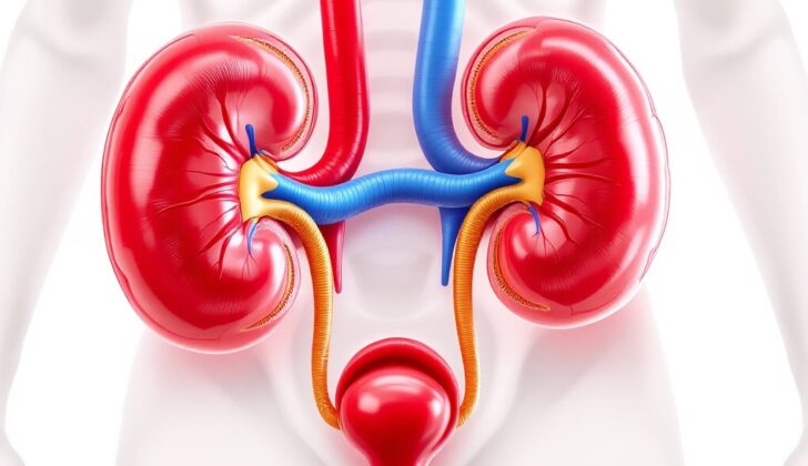
Our urinary system, made up of our kidneys, ureters, bladder, and urethra, is quite complex. Its main job is to keep our body balanced by regulating how much fluid it has, maintaining the right amount of salts (electrolytes), and getting rid of wastes through urine. Each kidney is made up of two main areas: an outer part called the cortex and an inner area known as the medulla. The medulla is shaped into pyramid-like structures that lead into a space called the renal pelvis, which then continues into the ureter.
Hydronephrosis and hydroureter can happen on their own or together and can affect anyone, regardless of age. The onset can be sudden or gradual, could be normal (like in pregnant women where these conditions are very common) or can be due to a disease, and could affect one or both sides of the urinary system.
What Causes Hydronephrosis and Hydroureter?
Urinary obstruction, or the blocking of urine flow, can happen because of two main types of problems: intrinsic, which are inside the urinary tract, and extrinsic, which are outside the urinary tract.
Intrinsic causes include things like kidney stones, cancer, narrowing of the where the kidney and ureter connect (ureteropelvic junction stenosis), scarring from past inflammation, kidney cysts, flaps inside the urethra (posterior urethral valves), an enlarged prostate (benign prostatic hyperplasia), and a disorder causing lack of bladder control (neurogenic bladder).
Extrinsic causes, or outside causes, include conditions like pregnancy, cysts around the kidney area, an abnormally positioned ureter (retrocaval ureter), cancer, trauma, fibrosis (thickened tissue) at the back of the stomach area (retroperitoneal), and a prostate abscess (an infected area filled with pus).
In children, most cases result from abnormal physical structures. These might include problems with the urethra, such as flaps or narrow passages, or narrowing of the joining areas between the bladder and ureter (ureterovesical junction) or kidney and ureter (ureteropelvic junction).
Risk Factors and Frequency for Hydronephrosis and Hydroureter
The reasons behind hydronephrosis, a condition where the kidneys swell due to a build-up of urine, vary depending on age groups.
- For newborns and children, it’s usually due to birth defects, particularly those that affect the kidneys and urinary tract. They make up about 1% of worldwide births. Hydronephrosis is increasingly common in this age group, primarily brought on by a blockage at the point where the kidney meets the tube that carries the urine from kidney to bladder (ureteropelvic junction). It is seen in 1 in 100 live births in the United States. But, it typically resolves itself by two years old. Less frequent causes include vesicoureteral reflux and obstructions which make up about 10-20% of hydronephrosis cases in newborns and infants.
- In young adults, the main cause is kidney stones, impacting approximately 600,000 US adults annually. Around 8.8% of adults in the United States experience kidney stones, with 10.6% men and 7.1% women being affected. Factors like being of white race, obesity, and diabetes increase the risk of kidney stones.
- In the elderly population, the causes often include issues like enlarged prostate, tumors in the pelvic and retroperitoneal area, and renal stones.
- About 80% of pregnant women experience hydronephrosis. It is because of the pressure on ureters due to the growing uterus and the effects of progesterone. It is usually identified in the second trimester and may last up to 6 to 12 weeks post-birth. If it leads to pain and renal failure, one of the preferred treatments is ureteral stenting.
- Between the ages of 20-60, Hydronephrosis is more common in women primarily due to pregnancy and gynecological cancers. However, in people above 60 years of age, it’s more common in men due to prostate disease and its related complications.
Signs and Symptoms of Hydronephrosis and Hydroureter
Congenital hydroureteronephrosis, a condition affecting infants, doesn’t usually show any symptoms. But, when the case is severe, there could be signs such as a lack of appetite and frequent urinary tract infections.
On the other hand, adults experiencing acute urinary outflow obstruction often feel a constant dull pain. This discomfort is usually the result of the renal capsule being stretched. Some adults experience moments of extremely severe pain when the genitourinary peristalsis momentarily increases pressure. Individuals with this condition might also feel sick and want to vomit, along with discomfort while urinating or a sudden urge to urinate. During a medical examination, these patients often show tenderness at the costovertebral angle and struggle to find a comfortable position. If the obstruction is farther along the urinary tract due to prostate enlargement, patients might feel a strong pressure in the lower abdomen and a constant urge to urinate.
Testing for Hydronephrosis and Hydroureter
If a baby is born with a condition known as hydroureteronephrosis, which is a swelling in the kidney and the tubes that carry urine from the kidney to the bladder, they should undergo a test called a voiding cystourethrogram. This test can identify if the baby has a condition called vesicoureteral reflux, where the urine moves backwards from the bladder to the kidneys.
If this baby shows symptoms such as fever, stomach pain, nausea, or is not eating properly, it’s recommended to have a urine test. This is because these babies are more likely to get urinary tract infections (UTIs), which are infections in parts of the body that produce, store, and get rid of urine. A basic metabolic panel, a type of blood test that measures your sugar level, electrolyte and fluid balance, and kidney function, should also be done. This gives a base level of kidney function that doctors can use to see if the swelling is decreasing or if the kidney function is getting worse, which might indicate the need for surgery.
Adult patients with intense pain in the back or side due to hydronephrosis should also have a basic metabolic panel and a urine test. These tests will help doctors assess kidney function.
When it comes to imaging, the choice of method depends on the patient’s previous medical history. Often, a computed tomography (CT) scan, which uses x-ray technology and powerful computers to create images of your body, is very effective. This can accurately identify if the pain is being caused by kidney stones or some form of cancerous growth. An ultrasound may be used instead if the patient has a history of frequent kidney stones and their symptoms match previous episodes. Ultrasound, which uses sound waves to create images, is also the preferred imaging method for pregnant patients with hydronephrosis.
Treatment Options for Hydronephrosis and Hydroureter
When a baby exhibits signs of hydronephrosis, or swelling in the kidneys, before birth, doctors base their treatment strategies on whether the swelling persists after birth, the severity of the swelling, and if both kidneys are affected. If the hydronephrosis is present in both kidneys, it generally means there is a blockage below the urinary bladder. A condition called posterior urethral valves is frequently the cause of this type of blockage. Doctors often take a special type of X-ray known as a voiding cystourethrogram to confirm the diagnosis.
In severe cases of hydronephrosis, wherein the diameter of the renal pelvis – the funnel-like part of the ureter in the kidney – measures more than 15mm in a newborn, it can lead to significant kidney damage. However, less severe forms of the condition usually resolve on their own by the time the child is 18 months old.
Babies born with hydronephrosis are more likely to develop a kidney infection, otherwise known as pyelonephritis, with girls being more at risk. However, there is limited evidence to show that giving a continuous course of antibiotics to infants with severe hydronephrosis will help prevent urinary tract infections in the future.
The specific treatment for hydronephrosis is determined by its underlying cause. Some signs that indicate an urgent need for intervention include a high potential for kidney damage, symptoms pointing towards a severe infection, and severe pain, nausea, and vomiting due to the hydronephrosis.
For blockages suspected to be at the bladder level, a urinary catheter, or inserted tube, can be used. If the blockage is in the ureter, the tube that carries urine from the kidney to the bladder, an internal stent can be placed via a procedure called cystoscopy. In situations where it’s not possible or advisable to use an internal stent, a less invasive procedure called a percutaneous nephrostomy tube placement can be done. This involves guided placement of a tube directly into the kidney, usually done by an interventional radiologist.
For kidney stones causing hydronephrosis, a non-invasive technique called extracorporeal shockwave lithotripsy is commonly used. It involves using high energy shockwaves outside the body to break the stones within the kidneys. For some cases, surgical intervention might be required, especially if the hydronephrosis is caused by external pressures from abnormal growths like tumors or aortic aneurysms.
What else can Hydronephrosis and Hydroureter be?
Doctors also need to consider other conditions which might have similar symptoms. These include:
- Peripelvic cysts (fluid-filled sacs around the kidney)
- Congenital megacalices (a birth condition causing unusually large spaces in the kidney)
- High urine flow
- Pyelonephritis (kidney infection)
- Renal calyceal diverticula (outpouchings from the kidney’s urine collecting system)
What to expect with Hydronephrosis and Hydroureter
Extended obstruction caused by conditions called hydronephrosis (a condition where the kidneys swell due to urine failing to properly drain from the kidney to the bladder) or hydroureter (a condition where the tube that carries urine from the kidneys to the bladder is enlarged or bulging), can lead to permanent damage to the kidneys. This damage occurs due to the wasting away of tiny tubes in the kidney and the development of scar tissue in the spaces between kidney cells.
The overall likelihood of the kidneys recovering after the obstruction is cleared, depends on how long the obstruction was there, and how severe it was.
Possible Complications When Diagnosed with Hydronephrosis and Hydroureter
The main complication of hydronephrosis, a condition where the kidney swells due to a build-up of urine, is urinary tract infection. This infection can escalate to pyelonephritis, which is essentially an infection of the kidney. Occasionally, when a long-standing blockage is resolved, it can result in post obstructive diuresis – a condition where the body produces a lot of urine after relieving a urinary blockage.
Common Complications:
- Urinary tract infection
- Pyelonephritis (kidney infection)
- Post obstructive diuresis (overproduction of urine after relieving a urinary blockage)
Preventing Hydronephrosis and Hydroureter
Hydronephrosis is a condition where the kidneys swell because urine cannot drain out correctly. Sometimes, a person might not even realize that they have it because there may be no symptoms. But it can cause pain in the lower stomach area, the back, or the areas around the genitals, especially when there’s a sudden blockage. This blockage may also make people experience frequent urination, trouble urinating, a weak flow during urination, or feelings that the bladder hasn’t fully emptied. This is often seen in cases where the prostate, a small male gland close to the bladder, grows larger than it’s supposed to (benign prostate hypertrophy).
It’s crucial to go to see a doctor without delay if you have these symptoms. If someone has had kidney stones, it’s recommended that they learn about specific changes to their diet to help stop more stones from forming in the future. A dietician could provide very useful advice to these individuals.


