What is Struvite and Triple Phosphate Renal Calculi?
The link between infections and kidney stones (renal calculi) has been understood for centuries. However, the exact connections were only identified in the early 19th century. It was discovered that a higher level of ammonia in urine, often caused by certain bacteria, can lead to the formation of a specific type of kidney stone known as a struvite stone.
Struvite stones were first discovered in bat droppings by the Swedish geologist Georg Ulex. These stones are named after his friend, Baron von Struve. Struvite is a type of crystal made up of magnesium ammonium phosphate. Despite its name, it’s often combined with different elements, including calcium. These combined stones are also known as “triple phosphate stones” even though pure struvite contains no calcium.
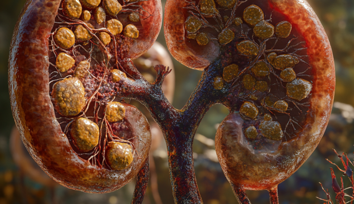
Struvite stones are typically a mix of different components including struvite and calcium phosphate. They can be found in the kidneys or bladder of patients who have catheters or a condition known as urinary stasis. This condition occurs when urine stays in the bladder for longer than it should. For example, about 8% of patients with spinal cord lesions will form stones, and 98% of these will be struvite. If not treated, these struvite stones can grow faster than other types of stones and can eventually fill up the entire kidney. This could lead to the formation of a large, branching stone known as a staghorn stone. Struvite stones make up around 5% to 15% of all kidney stones.
Struvite stones only form in conditions where the urine is more alkaline (pH >7) and are always associated with urinary tract infections caused by bacteria like Proteus. These bacteria produce a substance called urease. Almost all bacteria that can infect the urinary tract produce urease with the exception of E. coli, Citrobacter freundii, Streptococci, and Enterococci. Note that staghorn stones can also be made up of other compounds besides struvite or triple phosphate. For the purpose of this review, we’ll only focus on struvite staghorn stones.
Staghorn stones are also classified as either “partial” or “complete.” A “partial” staghorn stone involves at least two parts of the kidney (calyces), while a “complete” stone indicates that at least 80% of the kidney is involved.
What Causes Struvite and Triple Phosphate Renal Calculi?
Kidney stones primarily form due to metabolic issues and urinary tract infections. Struvite stones, also known as infection stones, are particularly linked to urinary tract infections. These stones can occur when bacteria that produce a particular enzyme, known as urease, infect or inhabit the urinary tract. Some of these bacteria include Proteus, Klebsiella, Staphylococcus, Pseudomonas, Providentia, Serratia, and Morganella. Not all bacteria are equal in producing urease. For instance, all Proteus bacteria produce urease, but only a tiny 1.4% of Escherichia coli bacteria do. Thus, Escherichia coli is least likely to cause Struvite stone formation.
Generally, patients with infection stones have fewer concurrent metabolic issues that could contribute to kidney stone formation than patients with non-struvite stones. That said, we still recommend 24-hour urine testing for dedicated patients with struvite stones, considering that at least about 50% of tested patients show metabolic abnormalities. The most commonly found metabolic issues are excess calcium in urine (hypercalciuria), low citrate in urine (hypocitraturia), and excess sodium in the blood (hypernatremia).
In a study involving 70 patients who had undergone surgical removal of staghorn stones, those who subsequently received metabolic testing and specific preventive therapy had a 98% lower kidney stone formation rate three years later than those who did not receive such testing or preventive treatment.
Risk Factors and Frequency for Struvite and Triple Phosphate Renal Calculi
Struvite stones, a type of kidney stone, are more commonly found in females due to their higher chances of getting urinary tract infections, which are closely linked with the formation of these stones. The ratio of first-time struvite stone formers is 3 females to 1 male. These stones are also common in older individuals with other medical conditions like urinary tract infections, hypertension, fever, and diabetes. Struvite stones can also occur if there is a blockage in the passage of urine resulting from various conditions like congenital urinary malformations and obstruction of the ureteropelvic junction, a place where the kidney and the ureter meet.
In the United States, struvite stones make up 5% to 15% of all kidney stone cases, and the global percentage is as high as 30%. It’s important to note that while these stones used to be considered relatively harmless if they weren’t causing symptoms, more recent studies have found that a significant percentage of people with untreated struvite stones died from serious complications like sepsis or kidney failure. Besides these, struvite stones can also lead to chronic inflammation and infections, which can sometimes result in a dangerous type of cancer called squamous cell carcinoma of the renal pelvis or collecting system. Although this cancer is rare, it has an average 5-year survival rate of less than 10%, making it particularly serious.
One other thing to consider about struvite stones is that even though most of them occur in one kidney, up to 15% may be found in both.
Signs and Symptoms of Struvite and Triple Phosphate Renal Calculi
Struvite stones, a type of kidney stone, don’t always have specific symptoms. However, some general signs may include pain along the side of your body (flank pain), discomfort while urinating (dysuria), and blood in your urine (hematuria). You may also experience symptoms of a urinary tract infection, such as fever and chills. Additionally, your urine might have a consistently high pH level, above 7.2.
- Flank pain
- Discomfort when urinating
- Blood in urine
- Fever and chills (signs of a urinary tract infection)
- Consistently high pH level in urine (>7.2)
Some people with struvite stones might have chronic kidney infections due to infected stones causing small scars in the kidney tissue. Chronic side pain and recurring urinary tract infections, especially with the same organism causing the infection, might indicate a large struvite stone called a staghorn stone. In some cases, a person may have these stones and show no symptoms at all, even when the stone takes up the whole kidney cavity (renal pelvis). People with advanced cases having large struvite stones may experience generalized symptoms. These include tiredness, feeling unwell, intermittent fevers, loss of appetite, and weight loss.
- Chronic side pain
- Recurring urinary tract infections
- Tiredness
- Feeling unwell
- Intermittent fevers
- Loss of appetite
- Weight loss
Even extremely large staghorn stones may not lead to severe kidney pain (renal colic) that is typically linked with acute blockages from urinary stones. This is because these large stones may not immediately block the urinary tract leading to a typical stretching and swelling seen with most other problematic kidney stones.
Testing for Struvite and Triple Phosphate Renal Calculi
If your doctor suspects that you have a kidney stone, they will order a urine test and a urine culture. The urine test usually shows a pH level higher than 7.2 and crystals that are shaped like a coffin lid. The urine culture can typically identify any bacteria in your urine that can create urease, an enzyme that promotes stone formation.
One of the best ways to diagnose kidney stones is a non-contrast Computed Tomography (CT) scan, especially for a particular type of kidney stone called a staghorn stone. The CT scan uses X-rays to create a detailed picture of your kidneys. If your doctor needs to look at the internal parts of your kidney more clearly, they might add a contrast dye to your CT scan.
If a CT scan isn’t available, your doctor can use an ultrasound and a plain X-ray image of your abdomen and pelvis. Struvite stones, a type of kidney stone, often show up on X-rays because of their calcium content. These stones typically have a density reading on a CT scan of 900 Hounsfield units or less.
A large struvite stone might appear like a tree branch or deer antler shape (called a staghorn) on an X-ray image. It fills up the part of your kidney where urine collects. Intravenous pyelography, an imaging test that uses a contrast dye to highlight the kidneys, ureters and bladder, is used less often to diagnose kidney stones. Magnetic resonance imaging (MRI), which uses a magnetic field and radio waves to create detailed images of the organs and tissues, isn’t usually recommended for visualizing staghorn stones as it isn’t as effective.
Passing a struvite stone naturally is rare. Sometimes, a small piece might break off and either be passed in the urine or found in the bladder. If a stone is passed or removed, it is usually analysed to identify its type, which can help guide further treatment.
Treatment Options for Struvite and Triple Phosphate Renal Calculi
Adjusting your diet doesn’t typically prove to be a successful way to break down a particular type of kidney stone known as struvite stones. Though there have been suggestions to follow a diet low in calcium and phosphorus, and add something like aluminum hydroxide to limit phosphorus absorption, or a urinary acidifier like ammonium chloride, these methods have their downsides and possible health risks. One example could be an overproduction of calcium in the urine and aluminum toxicity. However, it’s worth noting that a similar diet has had some success in treating cats who’ve developed these types of kidney stones. Some studies also suggest that modified diets can help reduce the risk of certain kidney stones in people.
There have been proposals to use L-methionine to increase acid in the urine and potentially lower the risk of struvite stones. Some studies show a significant reduction in the likelihood of these stones forming in people who took this supplement daily. However, we still need more research to confirm the benefits of L-methionine before we can start recommending its regular use.
If you have an infection stone, exact antibiotics would depend on details from personal cultures, but some general samples could be cefepime, amoxicillin-clavulanate, or ciprofloxacin. Although they might not dissolve the stone completely, antibiotics can significantly lower the risk of serious infections and help stop the stone from growing.
Managing struvite stones with medication alone tends to bring modest benefits and isn’t typically successful. Instead, it is mainly reserved for patients who are too sick for surgery, refuse surgery, or after maximum surgical removal has been done.
Acetohydroxamic acid (AHA), an inhibitor of a particular enzyme, has shown promise in decreasing the growth and recurrence of struvite stones. It works in tandem with precise antibiotics and lowers the alkalinity and ammonia levels in the urine. However, AHA should only be used once all surgical options have been exhausted due to significant side effects, such as nausea, tremulousness, and others.
It’s crucial to note that the best treatment for struvite stones is complete surgical removal because the stones tend to grow quickly. Removal ensures that no stone fragments are left behind, which otherwise could lead to the new stone formation.
Various surgical techniques are available. Extracorporeal shockwave lithotripsy or Percutaneous nephrolithotomy are two examples, depending on the stone’s size. In some cases, open surgery might be considered if intrarenal anatomy does not allow another approach, or the intention is to remove the kidney.
Lastly, after the surgical treatment of struvite stones, periodic check-ups involving imaging and urine cultures are recommended. They should be done every 6 to 12 months, though this could be more frequent for patients who’ve displayed rapid or frequent stone recurrences.
What else can Struvite and Triple Phosphate Renal Calculi be?
When experiencing flank pain (pain on the side of the body between the ribs and hip), it might be caused not only by a blood clot, but it could also be due to conditions such as a kidney stone, a urinary tract infection, ovarian issues – including cyst rupture or torsion, aortic aneurysm, gallbladder pain or inflammation, appendicitis, blocked blood supply to the intestines, obstructions in the area where the kidney meets the ureter, and even menstrual pain.
Tests such as CT scans can usually tell the difference between these conditions. For example, if the pain is caused by a gastrointestinal issue, there will usually be no blood in the urine. The CT scan can also give clues about the type of kidney stone you have based on how it looks and how dense it is, such as a large, antler-shaped stone that suggests a type of stone called struvite. Laboratory tests on a stone that has been passed can also help identify the type of stone.
What to expect with Struvite and Triple Phosphate Renal Calculi
Staghorn calculi, or large kidney stones that take the shape of a staghorn, can cause significant health issues and even death. In some cases, kidney function can worsen, with as high as 28% of individuals experiencing this outcome, regardless of whether they received treatment or not. This is especially true for patients with characteristics such as complete staghorn calculi (meaning the stone fills the entire kidney), urinary diversions (a new route for urine to leave the body), solitary kidneys (having only one functional kidney), and those with a history of kidney stones, high blood pressure, or a neurogenic bladder (a bladder that doesn’t function properly due to a nerve problem).
In the past, over a span of 10 years, there was found to be a 7.2% death rate in patients who underwent procedures to remove the stone, compared to a 28% death rate in those who were managed with less invasive measures. However, more recent studies have shown an extremely low mortality rate of less than 1% for procedures like Percutaneous Nephrolithotomy (PCNL, a procedure to remove large stones from the kidney) and Shock Wave Lithotripsy (SWL, a non-invasive procedure that breaks up the kidney stone). This small rate is typically associated with patients who already have cardiac dysfunction (heart problems).
Possible Complications When Diagnosed with Struvite and Triple Phosphate Renal Calculi
Struvite and triple phosphate kidney stones can lead to severe consequences if not treated adequately. Problems may include complete and continuous kidney obstruction resulting in irreversible kidney damage, sepsis (which is a life-threatening infection that can spread throughout your body), chronic pyelonephritis (a type of urinary tract infection that affects the kidneys), pyonephrosis (a severe, painful urinary tract infection where pus builds up in the kidneys) and kidney failure. This can be particularly problematic if these stones are present in both kidneys.
In addition, the treatment methods for these stones, which may involve Extracorporeal Shock Wave Lithotripsy (ESWL) and/or Percutaneous Nephrolithotomy (PCNL), can have its own set of complications. Patients could end up with kidney infection, a pocket of pus around the kidneys, bleeding, internal damage to the organs surrounding the kidneys, accidental puncturing of the renal pelvis during the procedure, water intoxication and low sodium levels in the blood, low body temperature, kidney failure, retroperitoneal hematomas (blood collects in the back of the abdomen), or strictures (narrowing of ureters or urethra).
Common Side Effects:
- Complete and continuous kidney obstruction
- Chronic pyelonephritis
- Sepsis
- Pyonephrosis
- Kidney failure
- Kidney infection as a side effect of ESWL or PCNL
- Perinephric abscess (pocket of pus around the kidneys)
- Bleeding or injury to adjacent organs
- Perforation of the renal pelvis (accidental puncturing during the procedure)
- Dilutional hyponatremia (water intoxication and low sodium levels)
- Hypothermia (low body temperature)
- Retroperitoneal hematomas (blood collects in the back of the abdomen)
- Stricture formation (narrowing of passages)
Preventing Struvite and Triple Phosphate Renal Calculi
It’s vital for patients to understand the risks and complications associated with struvite and kidney stones in order to prevent future health issues. If someone forms or passes a kidney stone, it’s important that they have medical imaging and lab tests done to analyze the stone. Moreover, patients who frequently get urinary tract infections (UTIs) should be informed that they might have struvite and triple phosphate kidney stones in their kidneys or urine collection system.
In such situations, merely treating the infection with antibiotics isn’t sufficient. It’s necessary to surgically remove the stone to protect the kidneys from long-term damage and lower the chances of getting further UTIs. So, understanding this condition and its treatment can go a long way in maintaining good kidney health.


