What is Ureterolithiasis?
Ureterolithiasis, a medical condition that affects millions worldwide, is the presence of stones in the ureter, the tube that connects your kidney to your bladder. It can happen to anyone but is particularly associated with other health problems like heart disease, diabetes, obesity, and a condition called metabolic syndrome.
You may also know ureterolithiasis for the severe and sudden pain often linked with it – a pain that starts in your side or back and radiates towards the groin. It’s not uncommon for this pain to come and go and for those experiencing it to feel a strong urge to keep moving. This type of pain is often described as “colicky.” Nausea, vomiting, and bladder symptoms can also happen when the stones approach the bladder.
When people with this condition come to the emergency room, they often have blood in their urine (a symptom found in about 85% of patients). Doctors can usually identify ureterolithiasis through this symptom but usually need an imaging test (like an X-ray, without any contrast dye) to make a definite diagnosis.
If you are diagnosed with ureterolithiasis, the standard treatments usually include pain killers, medicines to control nausea, intravenous (IV) fluids to keep you hydrated, and sometimes antibiotics. Your doctor may also prescribe medicine (such as α-blockers) to help pass the stones naturally without surgery, unless the stone is too large or hasn’t moved in a while.
If the stone is too big (more than 7mm in diameter) or hasn’t moved in a month or so, you might need surgery. The most common surgeries are ureteroscopy, which uses a thin tube equipped with a camera and a laser to break the stone into small pieces, and a technique called extracorporeal shockwave lithotripsy, which uses sound waves to break stones apart.
It’s crucial to treat ureterolithiasis promptly because a combination of kidney infection and a blockage caused by the stone can cause serious complications. If this occurs, a doctor will first treat the infection and then address the stuck stone. It’s difficult to tell the difference between a typical kidney infection and one caused by a blockage without appropriate imaging.
Unfortunately, complications can arise when dealing with ureterolithiasis. These can include severe urinary tract infection (urosepsis), kidney abscess, long-term kidney disease, blockages, urine leakage, scars in the ureter, and narrowing of the ureter due to physical damage or disease.
Finally, to prevent future stones, doctors recommend a urine test over 24 hours for high-risk patients. High-risk groups include people with recurrent stones, impaired kidney function, immune-compromised individuals, and those with a high surgical or anesthesia risk. Other patients may also take this test, but it’s a discussion to have with your doctor. Preventing stones from reoccurring requires commitment to long-term therapy, as ureterolithiasis has a tendency to return.
Now, a bit about the urinary system: It’s made of the kidneys, bladder, and tubes called the ureters and urethra. The kidneys filter the blood to produce urine, which travels through the ureters to the bladder where it’s stored until it’s eventually expelled from the body through the urethra. Kidney stones form when waste products in the urine become too concentrated and solidify. These stones can travel into the ureters and cause a blockage – a situation which can result in severe discomfort, especially if they get stuck in certain areas in the ureters which are known for their narrowness. Bunches of nerves around the ureters cause the severe pain you feel when the stone irritates them.
What Causes Ureterolithiasis?
It’s recommended that kidney stones, also called renal calculi, be removed when possible and sent for chemical analysis. It’s advised that all patients with kidney stones go through a 24-hour urine test. This test can help identify factors that may cause new stones to form and can guide the prevention treatment. There are several risk factors responsible for the formation of kidney stones. Here are some of them:
Chronic Infection
Chronic urinary tract infections can lead to the creation of a particular type of kidney stone called “struvite” or “infection stones.” These stones are composed of magnesium, calcium, and ammonium phosphate. Certain organisms, primarily Proteus or Klebsiella spp but not Escherichia coli, produce a substance, known as urease, that breaks down urea in the urine. This process leads to higher levels of kidney ammonia and raises urinary pH, eventually promoting the formation and growth of struvite stones. Treatment typically includes infection control with total surgical removal of all possibly infected stone material. A specific inhibitor, acetohydroxamic acid, may be used in select cases.
Cystinuria
Cystine stones make up only 1% to 2% of all kidney stones but these stones often recur and are resistant to a method of stone removal called shockwave lithotripsy. An inheritable, autosomal recessive trait can lead to the disease cystinuria which can cause the formation of cystine stones. Treatment aims to remove the stones using a procedure called ureteroscopy and laser lithotripsy. Preventive measures include drinking lots of fluids to produce more urine and ensuring a high urine pH (7.5). If these measures are not enough, a thiol-based agent may be used to increase the solubility of cystine.
Hypercalciuria
Hypercalciuria, or having too much calcium in your urine, is a common cause of kidney stones. In fact, about 80% of all kidney stones contain calcium. High calcium levels in urine might be due to a number of reasons, including: increased absorption of calcium in the gut, higher levels of calcium in the blood, and conditions like hyperparathyroidism or systemic acidosis. Hypercalciuria raises the levels of calcium salts like oxalate and phosphate in urine, which can cause stone formation. Treatment typically involves reducing daily oral calcium intake and using medicines such as thiazides to lessen calcium excretion in the kidneys.
Hyperoxaluria
Oxalate is a chemical that’s present in plants. When you eat vegetables, this chemical is absorbed in your gut and goal is to excrete it in your urine. However, when it’s excreted in the urine, it can form crystals and stones with calcium. Oxalate is regarded as one of the strongest chemicals that promote urinary stones, and to prevent stones, the ideal oxalate level in urine should be 25 mg/day or less.
Hyperuricosuria and Aciduria
Uric acid stones make up about 5% to 10% of all kidney stones. High levels of uric acid in urine can lead to the formation of both uric acid and calcium oxalate stones. Aciduria, or having high total urinary acid levels, usually causes pure uric acid stones. Aciduria results from excessive meat consumption or reduced kidney acid excretion. Treatment includes keeping the body’s pH levels around 6.5, medication to reduce uric acid, and reducing uric acid stone formation through use of potassium citrate or sodium bicarbonate.
Hypocitraturia
Low levels of citrate in urine can play a role in the formation of kidney stones. Patients with uric acid stone formation are often given potassium citrate supplements to increase urinary citrate levels, treat systemic acidosis, and optimize urine pH levels.
Inadequate Urinary Volume
Patients who have low urine volumes, typically less than 1 L per day, have increased urinary solute concentration which can lead to stone formation. Medical guidelines recommend increasing daily urine volumes to 2.5 L, with a minimum of 2 L, to prevent stone formation. Not drinking enough fluids and thus not producing enough urine is the most common single identifiable cause of kidney stones.
Risk Factors and Frequency for Ureterolithiasis
Ureterolithiasis, a painful condition affecting the urinary tract, is experienced by up to 12% of adults in the United States. This means about 1 out of 11 people will have this issue at some point. As for kidney stones (renal calculi), studies show that about 11% of men and 6-7% of women will have to deal with them in their lifetime, with the rates rising continuously. It’s important to note that while men are twice as likely as women to suffer from this condition, the rate of increase is significantly higher among females.
If we take a global perspective, the occurrence of urinary stones (urolithiasis) has risen worldwide from 1990 to 2019, continuing to increase. This growth rate is most significant in the Caribbean, followed by Central Asia and Africa (mainly Congo, Eswatini, Gabon, and Grenada). Racial differences also exist in prevalence, with Whites registering the highest numbers and Blacks the lowest. This difference is more connected with geographical, cultural, and socioeconomic aspects rather than genetic factors.
Kidney stones (nephrolithiasis) are most often diagnosed in people in their 40s. On average, symptomatic stones show up around the age of 45 for men and 41 for women. More than 70% of urinary stones occur in individuals aged 20 to 50 years old. Alarmingly, the fastest and most substantial increase in cases of urinary stones is among women and children, especially teenagers.
Location and climate also play a role in the prevalence of urinary stones. Warmer climates, known as the “stone belt”, have higher rates of this condition for both men and women. In Europe and North America, it’s estimated that 0.5% of the population will experience this problem each year. Half of those cases are likely to recur within the next decade if no preventive measures are taken. The relapse rates for symptomatic stones are estimated at 11% within 2 years, 26% within 5 years, 50% within 10 years, and 60-80% in a lifetime unless long-term preventive treatments are used.
Many medical conditions can increase the risk of developing urinary stones and their recurrence. They include:
- Aciduria
- Bariatric (Roux-en-Y) surgery
- Bowel and intestinal resections
- Calcium phosphate urolithiasis
- Calyceal diverticulum
- Cardiovascular disease
- Chronic diarrhea
- Cystic fibrosis
- Cystinuria or history of previous cystine stones
- Diabetes
- Distal renal tubular acidosis
- Enteric hyperoxaluria
- Family history of cystinuria or nephrolithiasis
- High oxalate diet
- Horseshoe kidney
- Hyperparathyroidism
- Hypertension
- Intestinal malabsorption
- Irritable bowel syndrome
- Kidney or kidney stone surgery previously
- Lesch-Nyhan syndrome
- Low urinary volume (especially if < 1000 mL daily)
- Malabsorption
- Medullary sponge kidney
- Metabolic acidosis
- Metabolic syndrome
- Nephrocalcinosis
- Obesity
- Pediatric urolithiasis
- Personal history of urolithiasis
- Primary hyperoxaluria
- Renal failure
- Sarcoidosis
- Short bowel syndrome
- Solitary kidney
- Struvite stones
- Ureteral strictures
- Ureterocele
- Ureteropelvic junction obstruction
- Uric acid urolithiasis
- Vesicoureteral reflux
Nonmedical factors such as being male or of White race are also linked with a higher chance of developing kidney stones.
Signs and Symptoms of Ureterolithiasis
Ureterolithiasis, or the process in which stones form in the ureter, is usually marked by sudden and severe pain in one side of the middle to lower back, which often spreads to the groin, lower abdomen, or testicles. Patients often describe the pain as unbearable and at its peak about 1 to 2 hours after starting. This is usually accompanied by restlessness, which is manifest by the patient continually moving or walking around. The pain is often compounded by bouts of nausea and vomiting. It’s interesting to note that, in comparison, people with a sudden severe stomach condition will prefer to stay still. Over time, the intensity of the pain becomes constant. It’s common for patients to wake up due to the pain and be able to recall exactly when the symptoms started.
- Sudden and severe back pain spreading to the groin, lower abdomen, or testicles
- Peak pain about 1 to 2 hours after onset
- Unbearable pain
- Restlessness
- Pain worsened by nausea and vomiting
Additional symptoms may include frequent urination, a strong urge to urinate, difficulty in starting urination, blood in urine and painful urination if the stone is located close to the bladder. Sometimes, patients may also have a history of kidney stones or related conditions, either personally or within the family. It’s also noteworthy that lower back pain, diarrhea, or recurrent urinary tract infections are other uncommon signs of obstructing ureteral stones.
If a patient presents with a fever and chills, this could indicate a urinary tract infection. Coupled with an obstructing ureteral stone, this can result in obstructive pyelonephritis or pyonephrosis, both are serious conditions requiring urgent surgical intervention.
Upon physical examination, a patient usually appears restless. While they might have a rapid heartbeat, they are not feverish and their blood pressure is normal unless they are also septic. On examining the abdomen, physicians usually find no abnormalities or only mild discomfort. However, there is usually tenderness in the costovertebral angle on the side of the stone. Extreme abdominal tenderness could suggest alternative conditions like appendicitis, diverticulitis, small bowel obstruction, gallbladder inflammation, herpes zoster, or an ectopic pregnancy.
Testing for Ureterolithiasis
When a patient is suspected of having kidney stones in the ureter, which are also known as ureterolithiasis, doctors will do a clinical exam, request some blood and urine tests, and order some scans or images to check for stones. This helps the doctors to confirm if there are stones, find their size and location, see if there are any complications, and decide on the best treatment plan.
Laboratory Studies
Doctors order a basic metabolic panel to check electrolyte levels and kidney function. If the kidney has been blocked too often or for too long by stones, this could have negatively affected its function. The test results can also provide clues as to where the stones in the kidneys came from, and what they are made of. Patients with calcium in the kidneys who show a high calcium level in their blood will also get a parathyroid hormone check. Similarity checks for serum uric acid levels are done for individuals with calcium or uric acid kidney stones. An ordinary blood cell count can show if there are too many white cells, a condition known as leukocytosis. Mildly high white cell counts often accompany ureterolithiasis, but significantly higher counts could indicate an infection.
Urinalysis, which is a test that checks your urine, can often find some blood in the urine. This is observed in about 85% of patients with ureterolithiasis, either visible to the eye or seen under a microscope. Sometimes crystals may be present in the urine which can give an idea about the stones’ chemical composition. Urinary pH is important to check as it can affect the type of substance that forms kidney stones. A microscopic urinalysis is the recommended method as dipstick testing can be unreliable.
A specific gravity that is high can be a sign of dehydration and signal the need for intravenous (IV) fluids. If there are white blood cells or bacteria in the urine, this may suggest a urinary tract infection or a more severe condition called pyonephrosis, especially in patients with diabetes, anesthesia or surgical risk factors, and impaired immune responses. Imaging scans can help tell the difference between pyelonephritis, which is medically treated, and pyonephrosis, which needs immediate surgery. Urine cultures should be done and suitable antibiotics given.
Imaging
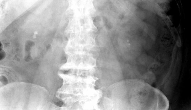
A kidney, ureter, and bladder (KUB) x-ray is an abdominal x-ray that mainly focuses on the kidneys, ureters, and bladder. However, it can only identify most but not all types of urinary stones. The KUB x-ray is inexpensive, readily available, and it offers information on the size, shape, and location of the ureteral stone, making it easy to follow the progress of the condition.
Before a Computed tomography (CT) scan is done for suspicion of ureterolithiasis, it is recommended that a KUB x-ray is done to avoid obscuring the stones, especially if an intravenous (IV) contrast is planned to be used. However, the KUB x-ray does also have a few limits. For example, it may have challenges in telling the difference between ureteral calculi and other nearby pelvic stones, or between hydronephrosis and simple obstruction. And it cannot provide information on kidney function or visualize the internal aspect of the kidney.
A KUB ultrasound can identify larger renal stones and determine the degree of hydronephrosis. This method cannot dependably pick up stones that are smaller than 3 mm or measure their size or place them in their exact location. KUB ultrasound is preferred during pregnancy and is often the first imaging study done in the emergency department in patients with abdominal pain or suspected acute renal colic due of its low cost, availability, easy use, and no radiation exposure.
A CT scan of the belly and pelvis without contrast is considered the gold standard for diagnosing ureterolithiasis. It can give information about the size, number, and location of any urinary stone, with only a few rare exceptions. But it is important to get a KUB x-ray before a CT scan to monitor and follow up on the passage of the stone.
In certain situations such as a urinary infection resulting in sepsis (urosepsis), which may lead to a life-threatening condition called obstructing pyelonephritis or pyonephrosis. This condition is a result of an infected kidney due to an obstructing stone. This condition may initially present similarly as acute pyelonephrosis, and thus can be challenging to tell the two apart without imaging. In such cases, emergency drainage of the blocked infected kidney is necessary.
Risk factors for developing pyonephrosis in patients with stones in the ureter include several such as diabetes, greater number and severe hydronephrosis, large stones in the ureter (more than 5mm in size), older patients, the presence of pus in the urine, kidney failure, and a weak immune response system.
Treatment Options for Ureterolithiasis
The first step in managing a sudden (acute) kidney stone (ureterolithiasis) involves controlling pain, using an IV to provide fluids and anti-nausea medication. Patients are typically not allowed to eat or drink, especially if they are hospitalized, until it’s determined that surgery isn’t needed.
Pain relief is a critical part of the early management of symptomatic kidney stones. There are several pain relief options available, including drugs administered through an IV such as ketorolac (a type of anti-inflammatory drug), opioids, or acetaminophen. Some patients may also benefit from IV lidocaine.
One standard medication to relieve kidney stone pain is ketorolac. This drug doesn’t have the stomach or breathing side effects of opioids and can be just as, or even more, effective at relieving pain, based on various studies. However, ketorolac should not be used during pregnancy or in patients with kidney failure, a history of significant upper gastrointestinal bleeds, or allergies to anti-inflammatory drugs.
Other drugs that can be given as an alternative to ketorolac or used for extra pain relief include opioids and acetaminophen. Morphine is the preferred opioid for kidney stone pain due to its effectiveness, especially when combined with ketorolac.
Anti-nausea medications and IV fluids are usually recommended for patients showing symptoms. Ondansetron is considered the most effective anti-nausea medication based on a few studies. IV fluids are generally recommended for patients with symptoms of acute kidney stone pain to prevent dehydration due to vomiting and lack of oral intake.
Medical expulsive therapy, a treatment that helps stones pass out of the body, is generally recommended for patients treated outside the hospital. Various α-blockers can help by reducing muscle tension in the part of the ureter closest to the bladder, which helps the kidney stone pass more easily. However, this treatment is not as effective for larger stones located closer to the kidneys.
The decision to have surgery is determined by several factors, but typically patients with small stones and manageable symptoms can be treated outside the hospital and will not require immediate surgery.
Many factors such as a patient’s previous experience with stones, gender, age, level of pain, nausea and vomiting, dehydration, and the size or shape of the stone do not reliably indicate if a patient needs surgery. However, the bigger the stone and the closer it is to the kidney, the more likely it is that a procedure related to the stone will be needed.
Case for having surgery may include complete blockage of urine, blockage of both ureters, failure of the kidney stone to pass or progress after 4 to 6 weeks, inability to control symptoms, known or suspected infectious kidney inflammation, the patient’s preference, existing kidney failure, progressive damage or loss of kidney function, recurring or ongoing urinary infections, thinning of the kidney cortex due to long term obstruction, a serious condition known as sepsis, significant other medical conditions, urinary tract infection at the same time, having only one kidney, stone size of 10 mm or larger, and uncontrollable or long-lasting severe pain.
Surgical intervention for kidney stones typically involves either a treatment that uses shock waves to break up stones from outside the body (extracorporeal shockwave lithotripsy) or a type of surgery in which a small telescopic instrument (ureteroscope) is used to remove or break up the stone.
For treating kidney stones, ESWL and ureteroscopy have similar overall rates of success. ESWL offers comparable success rates to ureteroscopy, with lower cost, greater safety, reduced anesthesia needs, and fewer complications, but may sometimes require a second treatment.
Meanwhile, ureteroscopy, a procedure to remove kidney stones using a thin tube, is preferred over open surgery. It is recommended to start an α-blocker medication before the ureteroscopic procedure, but the best drug and when to start taking it have not been determined. Ureteroscopy can be safely performed on individuals with kidney stones and is recognized to result in a lower rate of stone-free status and additional postoperative blood in the urine.
When considering ESWL and ureteroscopy for kidney stone treatment, decisions generally depend on the availability of the instrument, surgeon’s experience, anesthesia considerations, patient preferences, estimated stone hardness and composition, radiance, stone size and location, the distance from the skin to the stone, and the patient’s individual clinical factors. Open discussions with patients are crucial for reviewing relative advantages and disadvantages of these procedures. Both procedures offer equivalent rates of success for ureteral stone removal.
Should the renal colic episode in high-risk patients be successfully treated, a 24-hour urine analysis is recommended for kidney stone prevention. This test evaluates levels of various substances in the urine that can contribute
What else can Ureterolithiasis be?
Sudden, intense pain in the side of your abdomen, nausea, and vomiting can be symptoms of many things, not just a condition known as ureterolithiasis (stones in the ureter, a tube that carries urine from your kidney to your bladder). These symptoms could be especially tricky to pinpoint in people who have a history of kidney stones and other health issues, like heart disease. The pain can be due to a variety of problems, some harmless and others quite serious. Here’s a list of possible causes:
- An abdominal aortic aneurysm (this is when the large blood vessel that carries blood from the heart to the rest of your body tears or bursts)
- Angiomyolipoma (a benign tumor in the kidney)
- Appendicitis (inflammation of the appendix)
- Gallbladder inflammation (cholecystitis)
- Chest wall pain (costochondritis)
- Skin conditions like shingles (Herpes zoster)
- Problems related to a kidney tube (ureter) stent
- Intestinal inflammation (diverticulitis)
- Ectopic pregnancy (a pregnancy that develops outside the womb)
- Endometriosis (a condition in which tissue similar to the lining of the womb grows outside it)
- Epididymitis (inflammation of the tube that carries sperm)
- Ureter compression due to surgical items or cancers
- Stomach disorders like irritable bowel syndrome or Crohn’s disease
- Liver inflammation (hepatitis)
- Complications from medical procedures
- Groin hernia
- Nerve disorders and nerve pain
- Complications from blood vessels pressed by arteries (Nutcracker syndrome)
- Mesenteric ischemia (a condition causing poor blood flow to the intestines)
- Muscular issues
- Ovarian problems like cyst bursts or twisting of the ovary
- Necrosis in kidney tissue
- Pelvic inflammatory disease or pelvic pain syndrome
- Chest pain due to inflammation of the covering of the lungs (pleurisy)
- Prostate inflammation (Prostatitis)
- Kidney infection (pyelonephritis)
- Pain referred from back complications
- Kidney abscess, blockage, or clot
- Fibrous tissue behind the abdomen (retroperitoneal fibrosis) or other related conditions like abscess, hematoma, or tumor
- Bleeding under the kidney capsule (subcapsular spontaneous renal hematoma or Wunderlich syndrome)
- Torsion or twisting of the testicles
- Spasms or strictures in the ureter
- Cyst in the ureter (ureterocele)
- Blockage at the junction between the kidney and ureter
- Complications from a ureteroscopy procedure
- Urinary tract infection
It’s really important for your doctor to take a detailed history, perform a physical exam, and conduct the right diagnostic tests to find out whether you have ureterolithiasis or one of these other conditions.
What to expect with Ureterolithiasis
Generally, people with ureterolithiasis, a condition where stones are formed in the ureter (a tube that carries urine from the kidney to the bladder), have good chances of recovery. However, studies have shown that this condition is often linked with other health disorders like diabetes, heart diseases, and obesity. There are high chances, around 50%, of the stones recurring within a span of 10 years.
If the stones keep coming back, it’s important that a thorough health check-up is done to find out the root cause. Once understood, changes in lifestyle and suitable medication can be prescribed to lessen the chances of the stones forming again.
It’s worth noting that this condition can have a significant financial impact, especially when it keeps recurring. This is due to lost wages from taking time off work and the cost of visits to the emergency department.
Possible Complications When Diagnosed with Ureterolithiasis
Kidney and ureter stones can sometimes lead to serious complications. These may include infected kidney stones, bloodstream infections, scarring in the ureter, ruptures, abscesses in the kidney, bleeding, compromised kidney function, and forniceal rupture, which is breaking of the kidney’s outer surface. They can even cause Chronic Kidney Disease (CKD) due to a medical procedure, blockage, or a condition causing the stone.
However, the chance of these stones leading to end-stage kidney failure is low, even if the stones keep coming back.
Kidney and ureter stones can cause additional complications in pregnant women. These complications have been linked with an increased risk of spontaneous abortions, low birth weight infants, miscarriages, high blood pressure during pregnancy (preeclampsia), and diabetes during pregnancy (gestational diabetes). Therefore, certain treatments like Extracorporeal Shock Wave Lithotripsy (ESWL), which breaks down stones, is avoided in pregnant women, and ureteroscopy, which uses a thin tube to remove or break the stones, is used very selectively.
Additionally, ESWL can lead to a number of complications:
- Bruising in the area where the treatment is applied
- Inability to locate the calculus (stone)
- Injury to the digestive system, usually a bruise that disappears on its own
- Blood in the urine
- Large bruise near the kidney
- Contusion or bruise-like injury to the kidney
- Large bruise under the kidney’s outer layer
- Steinstrasse, a complication where multiple small stones block the ureter
- Obstruction of the ureter due to a piece of stone
Usually, Steinstrasse resolves on its own. But if it doesn’t get better within two weeks or starts causing symptoms, it is recommended to go through ESWL or ureteroscopy again.
Ureteroscopy can also lead to several complications:
- Failure due to abnormal ureteral structure or inability to reach the obstructing stone
- Inability to locate the calculus (stone)
- Fever
- “Forgotten” stents, which are small tubes used to keep the ureter open
- Blood in the urine
- Ileus, a type of bowel obstruction
- Injury to the kidney
- Perforation or hole in the kidney
- Segmental ureteral necrosis, death of a segment of the ureter
- Discomfort due to the stent
- Movement of the stone to a different location
- Perforation or hole in the ureter
- Spasm or cramp in the ureter
- Ureteral stricture, narrowing of the ureter
- Urinary extravasation, leakage of urine into the body
- Urinary tract infection
It’s crucial that patients are told to immediately seek medical help if they experience symptoms.
Preventing Ureterolithiasis
To prevent the formation of kidney stones, also known as ureterolithiasis, changes in lifestyle and diet can be very helpful. Some ways to reduce the chance of developing kidney stones include drinking more fluids, limiting intake of foods that are rich in compounds called oxalates, sodium, and proteins from animals, and eating more fruits and whole grains. Regular exercise also helps, as well as avoiding medicines known to increase the risk of kidney stones.
It’s important to inform those who are experiencing kidney or ureteral stones for the first time that there’s a high chance these stones will reoccur. For people at risk of repeated stone formation, increasing the intake of dietary calcium can be considered. Despite popular belief, eating more calcium can actually lower the risk of forming calcium oxalate stones. This happens because more calcium in the gut can bind to oxalates, limiting their absorption into the body and thereby reducing the amount that can end up in the urine where stones form.
Tests, such as a 24-hour urine analysis, can be helpful in finding out the cause of kidney stones. These tests are especially important for those who recurrently have stones or are at a higher risk. However, the effectiveness of preventive methods largely depends on the patient’s commitment to long-term treatment recommendations. It’s important to note that most patients at high risk of kidney stones do not get tested. Many may not even be aware that they have the option to get these tests done for their own benefit.
Patients can also be encouraged to use tools like a strainer or sifter to collect stone material for chemical analysis, directly from the urine. Additionally, signs to watch out for that might require treatment include fever, chills, feeling light-headed, low energy, or difficulty in urinating.
Doctors should also discuss the benefits of medical treatments designed to help pass kidney stones. Routine use of antibiotics is typically not advised unless the patient shows signs of a urinary tract infection, has an unexplained fever, or is at high risk of a serious infection called sepsis. It’s also normal for kidney stone patients to have slightly elevated white blood cell counts, often associated with infections, even if they do not have one. A follow-up with their primary doctor or a urologist should be recommended for continuing care.


