What is Ureteropelvic Junction Obstruction?
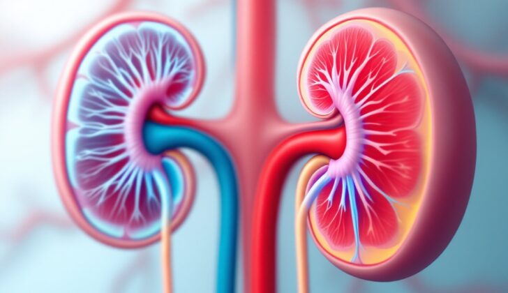
The kidneys are very important organs in our body. Their main functions are to control the balance of fluids and various minerals, help correct the balance of acids and bases in the body, regulate blood pressure and control the secretion of a hormone called erythropoietin.
During fetal development, the kidneys grow from a layer of tissue called the metanephric mesoderm, which forms the part of the kidneys up to the distal tubules. Other parts of the kidneys such as the collecting duct, major and minor calyces, renal pelvis, and ureters develop from something called the ureteric bud. This bud starts growing from the mesonephric duct during the fifth week of pregnancy. Therefore, the connection between the kidney and the ureter (ureteropelvic junction or UPJ for short) is actually made entirely by the ureteric bud, not by the joining of two different tissues.
There is a medical issue referred to as ureteropelvic junction obstruction (UPJO) that’s important to know about. This is a condition where the flow of urine from the kidney to the ureter is blocked. If it’s not found and treated properly, it could lead to complete loss of the affected kidney. UPJO mainly occurs at birth and can often be detected during pregnancy in the second trimester through an ultrasound.
What Causes Ureteropelvic Junction Obstruction?
Ureteropelvic junction obstruction (UPJO) is a condition where the flow of urine from the kidneys to the bladder is blocked. This condition can occur from birth (congenital) or develop later in life (acquired), with the first type being the most common cause.
Congenital Causes
1. A condition known as ureteral hypoplasia, where many parts of the ureter don’t fully develop, can cause the muscles that help push urine down the tube to not work correctly. This can cause a functional blockage, which is due to the tubes not working properly rather than being physically blocked.
2. If the ureter (the tube that carries urine from the kidneys to the bladder) enters the kidney at a high point, it can cause issues with emptying urine. Normally the ureter should enter the kidney in a way that allows free urine flow, but if it enters at a high point, it can cause a kink that leads to a blockage. This often leads to a condition called hydronephrosis, where the kidney swells due to urine backup.
Aside from these, the ureter can also get trapped by an extra blood vessel from the kidney, which can twist and kink the tube, hindering the free flow of urine. This is most commonly seen in kids who have symptoms and need surgery. Rarely, a kidney that is rotated improperly can also cause UPJO.
Acquired Causes
Acquired causes of UPJO are divided into two categories – extrinsic (external) and intrinsic (internal).
Extrinsic
This is caused mainly by something pushing on the ureter from the outside.
1. Retroperitoneal fibrosis, which is a hardening of tissue in the back of the abdomen.
2. Retroperitoneal lymphadenopathy, which is swelling of the lymph nodes in the back of the abdomen (this can be seen in testicular cancer or lymphoma).
3. Retroperitoneal mass, which is a growth in the back of the abdomen (e.g., a sarcoma).
4. A freely moveable kidney, which can move around and press on the ureter depending on the patient’s position.
Intrinsic
These causes are due to changes inside the ureter itself.
1. Scarring of the ureter wall due to a stuck stone, long-standing inflammation, or radiation treatment.
2. Tumors, such as transitional cell carcinoma, developing in the lining of the ureter.
3. Iatrogenic, problems that arise as a result of medical procedures, like after having a ureteroscopy or a failed repair of a primary UPJO.
Risk Factors and Frequency for Ureteropelvic Junction Obstruction
Ureteropelvic junction obstruction, or UPJO, is a medical condition often found in young children more than adults. More boys have this issue than girls, with twice as many cases in males. The left side of the body tends to be affected twice as often as the right. It’s the main reason for detecting hydronephrosis (a condition involving kidney swelling due to urine not draining properly) before birth, accounting for about 80% of all such cases.
- For every 1,000 to 1,500 people, approximately one person has UPJO.
- Even though it is more common in children, adults can also have it, so it’s not rare in adults.
Signs and Symptoms of Ureteropelvic Junction Obstruction
The condition being discussed is usually found during pre-birth scans, and doesn’t seem to be associated with complications before birth.
In children, a blockage at the point where the urinary tract meets the kidney may come along with other birth defects like a closed anus, a kidney with many cysts, and backflow of urine from the bladder to the kidney. For such patients, doctors typically tackle the blockage first since the problems in the lower part of the urinary tract are usually less severe.
In rare cases, there can also be a double urinary system with the lower part more likely to get affected by this condition. When this happens, urine may flow back up to the kidney. This can be identified using a specific type of X-ray called a voiding cystourethrogram (VCUG).
Older children may experience the following symptoms:
- Abdominal pain, especially after urination
- Vomiting
- Repeated kidney infections
- Fever
- Rarely, an abdominal mass or blood in urine due to infection
In adults, symptoms are comparable to those in children. However, blood in urine and chronic lower abdominal pain are common. They are associated with drinking lots of fluid or beverages that increase urination, like tea and coffee.
When examined, patients will often have long-standing tenderness in the lower abdomen coupled with blood in urine. Other signs of kidney infection may also be present, like high temperature and shaking chills.
Testing for Ureteropelvic Junction Obstruction
If you have symptoms of a blockage at the point where the kidney connects to the tube that carries urine from the kidney to the bladder (a condition known as ureteropelvic junction obstruction or UPJO), your doctor will need to carry out some tests. These may include a full blood test, including a measure of your complete blood count, as well as tests to check how well your kidneys are functioning. These kidney function tests might include tests for creatinine, Glomerular Filtration Rate (GFR), and Blood Urea Nitrogen (BUN). High levels of creatinine and a decreased GFR can be signs of UPJO. In cases where there is an infection as well, you may also have a high white blood cell count. Your doctor may also take a urine sample to check for recurrent urinary tract infections, which are common in people with UPJO.
If a blockage is suspected, your doctor may also use imaging techniques to get a clearer picture of what is going on. One of these techniques is an ultrasound scan. In some cases, swelling of the kidney (also known as hydronephrosis) can be detected in babies before they are born during routine antenatal ultrasounds. This can suggest that there’s a blockage.
If a baby has been diagnosed with mild to moderate hydronephrosis before they were born, doctors usually recommend a follow-up scan 48 hours after birth. However, in severe cases, a scan should be performed within the first 48 hours as immediate intervention might be needed.
Using ultrasound, doctors can grade the severity of the hydronephrosis, based on a grading scale set by the society for fetal Urology (SFU). This scale ranges from grade 0 (no hydronephrosis and the central part of the kidney is clearly visible) to grade 4 (severe swelling of the kidney and central parts of the kidney with thinning of the kidney’s substance).
Other tests may include a Voiding Cystourethrography (an x-ray test of the bladder and urethra), an Intravenous Pyelography (an x-ray test that provides pictures of the kidneys and bladder), or a CT or MRI scan of the urinary tract. These imaging techniques provide doctors with detailed images of your urinary tract and can help identify the exact location and severity of the blockage.
Finally, a test called a Diuretic Renography may be done. This is a type of imaging that examines how well the kidneys work, and can even tell how each kidney is functioning individually. The most common agent used in this test for children is Technetium 99m mercaptoacetyltriglycine (99m Tc-MAG3). If one of your kidneys’ function is less than 40% of the total, it could mean that the kidney has been significantly damaged.
Treatment Options for Ureteropelvic Junction Obstruction
For younger children under 18 months experiencing poor kidney drainage, there’s a chance that the problem may resolve itself after a few months as long as they have normal kidney function. However, older patients whose kidneys are functioning at 40% or higher should have their kidneys periodically checked every 3, 6, and 12 months. If their kidney function worsens, surgery may be needed.
Surgery is typically the best treatment for a condition known as ureteropelvic junction obstruction (UPJO), which occurs when the area connecting the kidney and the ureter gets blocked. Surgery is considered when certain indicators are present. These include:
- UPJO along with less than 40% kidney function when undergoing a diuretic renogram test (a special type of imaging test).
- Serious kidney tissue damage as a result of severe UPJO impacting both kidneys.
- Repeated infections despite taking preventive antibiotics.
- UPJO causing symptoms or paired with an abdominal mass.
The surgical options available are:
- Endourology (a technique involving the use of small cameras and instruments inserted into the urinary tract).
- Endopyelotomy: This is performed via a retrograde (backward) or antegrade (forward) method, involving a small incision in the area of the blockage, usually performed with a knife or laser. It’s often recommended for those patients who have experienced recurrent disease after pyeloplasty (another surgical procedure) or for older patients dealing with a condition called hydronephrosis (swelling of a kidney due to a build-up of urine). It should be noted this procedure has a high recurrence rate.
- Pyeloplasty: This procedure can be performed by the following methods:
- Open pyeloplasty: This is the most common procedure. It can involve dismembering or non-dismembering techniques. Dismembered pyeloplasty is preferred if the blood vessels around the area are tangled, while non-dismembered is used when there is no tangled vessel present.
- Laparoscopic pyeloplasty: This involves making small incisions in the belly to perform the surgery and can be performed via a transperitoneal (across the stomach lining) or retroperitoneal (behind the stomach lining) approach. The retroperitoneal approach is considered safer due to a lower risk of complications, like injury to the colon. Plus, it enhances the speed of recovery.
- Robotic-assisted pyeloplasty: This is ideal for complicated or challenging cases. It uses a surgical robot to enhance precision, flexibility, and control. However, surgeons should be aware of potential complications with the procedure, such as injury to the colon.
Alongside surgical treatments, medical treatments are used to ensure the urine remains sterile, treat urinary tract infections, and regularly evaluate the kidney’s function as well as its degree of swelling. However, medical treatments alone cannot reverse UPJO.
Lastly, for patients with kidney function less than 10%, observation is usually sufficient as long as they show no symptoms. However, if the patient experiences repeated urinary infections, persistent lower back pain or blood in urine, kidney removal may be needed.
What else can Ureteropelvic Junction Obstruction be?
The manner in which hydronephrosis at the ureteropelvic junction is diagnosed differs based on the age of the patient.
For babies and children, the possible reasons behind the condition could be:
- Vesicoureteral reflux (abnormal flow of urine)
- Multicystic Dysplastic Kidney (MCDK – an issue with the kidney’s development)
- Duplication anomalies (abnormalities in the structure of the urinary tract)
- Megaureter (an enlarged ureter)
- Posterior urethral valves (extra flaps of tissue in the urethra)
For adults, the causes could include:
- Damage to the kidney from an injury
- Cancer in the urinary tract
- A stone blocking urine flow further down from the ureteropelvic junction
- Outside pressure on the ureter caused by scar tissue, a tumor, or a blood vessel crossing over it
What to expect with Ureteropelvic Junction Obstruction
In most cases, newborns with ureteropelvic junction obstruction (UPJO) and hydronephrosis – conditions relating to the kidney and urinary tracts – get better without needing surgery. It has been shown that how severe the hydronephrosis is can predict the likelihood of the condition resolving on its own.
The Society for Fetal Urology came up with a ranking system for hydronephrosis severity, divided into four grades. About half of the babies with grade I (the least severe) hydronephrosis see their condition clear up on its own, while those with more severe grades witness lower rates of self-resolution. Specifically, grades II, III, and IV spontaneously resolve in 36%, 16%, and 3% of cases, respectively.
Possible Complications When Diagnosed with Ureteropelvic Junction Obstruction
Ureteropelvic Junction Obstruction (UPJO) can lead to various complications. The following problems may arise:
- Recurrent urinary tract infection, accompanied by inflammation around the kidney
- Chronic pain in the lower back
- Creation of extra kidney stones
- Loss of some or all kidney function if the obstruction isn’t resolved quickly
Surgical intervention for UPJO can also have potential complications including:
- Urinary tract infection
- Pyelonephritis, a type of kidney infection
- Escape and leakage of urine
- Reappearance of UPJO
- Bleeding
- Injury to organs near the surgical site
Preventing Ureteropelvic Junction Obstruction
It’s essential for expectant mothers to attend all of their prenatal clinic appointments, as certain complications like the obstruction at the point where the kidney meets the ureter (known as ureteropelvic junction obstruction or UPJO) can be detected before the baby is born. Having this information early allows your healthcare team to better manage any potential issues. In addition, anyone experiencing symptoms of urinary tract infections should seek immediate medical attention to avoid any further complications.


