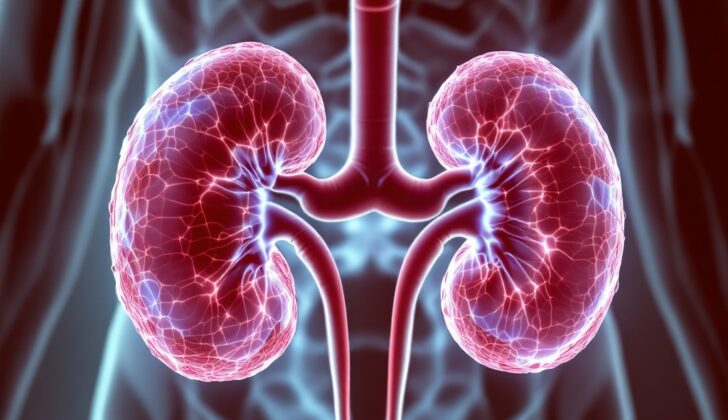
What is Xanthogranulomatous Pyelonephritis ?
Xanthogranulomatous pyelonephritis is a rare but serious type of chronic kidney inflammation that can lead to a non-working kidney. This variation often goes hand in hand with chronic blockages and kidney stones, along with ongoing or repeated infections.
This disease is often mistaken for more frequent conditions because the symptoms can be very similar. For this reason, it’s sometimes called a pseudotumor, a false tumor, as the affected kidney increases in size and acts like a tumor, damaging nearby tissues.
The disease destroys and replaces a portion of the kidney or nearby tissue with a type of tissue called granulomatous. This tissue contains specialized cells, called lipid-laden macrophages, filled with fats. The name of the condition, xanthogranulomatous, is derived from the Greek word xanthós, which means “yellow”, referring to the yellow fatty cells seen under microscopy. This kidney condition was first identified in 1916 but only got its official name in 1944.
Xanthogranulomatous pyelonephritis can often be wrongly thought to be a type of kidney cancer, due to its similar symptoms and the way it appears on medical imaging. As it can affect nearby organs, it’s essential for it to be identified and treated as early as possible to lower the risk of further health issues. Acute infections can be treated with antibiotics but the typical treatment involves surgery to remove partially or completely the affected kidney.
What Causes Xanthogranulomatous Pyelonephritis ?
The exact cause of a kidney condition called xanthogranulomatous pyelonephritis is still unknown. However, it’s often seen in people who have chronic urinary blockages and urinary tract infections. Common bacteria linked to this condition include Escherichia coli and Proteus mirabilis, along with Pseudomonas, Enterococcus faecalis, and Klebsiella.
A common cause of urinary blockages in people with this condition is the formation of kidney stones, especially a type called staghorn calculus. This is found in almost 80% of patients with this condition. This stone can serve as a site of infection that gradually damages the kidneys. But in children, birth defects that disrupt normal urine flow can lead to chronic urinary blockages and increase the risk of this condition because kidney stones are less common in children.
Other reasons for urinary blockages, such as abnormal formation of the urinary tract, a backward flow from the bladder to kidneys, and bladder cancer, have also been linked with this condition. These problems, along with having kidney stones and frequent urinary tract infections, are seen as major risk factors for this kidney condition. Other kidney conditions associated with it include chronic inflammation of the kidneys, kidney cancer, and a type of cancer that begins in the cells that line the urinary tract.
There are also several other factors that can increase the risk of getting this kidney condition. These include abnormal fat metabolism, a genetic condition called brachydactyly mental retardation syndrome in children, diabetes, Hepatitis C, high blood pressure, a weakened immune system, metabolic syndrome, kidney stones, recurrent urinary tract infections, kidney transplants, rheumatoid arthritis, blockages of the connection between the kidney and the tube that carries urine to the bladder, and a condition where urine flows backward from the bladder into the kidneys.
Risk Factors and Frequency for Xanthogranulomatous Pyelonephritis
Xanthogranulomatous pyelonephritis is a kidney infection that makes up about 0.6% to 1% of all renal (kidney) infections. Although it can appear in any age group, it is more likely to affect women aged 45 to 55. It isn’t associated with any particular race. Typically, only one kidney gets affected, but in rare cases, it can occur in both kidneys equally.
This disease is extremely rare in children, yet it’s responsible for 16% of all kidney removal surgeries (nephrectomies) in this age group. Children below the age of 8 are more prone to this infection than older children.
- Xanthogranulomatous pyelonephritis accounts for 0.6% to 1% of kidney infections.
- It tends to affect women between the ages of 45 and 55 more than men.
- No specific racial group is more prone to this infection.
- Usually, only one kidney gets affected but in rare cases, both kidneys could be infected.
- Even though it is really rare in kids, it still accounts for 16% of all kidney removal surgeries in children.
- Children under 8 are more likely to be affected than older ones.
- The general annual incidence is reported as 1.4 cases per 100,000 population.
- Around 4% of patients with chronic pyelonephritis develop xanthogranulomatous pyelonephritis.
Signs and Symptoms of Xanthogranulomatous Pyelonephritis
Xanthogranulomatous pyelonephritis typically occurs in middle-aged women who have a history of repeated urinary tract infections, commonly brought on by bacteria like Escherichia coli or P mirabilis. Kids with this condition may experience symptoms like fever, abdominal pain, and slower development. Around 50% of people with this condition present with a noticeable lump in the abdomen.
It’s important to note that this condition can look a lot like kidney tuberculosis, with similar symptoms. Therefore, doctors might also factor in any travel to areas where kidney tuberculosis is prevalent or any other risk factors associated with the disease.
For adults, the main symptoms of xanthogranulomatous pyelonephritis are:
- Dull and continuous pain in the side
- Tiredness
- Fever
- Painful urination
- Blood found in urine
- Increased need to urinate
- Loss of appetite
- Chills
- Unexplained weight loss
In children, the condition can manifest itself in two ways:
- A more common form involves the entire kidney and affects boys and girls equally.
- A less common form involves a localized area of the kidney, primarily affecting girls.
Physical signs may include:
- Pale eyes due to anemia
- Sensitivity in the side on palpation
- Fever
- A noticeable lump in the kidney area
In severe cases, the disease may begin to involve other parts of the body, leading to symptoms such as:
- Cutaneous fistula that drains, often mistaken as a minor abscess
- Enlarged liver or abscess near the liver resulting in abdominal pain, nausea, fever, fatigue, or cough
- Engagement of the spleen leading to abdominal pain, fever, and chills
- Gluteal or psoas abscess causing abdominal pain, fever, or swelling
Testing for Xanthogranulomatous Pyelonephritis
If your doctor suspects you may have xanthogranulomatous pyelonephritis, a type of kidney infection, they’ll need to examine you thoroughly and order several tests. However, correctly diagnosing this condition before surgery is only possible about 40% of the time, even with these tests.
Initial tests typically involve taking a sample of your blood and urine. The blood test might show low levels of red blood cells (anemia) and high levels of white blood cells (leukocytosis), which can suggest an infection. Other markers of infection, like C-reactive protein and lactic acid, might also be high. In the urine, signs of a urinary tract infection like bacteria, blood, and white blood cells could be present. The urine pH (acidity level) might also be elevated.
Your doctor may also perform tests to check your liver and kidney function. People with xanthogranulomatous pyelonephritis often have issues with both. In fact, even treating the condition may not improve kidney function. As for liver function, it might only be mildly impacted in some patients.
Imaging tests are also a crucial part of diagnosing this condition. These can involve an X-ray, ultrasound, CT scan, MRI, and others to get a detailed look at your kidneys. A common finding in xanthogranulomatous pyelonephritis patients is a larger-than-normal kidney, kidney stones, and a poorly functioning kidney.
An X-ray can detect kidney stones, and ultrasound might show things like enlarged kidneys, kidney stones, and other changes. However, ultrasound doesn’t always distinguish between this condition and other similar ones very well.
A CT scan is quite helpful in diagnosing xanthogranulomatous pyelonephritis but may show better results when used with a contrast solution. It can help identify kidney stones as well as specify the presence and spread of the infection. MRI and scintigraphy, a nuclear medicine scan, may also be used but are often considered secondary options.
If other tests don’t provide clear results, a kidney biopsy may be necessary. This test can be tricky, as it involves distinguishing between this condition and a certain type of kidney cancer. If your treatment won’t change based on the outcome (i.e., if you’ll require kidney removal either way), a biopsy may not be necessary.
Treatment Options for Xanthogranulomatous Pyelonephritis
If you have focal or segmental xanthogranulomatous pyelonephritis – a severe kidney infection where the kidney tissue gets destroyed and replaced by other types of tissue like fat – your initial treatment is likely to be antibiotics and drainage of the affected area. Urine cultures will probably be ordered to assist in tailoring your antibiotic treatment which will start broad, and possibly be adjusted based on the culture result. You might also want to speak to an infectious disease specialist during this time.
After you have started to show signs of improvement, your doctor may begin considering if removal of part or all of your kidney – a procedure known as nephrectomy – is necessary. If, despite this initial therapy, your situation hasn’t shown any improvement, surgery might then be performed.
Complete recovery from this infection without recurrence is usually possible, so if it’s safe, a partial nephrectomy is recommended. This is especially so in children, where it’s always preferable to try and spare as much kidney function as possible where feasible. However, it’s important to note that in most cases, by the time the diagnosis of xanthogranulomatous pyelonephritis is made, a lot of the kidney function may not be salvageable, and most patients end up going through a nephrectomy.
For patients with advanced stage xanthogranulomatous pyelonephritis, the standard treatment is nephrectomy. Though antibiotics may be used pre and post-surgery to control the infection and avoid complications, they are typically not enough for treating this condition. You will likely be put on an antibiotic regimen for about 2 weeks after you have had your nephrectomy.
The drainage treatments we mentioned earlier might show limited value for patients with nonfunctioning kidneys. Bowel preparation will be done before the surgery because it’s not uncommon for such procedures to get complex and difficult.
The surgery aims to get rid of any stones in the kidney and all infected tissue so as to reduce the risk of complications. It’s also worth noting that during this procedure, the surgeons may also have to fix fistulas or sinus tracts that have formed due to the infection. This would involve the removal and draining of infected fluid from any abscesses that have formed, as well as repairing damaged tissue.
During the surgery, the surgeons will also ensure any associated tissues that might become cancerous are removed. Your doctor might also recommend thorough antibiotic irrigation to keep the area free from infection and help it heal.
If the infection has spread to both kidneys, you might have to go through a bilateral nephrectomy, essentially resulting in the loss of both your kidneys. Such a scenario would entail you going on long-term dialysis, but it is possible to spare some kidney function particularly in children.
Laparoscopic nephrectomy, a minimally invasive surgery technique, is the preferred method for patients where the kidneys have ceased to function completely. However, this technique might come with its own difficulties due mostly to significant inflammation. Even then, it is a preferred choice because it offers lesser blood loss, fewer complications, reduced hospital stays, and better outcomes compared to other open surgeries.
This conversion rate of laparoscopy to open surgery is expected to decrease over time as surgeons improve their skills and techniques. But right now, it’s crucial that these are performed by surgeons who are experienced in high level laparoscopic procedures because they can be pretty complicated.
It’s also important for your surgeon to consider several factors during the surgery from planning based on CT scans, preserving important landmarks, clipping off the ureter to prevent spillage, to even whether a laparoscopic or open surgical approach is best for your case. All these are done to ensure the best possible outcome for you.
If the situation demands, a general surgeon might be needed on the team since the infection could have involved other organs beyond your kidneys. It’s also important for your medical team to be cautious about vascular injury which can occur easily, especially while dissecting an inflamed renal hilum, or the area where the vessels and nerves enter the kidney.
Research involving over 1000 cases of xanthogranulomatous pyelonephritis has revealed some important insights – fistulas were present in 8% of the patients, the accurate diagnosis before the operation was only made in 45% of the cases and only 59% of cases had urine cultures that tested positive. This same study found that laparoscopic nephrectomy was performed in just 34% of the patients, whilst a partial nephrectomy was performed in a minor 2% of cases.
What else can Xanthogranulomatous Pyelonephritis be?
When trying to diagnose xanthogranulomatous pyelonephritis, doctors will need to consider various other diseases or conditions. These include:
- Clear cell renal cell carcinoma
- Complex renal cyst
- Leiomyosarcoma
- Lymphoma
- Malakoplakia
- Megalocytic interstitial nephritis
- Oncocytoma
- Papillary renal cell carcinoma
- Renal abscess
- Renal angiomyolipoma
- Renal oncocytoma
- Renal tumor
- Renal tuberculosis
- Sarcomatoid renal cell carcinoma
- Squamous cell carcinoma of the kidney
- Wilms tumor
The symptoms and x-ray findings for xanthogranulomatous pyelonephritis and renal cell carcinoma might look very similar. However, fever and increased levels of inflammation indicators, like C-reactive protein and the speed at which red blood cells settle at the bottom of a test tube (erythrocyte sedimentation rate), are usually linked to xanthogranulomatous pyelonephritis.
Something called the ‘bear paw’ sign can be seen in xanthogranulomatous pyelonephritis cases. If there’s still some doubt about the diagnosis, a tissue sample (histological) examination can provide certainty.
What to expect with Xanthogranulomatous Pyelonephritis
The overall outlook for xanthogranulomatous pyelonephritis, a severe form of kidney inflammation, significantly improves with prompt treatment. Cases affecting only one kidney generally fare much better than those affecting both kidneys, which can often be fatal.
A laparoscopic radical nephrectomy, a surgery for removing a kidney, is the preferred treatment option, and it doesn’t typically result in a recurrence of the illness. However, nephron-sparing surgeries, operations that try to preserve as much kidney function as possible, may also be performed when it’s feasible. A study reported improved outcomes with minimal invasion techniques, i.e., surgeries that make smaller cuts into the body, however, despite these advancements, surgery still has a mortality rate of 7% to 10% due to the complications of surgical removal.
Different surgical approaches may result in different complications. For example, a transperitoneal approach, a type of surgical method, may cause more blood loss than a retroperitoneal one. Pay attention that a positive urine culture might be an indicator for a worse prognosis, or prediction of the likely course or outcome of the disease.
Possible Complications When Diagnosed with Xanthogranulomatous Pyelonephritis
A large-scale study found that certain patients suffering from xanthogranulomatous pyelonephritis are more likely to experience serious complications. These include those with a quick Sepsis-related Organ Failure Assessment (qSOFA) score of 2 or more, signs of kidney failure before surgery, disease spreading to the surrounding kidney tissues, positive urine cultures, or an enlarged kidney.
Around 30% to 40% of patients with xanthogranulomatous pyelonephritis experience complications. These can include sepsis (a severe infection), wall abscess (a pocket of pus in the wall of an organ), psoas abscess (a pocket of pus in the psoas muscle), and hemorrhage (abnormal bleeding).
Complications could also occur due to the disease affecting nearby organs and might include:
- Skin fistula (an abnormal connection between an organ and the skin)
- Injury due to medical intervention
- Infections such as abscesses, wounds, or urinary tract infections
- Nephrocolonic or pyeloduodenal fistula (an abnormal connection between the kidney and colon or between the kidney and the first part of the small intestine)
- Nephrocutaneous fistula (an abnormal connection between kidney and skin)
- Perinephric abscess (a pocket of pus around the kidney)
- Postoperative abscess (a pocket of pus after surgery)
- Psoas abscess
- Kidney failure
- Secondary amyloidosis and nephrotic syndrome (severe kidney diseases)
- Sepsis
- Vascular injury (damage to blood vessels)
Recovery from Xanthogranulomatous Pyelonephritis
Doctors recommend yearly imaging of the other kidney and encourage swift and thorough treatment of urinary tract infections. Abnormalities or issues with emptying the lower urinary tract should be corrected, if possible.
After surgery, if patients develop infections in the wound area, form an abscess (a pocket of pus in the body), or cutaneous fistulas (abnormal passages between a hollow or tubular organ and the body surface), they may require a CT scan. This is a type of detailed x-ray. They may also need to have any collected pus drained with a needle and given antibiotics that go straight into the vein (referred to as intravenous antibiotics).
If the patient has had kidney stones before, or the other kidney shows signs of kidney stones (calculi), patients who are at risk of developing more kidney stones (nephrolithiasis) should have tests done on their urine collected over a 24-hour period. They should also be given specific treatment to help prevent any more kidney stones from forming.
Preventing Xanthogranulomatous Pyelonephritis
When you’ve been treated for a medical condition called “xanthogranulomatous pyelonephritis”, which is a severe infection of your kidneys, there are several post-treatment care steps to follow:
* Continue taking antibiotics for at least 2 weeks after your procedure. This helps to ensure all germs are completely cleared from your body.
* It’s recommended to have annual imaging tests, similar to x-rays, on the healthy kidney to ensure it is working well and not getting the same infection.
* If you have recurring urinary tract infections (UTIs), further investigations will be done to find and sort out any conditions, wounds or blockages that might be causing these infections.
* Any new UTI should be promptly and thoroughly treated with antibiotics to prevent any complications.
* If you have been diagnosed with “nephrolithiasis”, which is the medical term for kidney stones, you should have a special urine test called a “24-hour urine test”. This allows your doctor to find out more about your kidney stones and discuss any necessary preventative actions with you.


