What is Hypoplastic Lung Disease?
Pulmonary hypoplasia is a rare condition that babies can be born with, where the lung tissue isn’t fully developed. Because of this, the baby may have trouble breathing due to fewer airways and alveoli, which are the tiny air sacs in the lungs where oxygen and carbon dioxide are exchanged.
This conditions is seldom seen without the presence of other abnormalities in the baby’s development. However, in 1912, Schneider offered a classification for abnormal lung development, which was later updated by Boyden in 1955. There are three types:
* Type 1 (Agenesis): The lung tissue, bronchus, and blood vessels are completely missing.
* Type 2 (Aplasia): The lung tissue is completely absent, but a basic bronchus is present on the affected side.
* Type 3 (Hypoplasia): There are varying amounts of lung tissue present with fewer lung cells, airways, and alveoli.
The severity of a case of pulmonary hypoplasia depends on when during the baby’s development the issue arises. Interruption in the lung’s development happens during a stage called the pseudo glandular stage, which takes place from the 5th to the 17th week of pregnancy. At this stage, the baby’s lung development relies on mechanical stimuli. Any pressure imbalance between the space outside the airway and the space within the airway could result in compression of the lung(s) and therefore hypoplasia.
This condition can range drastically in severity. In its most severe form, it can cause fatal breathing difficulties in newborns. Conversely, in its mildest form, it can lead to long-term lung disease with repeated respiratory infections in adults.
What Causes Hypoplastic Lung Disease?
Primary pulmonary hypoplasia, a condition where the lungs don’t develop properly, is believed to be caused by several factors. These include genetic and environmental influences, as well as maternal and nutritional aspects. Instances of this condition are frequently found in medical issues like congenital acinar dysplasia and genetic disorders like trisomy 21.
Secondary causes of pulmonary hypoplasia, or underdevelopment of the lungs, occur due to problems affecting the chest cavity or the amount of amniotic fluid, which is the fluid that surrounds the baby in the womb.
Certain health conditions can cause the unborn baby’s lungs to not develop fully. Examples of this are congenital diaphragmatic hernia (CDH), which is a hole in the diaphragm, and congenital pulmonary airway malformation (CPAM), which is a type of lung cyst. These issues, along with deformities of the chest wall, can disrupt the development of the fetal lungs before 16 weeks of pregnancy.
On the other hand, possible complications such as inadequate amniotic fluid (oligohydramnios), due to the unborn baby’s kidneys not functioning properly or other urinary tract problems, can affect lung development after 16 weeks of pregnancy. Prolonged premature rupture of membranes (PPROM) which means the amniotic sac breaks open before 37 weeks of pregnancy, can also hinder lung development.
An early rupture of membranes, coupled with low amniotic fluid, especially if it occurs before the 25th week of pregnancy and the pocket of fluid is less than 1 cm, has been linked to a higher risk of fatal pulmonary hypoplasia. They are considered independent risk factors that might lead to this severe lung condition.
Risk Factors and Frequency for Hypoplastic Lung Disease
The exact number of people affected by pulmonary hypoplasia, a lung condition, isn’t known for sure. However, estimates indicate roughly 1.4 out of every 1000 births, and about 0.9 to 1.1 out of every 1000 live births. Some think these numbers may be too low because babies with less severe symptoms might survive as newborns only to experience breathing symptoms later in life.
- A research study in the United States found that of 163 births where the mother’s water broke early (between 15 to 28 weeks of pregnancy), 12.9% (or 21 births) resulted in babies with pulmonary hypoplasia.
- In that group, the mortality rate was very high – 95.2% (20 out of 21 babies) did not survive.
- This compares to a survival rate of 48.2% (68 out of 141) in babies whose mothers did not experience early water breakage and were not diagnosed with pulmonary hypoplasia.
- Another study done in Spain found that the mortality rate was 47% within the first 60 days of life.
- In addition, 75% of those deaths occurred on the very first day of life.
Signs and Symptoms of Hypoplastic Lung Disease
Pulmonary hypoplasia, or underdeveloped lungs, can cause various symptoms based on how severe the condition is. The majority of cases are life-threatening. In cases that aren’t lethal, symptoms usually appear in early childhood and range from mild to severe difficulty breathing, depending on how much the condition has progressed. During prenatal care, doctors might note less fetal movement, prematurely ruptured fluid membranes, and a lack of amniotic fluid. Severe cases can lead to additional complications like high blood pressure in the lungs, lung disease in newborns, and bleeding in the small airways of the lungs. Mild cases may go unnoticed until adulthood, when they may be found during a chest X-ray or because of repeated lung infections. Sometimes, the only symptom may be lowered oxygen levels during physical activity.
In cases where only one lung is underdeveloped, there could be a noticeable difference in the size of the two halves of the chest, limited chest wall expansion, and weaker breathing sounds on the affected side. The organs from the other side of the chest may also push into the underdeveloped side. When both lungs are underdeveloped, it typically causes deformities in the chest’s shape. Most of these patients often breathe rapidly and struggle for air with shallow breaths. Sometimes, pulmonary hypoplasia may come along with other birth defects, which could be the first clue that leads doctors to suspect the presence of this condition.
Other defects related to the heart, digestive tract, urinary system, and skeletal system may also coexist. Cases with a congenital diaphragmatic hernia will present with a sunken abdomen and difficulty in breathing right after birth. These cases may also have association with birth defect in heart, neural tube defects, or kidney abnormalities. Potter syndrome, a condition that causes a specific set of physical abnormalities, can also present with underdeveloped lungs due to lack of amniotic fluid. The other features of Potter syndrome may include facial abnormalities, a broad distance between the two eyes, large and low-set ears, a flat nose, unusual hand shape, and inflexible joints.
Testing for Hypoplastic Lung Disease
Typically, regular lab tests don’t offer enough insight for cases of low amniotic fluid (oligohydramnios). However, your doctor may test your kidney health by checking your creatinine, blood urea nitrogen, and electrolyte levels. In cases where one lung doesn’t develop fully (unilateral lung hypoplasia), an x-ray of the chest often shows a lack of space on one side of the chest with overcrowd ribs and a shift in the heart and other structures towards that side. The other lung often expands more than normal to make up for the underdeveloped one. If air leaks into the chest, the x-ray will show this as well.
A CT scan of the chest can confirm whether a lung is underdeveloped and rule out other conditions like Swyer James syndrome or a partially collapsed lung. A CT scan typically shows the absence or incomplete development of a lung passage (bronchus), smaller blood vessels, and a collapsed lung on the affected side. Another test, called ventilation-perfusion scanning, checks how well air and blood are flowing through the lungs. It typically shows less air and blood flow in the affected part of the lung.
An ECG, which checks the electrical activity of your heart, can differentiate between conditions like dextrocardia (heart positioned on the right side of the chest) and dextroversion (heart pointing towards the right) in cases of right lung underdevelopment. An ECG typically shows a shift in the heart’s electrical axis towards the right, a positive wave pattern in a specific section of the ECG, and inverted waves in another specific section.
A heart ultrasound (2D Echo) can help rule out any congenital heart anomalies.
During pregnancy, the doctor might utilize 2D ultrasound to check measures like lung area, thoracic circumference (TC), and the ratio between thoracic circumference and abdominal circumference (TC:AC). These measures can give insight into the risk of the baby developing an underdeveloped lung (pulmonary hypoplasia). Volumetric assessments by MRI or 3D ultrasonography have also been tried for this purpose but aren’t used regularly due to their varied results.
A lung function test, performed in late childhood or adulthood, identifies any breathing difficulties due to lung underdevelopment. Pathological criteria used for diagnosing this condition include certain measures of lung weight, size of small air sacs in the lung (radial alveolar count), and a specific ratio of lung weight to body weight.
Other alternative methods include counting the number of air sacs per unit volume, counting airway branches, and checking the amount of lung DNA. Microscopic examination can show a reduction in lung cells, fewer branches of lung airway, immature lung cells, smaller and thicker blood vessels in the lung, and low amounts of a substance called surfactant, that helps keep the air sacs in the lungs open. These all are different criteria used by healthcare providers in diagnosing lung underdevelopment. However, there isn’t an established set of guidelines that are strictly used for this purpose.
Treatment Options for Hypoplastic Lung Disease
Treatment for this condition begins during pregnancy. In cases when the water breaks too early (preterm premature rupture of membranes), doctors often use medication to boost the development of the baby’s lungs. They may test the amniotic fluid (the fluid surrounding the baby in the womb) to check the baby’s risk of complications.
A combination of antibiotics, medication to stimulate lung development (corticosteroids) and medication to temporarily stop preterm labour (tocolytics) may be used.
One study of 49 patients found that the ongoing injection of fluid into the amniotic sac (known as serial amnioinfusions) may reduce complications for the baby and prolong pregnancy when the waters break very early (before 26 weeks). Another study found no significant differences in the health of the baby or mother with this treatment, suggesting that more research is needed to understand its benefits.
Another treatment option is an ‘amniopatch’. This is an injection of platelets (blood clotting cells) and a frozen blood product known as ‘cryoprecipitate’ into the amniotic fluid.
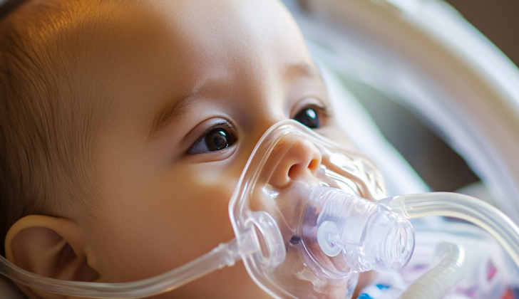
After birth, the newborn may need help with breathing. This can range from extra oxygen to more intensive interventions like using a specialised machine to support the lungs. There’s limited evidence to suggest an inhaled treatment called nitric oxide could be beneficial, but we need more research in this area.
In relation to congenital diaphragmatic hernias – a birth defect where there’s a hole in the diaphragm – a procedure called a fetal endoscopic tracheal occlusion can be used. This procedure blocks the baby’s windpipe, helping the lungs develop and reducing the risk of high blood pressure in the lungs. However, it also reduces certain cells that create a substance crucial for lung function (surfactant proteins). To restore these proteins, the blockage is removed just before birth.
The repair of the diaphragmatic hernia is usually postponed for 48 to 72 hours after birth to allow the newborn to become more stable.
Survivors of pulmonary hypoplasia (underdeveloped lungs) commonly have chronic lung disease. Conservative treatment options for these adults include medication to widen the airways, antibiotics for lung infections, chest physiotherapy, and preventative vaccinations. If the diseased area of the lung is confined to a small area and causes repeated lung infections, it might be recommended to surgically remove that part of the lung.
What else can Hypoplastic Lung Disease be?
When doctors see a lower than normal amount of air in one lung on an imaging scan, they consider several possible causes:
- Scimitar syndrome: This is an underdevelopment of the right lung, where the blood that should go to the right lung instead goes to a large vein called the inferior vena cava.
- Congenital pulmonary airway malformation: This is a birth defect where a cyst forms in either the windpipe or the air tubes going to the lungs. This can push other organs and tissues to the opposite side of the chest.
- Bronchopulmonary sequestration: This occurs when a section of the lung isn’t connected to the rest of the lung’s airways. Unlike other parts of the lung, it gets its blood supply from the body’s artery system instead of from the lungs. This can cause other organs and tissues to move to the side of the chest with the disconnected lung segment.
- Congenital lobar emphysema: This is another birth defect where a section of the lung gets too inflated. This can push other organs and tissues to the opposite side of the chest.
- Swyer James or Macleod syndrome: This condition occurs after an infection in the small airways of the lungs (bronchiolitis), leading to an overly bright-looking lung on an imaging scan.
- Persistent pulmonary hypertension of the newborn (PPHN): This is when a newborn’s circulation system does not adapt well to breathing outside the womb, putting extra pressure on the lungs.
What to expect with Hypoplastic Lung Disease
Primary pulmonary hypoplasia, a condition where the lungs don’t develop fully, is very rare but often deadly. Most cases are secondary, meaning they are caused by another condition or factor, and can result in serious health issues for survivors. Typically, up to 70% of newborn babies diagnosed with the condition might not survive.
Nevertheless, patients with pulmonary hypoplasia affecting only one lung usually live healthy lives and continue to grow and develop normally, provided there are no associated problems.
For patients with secondary pulmonary hypoplasia, the symptoms and outcomes can vary greatly depending on what’s causing the condition. For those born with a hole in the muscle separating the chest and the stomach (known as a congenital diaphragmatic hernia), mortality can be as high as 50% in the newborn period. The main factors affecting their survival are the under-development of the lungs (pulmonary hypoplasia) and high blood pressure in the lungs (severe pulmonary hypertension), with outcomes being worse if the hole is on the right side of the diaphragm.
Other factors that can affect the prognosis include complications related to the brain, digestive system, muscles and bones, or nutrition. Babies who overcome this condition may go on to experience chronic lung problems such as reduced capacity for exercise and increased vulnerability to infections.
Possible Complications When Diagnosed with Hypoplastic Lung Disease
Babies with underdeveloped lungs (or pulmonary hypoplasia) can suffer from several complications. This can range from sudden severe breathing difficulties (acute respiratory failure), softening of the windpipe (tracheomalacia), persistent high blood pressure in the lungs, and a condition called pneumothorax where air gets trapped in the space around the lungs. This condition can either happen spontaneously or can be due to using a mechanical ventilator.
Down the line, babies who survive pulmonary hypoplasia may experience delayed growth, chronic lung disease, frequent chest infections, lower physical strength capacity, and changes to the shape of their chest, like developing a curved spine (scoliosis).
Common Complications:
- Acute respiratory failure
- Tracheomalacia
- Persistent pulmonary hypertension
- Pneumothorax
- Delayed growth
- Chronic lung disease
- Frequent chest infections
- Lower physical exercise ability
- Chest deformities like scoliosis
Preventing Hypoplastic Lung Disease
If a patient has the condition pulmonary hypoplasia, which means underdeveloped lungs, they will require health check-ups and care over an extended period of time. To manage this, a team of different healthcare professionals is needed. This team might include children’s doctors (pediatricians), lung specialists (pulmonologists), critical care doctors (intensivists), heart surgeons (cardiovascular surgeons), and kidney doctors (nephrologists).
It’s important for the patient and their caregivers to start learning about the condition as early as possible, even before the baby is born, if possible. Understanding the usual course of the condition, the potential problems that might come up, and what the future might look like can help everyone involved in making decisions about care and treatment.


