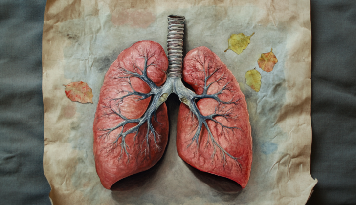
What is Lung Torsion?
Lung torsion is a rare condition where the lung twists, typically caused by changes in the chest cavity from surgery, lung transplants, or injuries. However, there have been cases where it happened without any apparent cause. This situation can be life-threatening and needs to be diagnosed quickly. The twisted lung can disrupt blood flow and block the airway, leading to death of lung tissue. However, with immediate attention and treatment, the affected part of the lung can get better. Unfortunately, prognosis for lung torsion is generally not good due to misdiagnosis and delays in treatment. If the twist is undone and the lung is stabilized, it can recover completely. This summary will address the causes, frequency, symptoms, diagnosis, and treatment of lung torsion.
What Causes Lung Torsion?
Lung torsion, also known as a twisted lung, may occur in patients who have previously had procedures relating to the chest area, though it can also occur naturally. A twisted lung in adults can be caused by several things, such as:
* Injuries to the chest or abdomen
* Lung transplant, which can involve one or both lungs
* A type of minimally invasive chest surgery known as VATS (video-assisted thoracoscopic surgery)
* Thoracentesis, a procedure to remove fluid from the space between the lungs and the chest wall
* Different procedures involving the chest, like transesophageal operations (procedures performed through the esophagus) or repairs to the aorta, the main blood vessel carrying blood from your heart to your body
* Corrections to a hiatal hernia, which occurs when part of your stomach bulges through the large muscle separating your abdomen and chest (diaphragm), carried out through the chest
* A procedure using a needle to remove fluid or cells from the chest, known as transthoracic needle aspiration
* Surgery to fix a diaphragmatic hernia, a birth defect that allows abdominal organs into the chest cavity
* Spontaneous causes can also occur, such as pneumothorax (a collapsed lung), pleural effusion (buildup of fluid between the layers of tissue that line the lungs and the chest cavity), lobar atelectasis (collapse of a lobe of the lung), pulmonary sequestration (a mass of non-functioning lung tissue), or a diaphragmatic hernia
In children, known causes of a twisted lung include:
* Blunt force trauma to the chest and abdomen
* Repair of a tracheoesophageal fistula, an abnormal connection between the esophagus and the trachea
* Surgery for a hiatal hernia
* Ductus arteriosus closure, a procedure to close an extra blood vessel found in newborns shortly after birth
Risk Factors and Frequency for Lung Torsion
Lung torsion is a rare condition that occurs in only 0.089% to 0.3% of patients, according to a case study. The majority of these cases (62.4%) were found in patients after surgery. A smaller percentage (8.3%) occurred following physical trauma, and in 29.4% of patients, the condition developed spontaneously.
Regarding specific surgeries, 21.6% of lung torsion cases took place after Video Assisted Thoracic Surgery (VATS), while a significant 78.4% happened after a thoracotomy, a surgical operation where the chest is cut open. The study also looked into the exact location where lung torsion often develops. It was found that the majority of lung torsions (74.4%) occurred after the removal of the right upper lobe (a type of lung surgery). Furthermore, in 29.4% of patients who had thoracic surgery, the right middle lobe was the most affected part.
The case study also indicated that lung torsion affects both genders relatively equally. Among the cases studied, it was seen in 58.3% of males and 41.7% of females.
Signs and Symptoms of Lung Torsion
Lung torsion, or the twisting of the lung, can be quite tricky to diagnose just based on physical examination or symptoms. People with this condition may experience fever, chest pain, breathing difficulties, and coughing. In fact, the most common symptoms include difficulty breathing, fever, and chest pain. These usually appear 4 to 14 days after a chest surgery, injury, or some other event. However, some people might not have any symptoms or signs at all.
Physical examinations may suggest some factors like low oxygen levels in the body (hypoxia), discomfort in breathing, and rapid breathing (tachypnea). But to correctly diagnose lung torsion, radiological imaging (like an X-ray or CT scan) is necessary.
Testing for Lung Torsion
Medical tests can often uncover signs of a health condition, like leukocytosis, which happens when your body has too many white blood cells. However, this may not always be present. An arterial blood gas test, which measures the amount of oxygen and carbon dioxide in your blood, might also appear normal and not display the low oxygen levels that can be associated with this condition. Because of this, doctors often use imaging techniques for diagnosis.
If your doctor suspects lung-related issues, they might use an x-ray to look for signs of worsening lung consolidation, when the small air sacs in your lungs fill up with a substance like pus or fluid. The x-ray might also show parts of your lung separating from the pulmonary artery, a blood vessel that carries oxygen-poor blood from the heart to the lungs. Images from x-rays can also reveal anatomical abnormalities or cloudy areas in the lung, which might indicate a disease such as pneumonia. However, a single x-ray might not be enough so doctors often take several x-rays over time to track the progression of the disease.
Bronchoscopy, which involves inserting a thin tube with a light and camera into your airways, can help detect lung torsion, a condition where part or all of the lung twists upon itself. This condition can cause symptoms such as difficulty breathing and chest pain. during the procedure, your doctor might find the entrance to the bronchus (the airway that leads from the trachea to the lungs) looking like a “fish mouth” or that the bronchus is narrowed or twisted.
However, bronchoscopy may not always provide a full diagnosis of lung torsion, as not all patients may exhibit these symptoms. In such cases, a CT scan – a type of x-ray that can provide a 3D image of your lungs – is often essential in confirm the diagnosis. This scan can show blocked bronchial arteries (the blood vessels supplying air passages in the lungs), areas of your lung that look opaque, or underinflated lungs and collapsed lung lobes. It can also reveal the narrowing of air passages in the affected lung or the bronchus, as well as different possible angles of its rotation. The position of any lesions may also change with progress of the lung torsion, as seen on follow-up CT scans.
CT angiography, which uses a contrast dye to take images of blood vessels, can show if the pulmonary artery is suddenly truncated or blocked. The imaging could also reveal increased tissue thickness between lobes of the lung as well as signs of venous congestion. In severe cases, if lung tissue has died (became necrotic), this test would show a loss of lung tissue and blood vessels.
Treatment Options for Lung Torsion
If a patient’s lung gets twisted (a condition called “lung torsion”), it’s crucial to act quickly before significant tissue damage happens. If treated promptly, the twisted lung can be straightened out (“detorsion”), and the lung can recover fully. To prevent the lung from twisting again, it’s stitched or stapled to the surrounding tissues.
However, if it’s not possible to straighten the twisted lung, or if the procedure to fix it doesn’t work, then part of the lung may need to be removed, a procedure called “lobectomy.” To save the lung, the treatment should be done within a few hours of diagnosis because, if too much time passes, the tissue could be permanently damaged due to insufficient blood supply. If there is a severe risk, doctors might decide to directly remove the twisted part of the lung instead of attempting to untwist it. This is to prevent the harmful substances built up during the torsion from spreading to the rest of the body, which could lead to multiple organ failure.
If the lung remains twisted for too long, a blood clot could form, leading to serious problems like a pulmonary embolism or stroke. To prevent this, a medication called heparin may be given to the patient. It’s worth noting that if left untreated or managed solely with medicines, patients could develop repeated episodes of pneumonia that can be fatal. However, surgeries to fix a twisted lung usually have a good outcome, with very few incident of pneumonia, air leaks, or emphysema afterwards and most patients recover without any complications during or after surgery.
What else can Lung Torsion be?
In some studies, it was found that the condition called lung torsion was wrongly diagnosed nearly 18.3% of the time. This is a list of conditions that could be mistaken for lung torsion:
- Blood accumulation in the chest cavity (Hemothorax)
- Bleeding inside the body (Hemorrhage)
- Lung infection (Pneumonia), either due to germs or by inhalation of substances
- Contusion (which can be seen after a specific type of lung surgery and may appear similar to lung consolidation but usually resolves within a few days)
- Death of lung tissue (Lung gangrene)
- Infection in the lung tissue (Parenchymal infection)
- Collapsed lung (Atelectasis)
- Lung tumor
- Trapped fluid in the chest (Loculated effusion)
- Destruction of lung tissue due to long-term lung conditions (Emphysema)
- Unintentional tying off of the lung pedicle (Inadvertent ligation of the hilum)
- Hiatus hernia (Diaphragmatic herniation)
- Leak from the surgical connection point (Leakage of the anastomosis site)
Specific types of scans such as a CT scan, CT angiography, or a bronchoscopy can rule out these other conditions. The correct diagnosis of lung torsion is usually confirmed by viewing the obstructions in the blood vessels and airways on the CT scans.
What to expect with Lung Torsion
If the treatment for lung torsion, a condition where the lung rotates on itself, is delayed, it can lead to serious complications, like poor blood flow to the lung tissue or severe infection called sepsis. These situations can increase the chance of death, up to as high as 8.3%.
It’s also important to note that the chance of death from lung torsion can be higher if the whole lung is twisted, rather than just a part of it, known as lobar torsion.
Both methods of managing lung torsion, which involve repositioning the lung or directly repositioning and cutting away affected tissue, have similar survival rates. However, a method called indirect resection, which involves removing lung tissue, has shown a higher chance of death.
Furthermore, patients with lung torsion who got this condition due to trauma have the highest mortality rate at 22.2%, followed by those undergoing chest surgery at 8.8% and those who experience lung torsion spontaneously, without any clear cause at 3.1%.
Possible Complications When Diagnosed with Lung Torsion
Correcting lung torsion, a severe condition where the lung twists on its axis, has several known risks. This is a surgical procedure that needs to be performed immediately. Here’s a simplified list of potential complications:
- Pneumonia – a lung infection
- Cerebrovascular accident – a stroke
- Necrosis of the lung tissue – death of lung tissue
- Hemorrhage – excessive bleeding
- Vocal cord injury – damage to the voice box
- Bronchopleural fistulae – abnormal connection between the bronchus and pleural cavity
- Pulmonary embolism – a blood clot in the lungs
- Post-thoracic surgery noncardiogenic pulmonary edema – fluid accumulation in the lungs after chest surgery that’s not related to heart problems
- Atelectasis – the collapse or closure of a lung
- Bronchospasm – contraction of the airways in the lungs
- Respiratory failure – inability of the respiratory system to maintain oxygen and carbon dioxide levels
- Air leak conditions like pneumothorax (air in the chest cavity), pneumomediastinum (air around the heart), pneumopericardium (air in the middle space of the thorax), and emphysema (lung damage causing shortness of breath)
Preventing Lung Torsion
After having chest surgery, patients are strongly advised to avoid any hard impact to the chest or belly area. This is necessary to lower the risk of the lung twisting after the operation, which is a complication that could arise.


