What is Lymphangioleiomyomatosis?
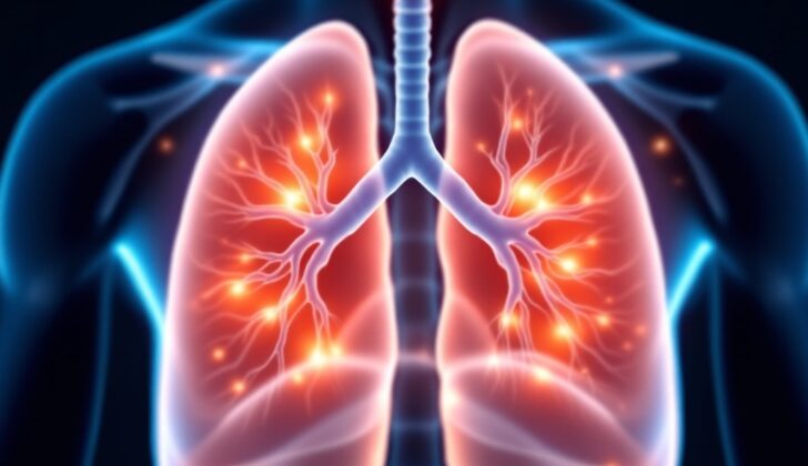
Lymphangioleiomyomatosis (LAM) is a disease that primarily affects the lungs. This condition occurs when unusual muscle cells grow abnormally in the blood vessels, lymph vessels, and tiny air sacs (alveoli) within the lungs. This growth causes multiple cysts, or fluid-filled sacs, to form in both lungs, leading to symptoms such as tiredness and shortness of breath during physical activity.
While LAM mostly affects the lungs, it can also have impacts on other parts of the body. For example, it can cause benign (non-cancerous) angiomyolipomas, a type of tumor in the kidneys, or sometimes result in the formation of cell tumors surrounding blood vessels and involving internal organs. This article simplifies the causes, prevalence, symptoms, and treatment options for LAM.
What Causes Lymphangioleiomyomatosis?
Lymphangioleiomyomatosis (LAM) can occur randomly or alongside a genetic disorder called tuberous sclerosis complex (TSC). Even though these two types can happen under different circumstances, they can’t be physically distinguished from each other.
We’re not exactly sure what causes LAM, but recent research has shed light on potential hormonal and genetic factors involved. Here are some key points:
- The condition can worsen when estrogen levels are high, which indicates that hormones may play a part.
- There’s evidence that certain genetic factors and mutations are associated with both kinds of LAM (S-LAM and SCL-LAM).
- The version of LAM that occurs randomly (S-LAM) almost exclusively affects women before menopause, though there have been rare reports of it happening in men.
- The most common type of LAM, TSC-LAM, is a genetic disease that runs in families and it’s the most frequent type of LAM.
There are certain genes associated with TSC that work to stop tumors from growing, called tumor suppressor genes. Two of these genes are TSC1, which creates a protein called hamartin, and TSC2, which creates a protein called tuberin. Both of these genes are part of a complex that controls a mechanism within cells called mTOR.
The issues arise when mutations occur in these TSC genes. For example, a mutation in TSC2 can cause the mTOR pathway to become overactivated. This leads to problems with the growth of smooth muscle cells and it can even cause benign tumors to form in various organs, a common occurrence in LAM.
So, how does this mTOR pathway work and why is it important? Well, it does a variety of jobs within cells but most importantly it helps regulate cell growth and proliferation – how cells divide and increase in number. That’s why interfering with the mTOR pathway is a key part of developing new treatments for patients with TSC. In fact, the majority of TSC cases (75%) are due to mutations in the TSC2 gene.
Risk Factors and Frequency for Lymphangioleiomyomatosis
S-LAM, or sporadic lymphangioleiomyomatosis, is a condition whose prevalence isn’t fully known, but some studies have provided estimates. These suggest that there are 3 to 7 cases per million women, while the sporadic form affects 1 in 400,000 adult women. This condition typically affects individuals around 35 years old.
Another form of this disease is LAM associated with Tuberous Sclerosis Complex (TSC-LAM), which is more common than S-LAM. It is estimated to affect 1 in 5000 to 10,000 live births. If a person has both Tuberous Sclerosis and LAM, they are more likely to develop angiomyolipomas, which are benign tumors that can occur in nearly 90% of these patients.
As people age, the rate of these conditions can increase. In fact, women older than 40 have the highest rates, which can be as high as 80%. Unlike S-LAM, which primarily affects women, TSC-LAM can affect both men and women with TSC. It is believed that 10 to 30% of men with TSC also have cystic lung disease.
- S-LAM affects 3 to 7 cases per million women, while the sporadic form affects 1 in 400,000 adult women.
- The typical age of those affected by S-LAM is around 35 years old.
- TSC-LAM affects 1 in 5000 to 10,000 live births.
- Those with both Tuberous Sclerosis and LAM are more likely to develop benign tumors known as angiomyolipomas.
- The incidence rate of these conditions increases with age, particularly in women older than 40.
- TSC-LAM can affect both men and women with TSC, whereas S-LAM primarily affects women.
- 10 to 30% of men with TSC also have cystic lung disease.
Signs and Symptoms of Lymphangioleiomyomatosis
Patients with Lymphangioleiomyomatosis (LAM) – a lung disease that affects primarily women – may display a range of symptoms. Each person’s case can look quite different, and this disease can impact multiple organs, including the brain, heart, skin, eyes, kidney, lung, and liver.
Common symptoms of LAM often resemble other respiratory issues and may be incorrectly diagnosed as chronic obstructive pulmonary disease (COPD) or asthma. These include:
- Shortness of breath, even at rest, and worsening with physical exertion.
- Fatigue and progressive breathlessness, which are initially noticed in about two-thirds of patients.
- Spontaneous ‘pneumothorax’ (a collapsed lung) felt by about one-third of patients.
- ‘Pleural effusion’ (fluid in the chest) witnessed in around a quarter of patients.
- Abnormalities in the lymphatic system, such as dilated thoracic duct, chylothorax, and chylous ascites.
- Other less frequently seen symptoms include a cough, chest pain, high blood pressure in the lung arteries (pulmonary hypertension), and coughing up blood (hemoptysis).
- Wheezing which can sometimes be detected during a physical examination.
In the case of patients with Tuberous Sclerosis Complex (TSC) – a rare genetic disease causing benign tumors to grow in different body organs – they may have skin problems like light colored patches, plaques on the forehead, and fibroadenomas, or nail issues like longitudinal nail grooves. Brain-related issues like epilepsy and cognitive impairment are also common In TSC patients.
Additionally, signs of brain disorders such as cortical hamartomas, low-grade brain tumour (astrocytoma), and misplaced collections of neurons (heterotopia) can be seen. These may not show any symptoms and may only be detectable via an imaging test. LAM can also be associated with benign kidney tumors (renal angiomyolipomas), and in some cases, polycystic kidney disease. Lung symptoms in women with LAM may get worse during menstrual cycles, and their lung function may deteriorate post estrogen treatment or during pregnancy.
Testing for Lymphangioleiomyomatosis
If you’re suspected of having Lymphangioleiomyomatosis (LAM), a rare lung disease, some tests might help your doctors figure it out. Your complete blood count or metabolic panel will not show any abnormality unless you have other health conditions. One possible test measures a substance called vascular endothelial growth factor D (VEGF-D). If your level is 800 pg/mL or above, there’s a good chance you have a form of LAM that affects your lungs. If there’s fluid in your chest, chances are it might be due to a type of fat blockage caused by unusual growth.
When performing lung function tests, the most common indication of LAM is obstructed airflow. Some patients show signs of asthma, with reversible blockages and increased resistance to airflow in their lungs. As a result, test results usually show a decrease in lung capacity and air exchange efficiency. A six-minute walk test can also help your doctor see if you’re not getting enough oxygen.
A chest X-ray might not show any clear signs of LAM as the disease effects are hard to see in this type of image. However, a High-Resolution Computed Tomography (HRCT) scan can provide a more precise picture. It has high sensitivity and is recommended for people suspected of having LAM. It typically shows multiple, thin-walled, well-demarcated, evenly distributed lung cysts. Over time, other lung changes might become visible.
An image scan of the kidneys is recommended too since many LAM patients also have kidney abnormalities called renal angiomyolipomas. A CT scan is a good way to look for these, along with growths in the lymphatic system. If young women have had unexplained lung collapses, they should also get a screening CT scan.
Usually, a biopsy, a test that examines a small piece of tissue from your body, is necessary for diagnosing LAM. The tissue shows abnormal growth of cells that look like smooth muscle under the microscope. These cells will usually test positive for some markers that are indicative of LAM. But, there’s no clear consensus on the best way to get the tissue for the biopsy.
Under a microscope, pathologists can identify LAM by looking for a specific type of cell growth in the walls of the tiny air sacs (alveoli) and blood vessels in lung tissue. Immune system tests will give positive results for certain substances. Diagnosing LAM can often be based on CT scan results along with certain factors such as a Tuberous Sclerosis Complex (TSC) diagnosis, presence of a specific type of effusion, lymphangioleiomyoma, renal angiomyolipoma, or an elevated VEGF-D. Historically, a surgical lung biopsy was needed to confirm diagnosis, but in many cases, the VEGF-D test has made biopsy unnecessary.
Treatment Options for Lymphangioleiomyomatosis
Treatment aims to alleviate symptoms, improve quality of life, and slow the progression of the disease. This may include a combination of medication, lifestyle changes like quitting smoking, and oxygen supplementation, if needed.
Bronchodilators, a type of medication that helps open up the airways, may be used to alleviate symptoms for patients where lung function tests indicate they might help. These usually include beta-agonists and anticholinergics, types of drugs that relax the muscles around your airways. Research has shown that these types of drugs can improve airflow in patients with this disease.
In cases where a patient’s functional capacity and respiratory symptoms are diminished, respiratory rehabilitation can help. This type of therapy encourages endurance and can improve quality of life.
Sirolimus, a medication that inhibits the abnormal growth of smooth muscle cells in the lungs, is approved by health agencies in the US and internationally. It can help prevent worsening of lung function and respiratory symptoms, and improve quality of life. Side effects can include nausea, diarrhea, mouth ulcers, acne, lower-leg swelling, and increased cholesterol levels.
Sirolimus can also help alleviate lymphatic manifestations of the disease, such as chylous effusions and lymphangioleiomyomas, disorders concerning the lymph vessels. It is usually considered the first-line treatment for patients with particular symptoms or conditions.
Everolimus, another drug, is an alternative for patients who can’t tolerate sirolimus, but isn’t officially approved for treating this disease. However, it has been used for treating certain types of tumors and epilepsy.
Potential treatments involving hormone modulation have been suggested due to the role estrogen plays in the disease, but official clinical trials have not been conducted. The safety of letrozole, a drug used to treat breast cancer, was assessed in postmenopausal women with this disease, though the results were inconclusive. Therefore, this hormonal therapy option isn’t recommended for patients outside of investigative clinical trials.
In severe cases where symptoms persist despite treatment, a lung transplant may be the best option, though there have been reports of recurrence after transplant. Consequently, the long-term effectiveness of this treatment is uncertain.
In very specific cases of recurring collapsed lung linked to this disease, a procedure called chemical pleurodesis, which uses talc to adhere the lung to the chest wall, is recommended. If the patient is taking sirolimus, it’s typically suggested to stop the medication for a few weeks after the resolution of the collapsed lung.
What else can Lymphangioleiomyomatosis be?
When determining if a patient has lymphangioleiomyomatosis, other possible health conditions with similar symptoms should also be considered. Here are some of them:
- Amyloidosis
- Asthma
- Benign metastasizing myeloma
- Birt-Hogg-Dube syndrome
- Diffuse pulmonary lymphangiomyosis
- Emphysema
- Eosinophilic granuloma
- Follicular bronchiolitis
- Interstitial myofibrosis
- Leiomyosarcoma
- Lymphoid interstitial pneumonia
- Light-chain deposition disease
- Pulmonary Langerhans cell histiocytosis
What to expect with Lymphangioleiomyomatosis
The lifespan of patients with Lymphangioleiomyomatosis (LAM), a rare lung disease, has been found to be longer than previously reported. To be more exact, patients without a lung transplant could survive, on average, 29 years from the time symptoms first appear and 23 years from the point of diagnosis.
Given the chance that the disease could come back even after a lung transplant, many experts now refer to it as a low-grade type of cancer. Diseases affecting the lining of the lungs (pleural disease) and collapsed lung (pneumothorax) are stubborn conditions in LAM and can return even after a surgical procedure (pleurodesis) meant to prevent them, which happens in one third of cases.
Because of the changes in air pressure during air travel, LAM patients have a higher risk of lung cysts rupturing and lungs collapsing spontaneously. So, patients should be warned about the risks associated with air travel and the possible need for extra oxygen supply during flights.
Possible Complications When Diagnosed with Lymphangioleiomyomatosis
Patients diagnosed with lymphangioleiomyomatosis (LAM) are recommended to stay away from oral contraceptive pills that contain estrogen. These pills can make their symptoms worse over time. However, it’s not all contraceptive pills they need to avoid. Oral contraceptives based on progesterone have been proven to be safe for these patients and can be considered an alternative.
Pregnancy might also worsen their symptoms and increase the risk of complications, but this isn’t always the case. Some women with LAM have been through multiple pregnancies without experiencing any complications.
Lastly, it’s important that LAM patients get their vaccinations for influenza and pneumococcal disease.
Key Facts:
- Avoid estrogen-containing oral contraceptive pills if you have LAM
- Progesterone-based oral contraceptives could be a safe option
- Pregnancy may increase the risk of worsening symptoms and complications, but not always
- Patients with LAM are advised to get flu and pneumococcal vaccines
Preventing Lymphangioleiomyomatosis
Patients with lymphangioleiomyomatosis, or LAM, and their families should understand their treatment plan and the potential complications linked to this disease. It’s important for them to know what to expect with regard to the future (prognosis) and the options available for treatment. If a woman with LAM is thinking about having a baby, she should discuss the potential risks with her doctor. These risks can depend on various elements, such as how well her lungs are working (pulmonary reserves).
Additionally, women should be advised to avoid medications and birth control methods that contain estrogen. This is important because these hormones can increase the chances of the disease worsening (LAM progression), especially during pregnancy.


