What is Pediatric Malignant Pleural Effusion?
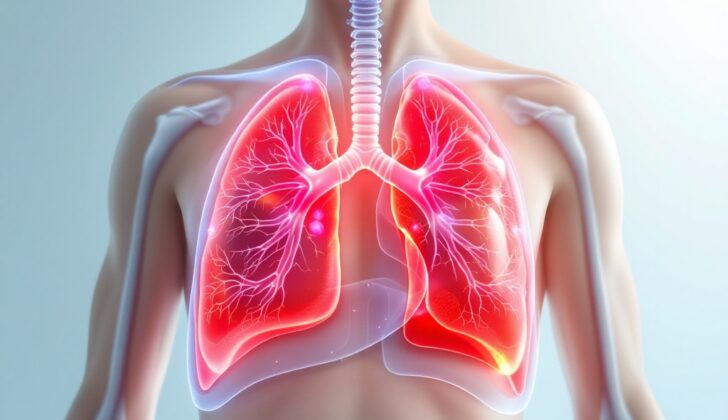
The pleural cavity is a thin sac that wraps around the lungs, separating them from the chest wall. This sac is lined with a smooth membrane. There’s a layer on the inside of this sac that covers the lungs called the visceral pleura, and an outer layer called the parietal pleura. A very small amount of fluid, around 10 microliters thick, is found between these two layers which helps the layers to slide easily over each other when we breathe in and out.
The volume of this fluid is kept in check by a delicate equilibrium of pressures between the pleural space and blood vessels, together with the natural fluid drainage process. The Frank-Starling law, a principle in physiology, helps manage this balance.
It’s important to note that up to 15 percent of patients with lung cancer may develop what are called malignant pleural effusions (MPEs). MPEs are a condition where there’s an excess amount of fluid in the pleural cavity, caused by cancerous cells. However, MPEs aren’t limited to lung cancers and can be an effect of cancers spreading from nearby or distant parts of the body. This could include cancers like malignant mesothelioma (a type of cancer affecting the membrane lining the lungs), metastatic cancers (cancer that has spread) affecting areas like the lungs, lymphoma (cancer of the lymph nodes), or cancers of the breast or ovary, as well as blood cancers. Generally, having an MPE indicates a serious prognosis or outlook.
In MPEs, cancer cells spread into the tissue lining the lung. These cancerous cells can usually be observed during a pleural biopsy, a procedure that involves removing a small sample of the pleural tissue to examine under a microscope.
Specific types of cancers are more likely to spread to the pleural tissue and cause malignant pleural effusion. This includes a group of cancers known as small blue-round cell tumors, of which a specific type known as rhabdomyosarcoma is seen to be common in children.
What Causes Pediatric Malignant Pleural Effusion?
Pleural effusion, a condition where fluid builds up in the space between your lung and chest wall, often happens in children because of cancer. This buildup of fluid can be caused by cancer spreading to the chest wall, problems with fluid draining from the chest area, or a blockage in the airways that leads to a condition called atelectasis. This situation can affect one or both lungs.
Cancer in this case can be either a primary cancer (which starts in the lung or chest area) or metastatic cancer (which starts somewhere else in the body and then spreads to the chest). Here are a few examples:
Lymphoma: This is the most common type of cancer linked to pleural effusion. It accounts for about 13% of all child cancers. Lymphoma often causes a lump in the chest, and about 5% of kids with lymphoma will get pleural effusion.
Leukemia: This is a type of blood cancer that can also cause pleural effusion. About half to 70% of kids with T-cell lymphoblastic leukemia (a type of leukemia) will develop pleural effusion. Doctors use bone marrow biopsy to tell the difference between lymphoma and leukemia.
Germ Cell Tumor (GCT): These account for 6% to 18% of chest lumps and can be cancerous. They can spread to nearby structures in the chest causing breathing problems. Pressure from these cancers on the airways and blockage of the fluid drainage can cause pleural effusion.
Neurogenic tumor: Neuroblastoma and ganglioneuroma make up about 90% of the tumors found in the back part of the chest. These don’t usually cause pleural effusion.
Chest wall and pulmonary malignancy: These can be either primary or metastatic but it’s hard to tell the difference. Examples include carcinoid tumors, pleuropulmonary blastoma, and Askin tumors.
Carcinoid tumor: This type of cancer is common in older children and teenagers. It makes up about 80% to 85% of primary cancerous tumors in children. They can cause compression atelectasis and pneumonia which can lead to pleural effusion.
Pleuropulmonary blastoma: This is a rare kind of cancer that originates from the lung and chest cavity. Diagnosis is often late and there are three types recognized.
Askin tumor: This is another rare cancer that can affect the bones of the rib or chest wall, or lungs and often spread to nearby structures. Prognosis is poor and it can cause pleural effusion.
Risk Factors and Frequency for Pediatric Malignant Pleural Effusion
In children, pleural effusion, which is when there’s too much fluid in the space around the lungs, is usually caused by one of three main conditions.
- Infection covers about 50% to 70% of cases
- Congestive heart failure accounts for about 5% to 15% of occurrences
- Malignancy, or cancer, is the cause in about 10% to 15% of cases
It’s worth noting that cancer is a rare cause of this condition in children. When cancer is the cause, it is often due to a type of cancer called non-Hodgkin lymphoma.
Signs and Symptoms of Pediatric Malignant Pleural Effusion
Malignant pleural effusion is a medical condition that can be diagnosed through a history and physical examination. During the history, the individual’s main health complaint is of primary importance.
- Weakness
- Tiredness
- Fever
- Weight loss
- Shortness of breath
- Chest pain
Other physical signs of malignant pleural effusion, a condition caused by cancer, are:
- Rapid breathing
- Fast heartbeat
- Excessive sweating
- Presence of a mass in the neck or the chest
During a physical examination, the patient is observed and examined in a comfortable position. Most people with malignant pleural effusion generally have a mass in the mediastinum, the central part of the chest, which may cause difficulty in breathing or discomfort. They may appear sweaty and have trouble breathing, with mild to moderate respiratory distress.
Listening to the chest can reveal a distinct sound in the early stages which may disappear as fluid accumulates in the pleural cavity. Too much fluid can shift the heart and windpipe towards the opposite side. The affected side may display certain characteristics, such as a dull sound when tapped, decreased vibrations felt when the patient speaks, and change in voice transmission. However, this may be difficult to identify, especially in a child who cannot cooperate with the examination.
Testing for Pediatric Malignant Pleural Effusion
If you have a malignant pleural effusion (MPE) – a condition where excess fluid builds up between the layers of the pleura, a thin tissue that lines the lungs and the chest wall – your doctor starts by asking about your symptoms and any family history of cancer, and performs a physical examination. Other tests might be used as well.
All these tests help your doctor confirm the size, location, and cause of the effusion:
- Chest X-ray – Shows the size and location of the fluid in your chest. It may not catch a small effusion, but it could show signs that suggest the presence of cancer.
- Chest Ultrasonography – Almost 100% accurate in detecting pleural effusion. Some features in this test like thickened pleural and diaphragmatic walls could highly indicate MPE.
- Computed Tomography (CT) Scan – A powerful imaging test that can help find the primary or metastatic (spread from another part of the body) tumor causing the effusion. It provides better imaging quality than an x-ray or ultrasonography.
- Magnetic Resonance Imaging (MRI) – Not often used for diagnosing pleural effusion because of issues such as lower spatial resolution (quality of the image).
The doctor may also use a test called Pleural Thoracentesis, where fluid is removed from the pleura and is then analyzed. If the fluid is bloody or contains a lot of protein (exudative), that may suggest a higher suspicion of cancer. The doctor may also check for other substances in the fluid like pH, glucose, lactate dehydrogenase, red and white blood cells, and more.
About half the time, cancer cells may be found in the fluid. Cancer in the MPE often presents as a fluid high in protein (exudate). Sometimes, it might present as a fluid low in protein (transudate), which is rare. In MPE, an infection-related effusion could also develop. The presence of cancer can be determined using a test parameter called Lights Criteria for pleural fluids, which compares the levels of certain substances in the pleural fluid and the blood.
Lastly, your doctor may opt for additional diagnostic tools, like a Pleural Biopsy where a small sample of the pleural tissue is taken to look for cancer cells or a Thoracoscopy that can be done under local or general anesthesia with high diagnostic accuracy. If they suspect the tumor originates from your lungs or if you’re coughing up blood (hemoptysis), they might perform a Bronchoscopy.
Treatment Options for Pediatric Malignant Pleural Effusion
Managing malignant pleural effusion, a buildup of fluid in the chest area that can lead to difficulty breathing, requires a team approach. This team usually includes a pediatric blood and cancer doctor, a pediatric surgeon, and a specialist in image-guided procedures, known as an interventional radiologist. Non-Hodgkin lymphoma is typically the major cause of this condition in children. Doctors will initiate a treatment program aimed at the cause of the fluid accumulation.
One of the procedures used to manage this condition is thoracentesis, which has three main goals: to remove the excess fluid, to improve symptoms, and to stop the fluid from accumulating again. This procedure is employed both for diagnostic purposes and as part of the treatment. If the patient has mild fluid accumulation, it can be monitored while they receive chemotherapy. For patients with a moderate to large amount of fluid, doctors can use thoracentesis to remove it. It is typically better to remove the fluid slowly to avoid further strain on the respiration system.
Despite this procedure, there are instances where patients may not notice immediate improvement. In such cases, doctors consider other possible causes of the symptoms. Even though some patients could experience immediate relief from symptoms, it’s crucial to keep on monitoring them for recurring symptoms like difficulty in breathing and chest pain, and to perform imaging tests if needed. If fluid buildup recurs, especially within the first month after thoracentesis, doctors may recommend placing a catheter in the chest to drain the fluid. This is a minimally invasive procedure that can be performed in an outpatient setting.
In cases where there are repeated instances of this condition, or in very young patients, a procedure known as tube thoracostomy can be performed under local anesthesia. However, this tube should not remain in place for too long due to the risk of infection, bleeding, or lung collapse. In some cases, doctors may drain the fluid using a chest tube or a small-bore catheter instead of performing thoracentesis, particularly when larger volumes of fluid need to be removed.
Once the fluid is evacuated, the next step is pleurodesis. This procedure involves introducing substances into the chest cavity to induce inflammation and seal off the space to prevent future fluid accumulation. It’s usually recommended for patients who are expected to live for more than three months. Several agents can be sued for pleurodesis, like bleomycin and talc. Talc can be applied in powder form to irritate the opposing surfaces and seal the pleural cavity. Alternatively, it could be mixed with 5% dextrose and a local anesthetic which then is introduced into the pleural cavity. This could be performed conventionally, or it could be performed using video-assisted thoracoscopy technique.
A different technique commonly used for palliative purposes is the insertion of a tunneled indwelling pleural catheter. This can be done under local anesthesia or moderate sedation and provides regular drainage of the fluid. The drainage can be done intermittently by a family member or a healthcare professional. It has grown popular because it’s less expensive, allows patients to be managed as outpatients, thus shortens hospital stays and speeds up recovery time. However, complications like lung collapse, bleeding, infection, or obstruction leading to tension fluid buildup can occur.
Pleuroperitoneal shunt is a palliative measure used for those who can’t undergo pleurodesis. A constant drainage system between the chest and belly cavity is created. It’s infrequently used in children and only as a palliative measure in adults.
To summarize, repeated thoracentesis can provide symptomatic relief for patients with a short lifespan. Tube thoracostomy is suitable if the patient’s survival is about a month or more, and the expected benefits outweigh the risks. Pleurodesis is a good option if the patient is expected to survive more than two to three months.
What else can Pediatric Malignant Pleural Effusion be?
When diagnosing a malignant pleural effusion, or fluid in the space between the lungs and chest wall, other possible causes need to be considered. For children, the most common cause of this fluid build up is an infection, usually pneumonia, which can lead to an effect known as parapneumonic effusion. It’s also possible for fluid to build up due to heart failure, which in children is most often due to congenital heart diseases.
In some cases, a condition called chylothorax can develop in children who have had surgeries for their congenital heart diseases. This can be caused by:
- Injury to the thoracic duct, a large vein in the chest
- Venous or lymphatic congestion, which is a buildup of fluid
- Central venous thrombosis, or a blood clot in a major vein
This can result in a milky-white liquid, known as chylous fluid, building up in the chest and causing significant fluid build up with breathing problems.
In order to correctly identify the cause, doctors may rely on a detailed patient history and physical examination.
One common test used in these cases is called a thoracentesis, which is where a needle is inserted into the chest to remove the fluid for testing. Analysis of this fluid helps to determine whether it’s a result of inflammation (exudate) or pressure-related leakage (transudate).
For a malignant pleural effusion (MPE), the fluid often has a glucose or sugar level below 60 mg/dl, and a pH lower than 7.3. Infections usually lead to exudative fluid, whereas heart failures lead to transudative. In around 95% of MPE cases, the fluid is exudative.
The fluid may also be inspected for cancer cells, which can sometimes lead to false positives. For adults, the chest fluid might be tested for tumor markers to help identify the primary tumor causing the fluid buildup.
What to expect with Pediatric Malignant Pleural Effusion
The outlook for Malignant Pleural Effusion (MPE), a condition where there is abnormal accumulation of fluid in the space between the lungs and chest wall due to cancer, greatly depends on the type of primary tumor causing the fluid build-up. In children, neuroblastoma and rhabdomyosarcoma, types of cancer that usually occur in the adrenal glands and muscles, respectively, are the most common causes of MPE.
If a patient experiences several episodes of MPE, their survival rate can be significantly impacted and unfortunately, the prognosis often isn’t positive. It’s important to note that ‘prognosis’ refers to the likely course or outcome of a disease.
Possible Complications When Diagnosed with Pediatric Malignant Pleural Effusion
The primary issues that people often come across after developing MPE (Malignant Pleural Effusion) usually involve problems with breathing, known as respiratory distress, and a serious condition called respiratory failure.
It’s also important to note that if MPE is caused by a mass in the mediastinum (the middle section of your chest) pressing on your windpipe and other important organs, there’s a high possibility of developing respiratory failure, or even experiencing cardiovascular collapse, which is when your heart can’t pump enough blood to your body.
Common complications:
- Respiratory distress
- Respiratory failure
- Cardiovascular collapse from a mediastinal mass


