What is Pneumatocele?
Pulmonary pneumatoceles, sometimes called pseudocysts in medical studies, are air-filled sacs with thin walls that form in the lung’s interstitial tissue, the network of tissue that supports the lungs. These sacs can occur as a single entity but are more commonly found in multiple sets. The term “pseudocyst” refers to the lack of a certain lining, called an epithelial lining, in these sacs. These sacs can vary in size, from being larger than 1cm in diameter to occupying half of the chest area. They typically have a uniform thickness with walls less than 4mm.
Sometimes, they may or may not have different levels of air and fluid inside. Although they usually don’t form at the very top of the lungs, they can appear anywhere within the chest cavity. In 1977, medical experts Fraser and Pare defined pneumatoceles as air-filled gaps that take up one-third of the lung’s volume. In certain trauma situations, these pneumatoceles can occur alongside lung bruising, pneumothorax (collapsed lung), and pneumomediastinum (air leak in the chest).
However, pneumatoceles do not occur in the space next to the mediastinum (the space in the chest between the lung sacs), nor does gas form in the lung ligament (a fold of tissue connecting the lungs). In a study of 4 cases, it was found that what looked like pneumatoceles in these regions were actually air spaces behind the lung ligament or pockets of air leaking into the space behind the lung.
Heamatopneumatocele is a specific type of pneumatocele that occurs after trauma and contains both air and blood.
It’s worth noting that pneumatoceles in the neck area of infants, known as cervical pneumatoceles, have been reported but they are rare and can mimic other conditions, like laryngocele (air sacs in the voice box), making them difficult to diagnose correctly.
A specific lung disease called desquamative interstitial pneumonia can result in the formation of multiple lung sacs or (pneumatoceles). In one reported case, these pneumatoceles resolved after treatment with a medication called cyclophosphamide.
What Causes Pneumatocele?
Pneumatoceles, or air-filled spaces within the lung tissue, can be formed through one or more of the following processes.
First, they can formed due to infections caused by various bacteria. Some examples include tuberculosis, Staphylococcus aureus (a common bacteria found on the skin and respiratory tract), Streptococcus pneumoniae (bacteria commonly involved in pneumonia), Proteus mirabilis (usually found in the gut), Escherichia coli (also found in the digestive tract), or Acinetobacter calcoaceticus (typically harmless, but can cause infections in weakened immune systems). This condition can also be seen in people who use drugs intravenously, like heroin.
Second, pneumatoceles can occur from non-infectious factors. This includes physical injuries, surgeries, mechanical ventilation (where a machine helps the patient breathe), hydrocarbon inhalation (breathing in chemical fumes), and burns. It may also be a result of certain medical treatments like the use of endobronchial valves (devices to lessen the effect of severe emphysema), kerosene ingestion, or persistent interstitial pulmonary emphysema (a lung condition where air pockets develop). Additionally, they can happen if positive pressure ventilation is used, which is when a machine helps push air into your lungs, such as with a ventilator or a CPAP machine (a device that helps people with sleep apnea breathe more easily during sleep).
Risk Factors and Frequency for Pneumatocele
Pneumatoceles, which are pockets of air that form in the lung tissue, are often seen amongst infants, children, adolescents, and people with weakened immune systems, such as individuals with AIDS. However, traumatic pneumatoceles, caused by chest injuries, are quite rare. They tend to affect children and young adults, and make up for less than 3% of lung tissue injuries. Interestingly, men appear to be at a higher risk mainly due to their involvement in situations like vehicular accidents and falls.
Pneumatoceles can also develop due to complications from pneumonia. About 2.4% to 8.3% of children admitted to hospitals with pneumonia have been reported to have pneumatoceles. In a Brazilian study, the incidence of pneumatoceles was found to be high, at 9.5%. This finding was connected to an elevated rate of malnourishment amongst the children studied. It was proposed that poor nutrition might hinder the development of certain lung structures, increasing the risk of pneumatocele formation.
When traumatic pneumatoceles occur, it’s usually due to chest trauma that doesn’t pierce the skin, or from pressure changes during mechanical ventilation. In most documented cases, 85% of patients were under 30 years of age. Younger individuals have more elastic chest walls than adults, and any injury to the chest can lead to tears in lung tissue. Pneumatoceles often form as a result and continue to grow until an equilibrium of pressures is reached.
An examination of over 10,000 trauma admissions found that of the 204 children with lung contusions, 12.3% had a pneumatocele. About 76% of these patients were male with an average age of 13 years. The majority of these pneumatoceles were identified through CT scans of the chest. It was also found that some patients had multiple pneumatoceles, but they all healed on their own without further treatment.
Moreover, a few unique cases have been reported in medical literature. A person with recurrent cervical cancer developed multiple pneumatoceles after treatment, and another case was reported during the H7N9 influenza pandemic where about 10% of survivors developed pneumatoceles, as shown by chest X-rays, after a year.
Signs and Symptoms of Pneumatocele
Traumatic pulmonary pseudocysts are typically diagnosed within a day or two after suffering a blunt force injury to the chest. The severity of this condition can vary greatly – some people might not exhibit any symptoms at all, while others might experience severe breathing difficulties that require the use of ventilators.
People who have these pseudocysts usually cough up blood in more than half of the cases. Chest pain and coughing are also common symptoms. However, these symptoms are usually caused by the injury to the lung tissue, not the pseudocysts themselves. Mild fever or a high white blood cell count can happen within 12 to 36 hours following the injury. This is usually due to the body absorbing damaged lung tissue or blood clots and should not be confused with infection.
Toxic Shock Syndrome caused by the bacteria called “Staphylococcus aureus” can sometimes cause symptoms like rash, fever, chills, mucositis, and also lead to the development of pneumatocele during the disease’s course.
In cases where a traumatic pneumatocele causes acute respiratory failure, it is typically because the condition has also resulted in another related lung injury known as pulmonary contusion.
The following are risk factors for the development of pneumatocele:
- Being a child or young adult, as they are at a higher risk
- Having had a recent infection
- Recovering from trauma
- After pneumonia (post-pneumonic)
- Having HIV
- Using intravenous drugs
- Having Hyper IgE syndrome, a rare genetic disorder
Testing for Pneumatocele
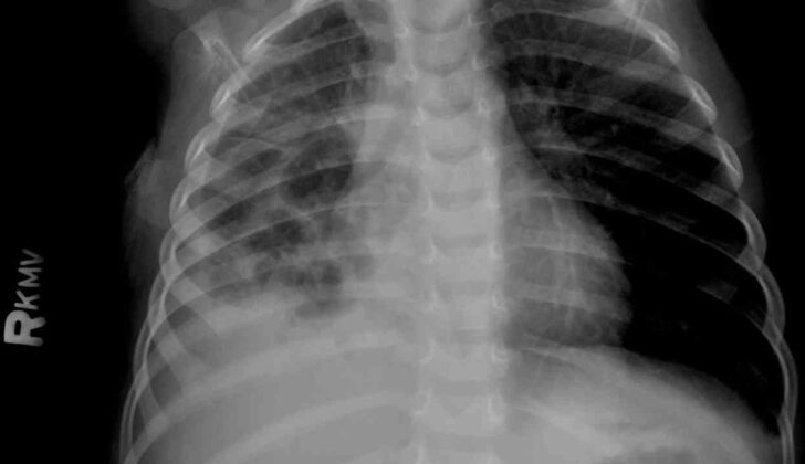
If your child is suspected of having pneumonia, a chest x-ray will most likely be the first procedure used. This test can usually identify pneumonia in 90% of cases on the first try. However, a clear image typically won’t become evident until around the fifth to sixth day of being in the hospital.
When trying to diagnose traumatic pneumatoceles – a specific type of lung injury that leads to air-filled spaces in the lung, chest x-rays can be a bit less reliable, especially if the individual is lying flat during the scan or if the lesion is smaller than 2 cm. In fact, the accuracy of a chest x-ray can range from 24% to 50%. To help improve the accuracy, chest x-rays may be repeated over several days. Most of these types of injuries occur in the lower lobes of the lungs and may not be noticed until days after the injury occurs. Despite the potential for false-negative results, chest x-rays are often the first test done in patients with chest injuries.
Pneumatoceles usually show up on x-rays as round or oval shadows, often surrounded by signs of lung tissue injury. Sometimes, there may be visible air-fluid levels, indicating bleeding into the pneumatocele.
A CT scan is a more precise imaging tool for diagnosing traumatic pneumatoceles as it has a reported accuracy rate of 96%. It gives detailed information about the size and location of the cyst and can help detect and differentially diagnose it earlier. It’s helpful specifically for spotting smaller lesions and identifying a pneumatocele when lung tissue injury is present. On a CT scan, a pneumatocele is seen as a round or oval thin-walled cavity in the lung with air-fluid levels.
An increase in the reported cases of pneumatoceles, up to 10% according to recent studies versus 3% in earlier research, is more likely due to the increased use of CT scans for diagnosis rather than a rise in cases.
In some cases, lung ultrasounds used in conjunction with chest x-rays have proven useful, particularly in children, based on two case reports.
The size of traumatic pseudocysts, another term for pneumatoceles, can vary greatly, from 1 to 14 centimeters in diameter. It’s unclear if the size of the lesion has any relation to the severity of the chest injury that caused it. However, larger pseudocysts are more commonly found in patients with multiple injuries, damage to both lungs, or significant breathing problems. Those with larger pneumatoceles may be at a higher risk of complications.
Since the size, shape, and makeup of pneumatoceles can change within days of the injury, doctors may need to perform repeated imaging tests to differentiate these lesions from other types of injury.
Treatment Options for Pneumatocele
Diagnosing a condition known as pneumatocele accurately and early is incredibly important for achieving a successful outcome for a patient. A pneumatocele is a pocket of air that forms in the lung, often after lung disease or injury. Confusing it with other types of lung lesions, such as an abscess, a fungal infection, or even cancer, could lead to unnecessary and potentially harmful treatments. For example, mistakenly identifying a pneumatocele as a more serious condition could lead to extra tests or the insertion of a chest tube or drainage catheter. Even worse, if a simple pneumatocele is wrongly identified as an abscess, it could be subject to invasive procedures, potentially leading to infection.
Treatment Options
There are various treatment options available for pneumatocele. Sometimes, simply monitoring the condition (known as “observation”) is enough, especially since most pneumatoceles disappear on their own without medical intervention within a few weeks to 6 to 12 months. However, for larger or more complex cases that cause instability in the heart and lungs, further treatments might be needed. These could include different types of specialist breathing (ventilation) techniques, precise positioning in bed (decubitus positioning), selective intubation (inserting a tube into the bronchus – one of the main airways), or direct drainage via a needle or catheter.
Surgical interventions might also be considered in some cases. These can include the injection of a substance called fibrin sealant into the lung’s lining, chest tube insertion, or operations to remove diseased parts of the lung (called resections). These might range from removing small parts of the lung (segmentectomy) to removing a whole lung (pneumonectomy). Advanced surgical techniques such as Video-Assisted Thoracoscopic Surgery (VATS) can also be used.
When treating pneumatocele, the patient’s overall clinical condition should be considered, not just the size of the pneumatocele.
In children, 90% of pneumatoceles resolve by themselves. There are cases where the pneumatocele forms a large air-filled cavity in one side of the chest, sometimes mistaken for a lung abscess. In these cases, treatment is usually with antibiotics and time, allowing the pneumatocele to resolve naturally.
Some advanced non-invasive treatment strategies might be needed when the pneumatocele is located deep within the lung, at the back near the heart. Percutaneous needle decompression, a procedure that involves using a needle to remove excess air from the lung pockets, is considered under certain conditions such as if the pneumatocele occupies more than half of the chest space, is causing the lung to collapse or is causing persistent signs of chest infection.
Decompression of a pneumatocele can be managed successfully by injecting a substance called fibrin sealant through a catheter. This procedure is rapid and safe, and may shorten the overall length of hospital stay.
Sometimes, especially in infants, surgical approaches aren’t suitable, due to extreme lung disease and a higher chance of complications. However, for those whose condition doesn’t improve with more conservative treatments, surgical approaches like resection or removal of a part of the lung may be considered.
It’s important to note that surgical complications, such as infection, decay of lung tissue, or needing further surgery, tend to be lower following lobectomy, a procedure in which an entire lobe of the lung is removed. This procedure is not recommended if there’s not enough healthy lung tissue left, or if it would leave decayed tissue within the chest.
Post-traumatic pneumatoceles, or air pockets that form in the lungs following injury, typically resolve on their own, but in some instances may need surgical intervention. The time until these lesions fully heal varies considerably, typically averaging about 3 months but sometimes taking as short as a month or as long as six months.
Bronchoscopy, a procedure in which a doctor visually examines the patient’s airways using a flexible tube, might be needed in cases where there is significant endobronchial bleeding, thick sputum, large air leaks, air leaking into the mediastinum (the central compartment of the chest), or lobar collapse (collapse of a part of the lung.)
Lastly, in rare cases where there is no improvement despite drug treatment and drainage, thoracotomy (a surgical operation in which a cut is made into the chest) or thoracoscopy (a procedure which allows the doctor to look at the chest cavity) might be considered. In children, most long-term complications from pneumonectomy, such as changes in the spine (scoliosis) and decreased lung function are typically mild, and most children do not experience marked breathing difficulties or difficulty exercising. However, there has been a decrease in pneumatocele cases in newborns due to advancements like protective lung ventilation and surfactant replacement therapy.
What else can Pneumatocele be?
In order to help understand some conditions related to the lungs, we’ll focus on a few specific ones: lung abscess, bronchial cyst, pulmonary sequestration, tuberculosis, granulomatosis with polyangiitis, cystic fibrosis, and several others. Each of these conditions might cause changes in the lungs, bringing about symptoms and signs that need medical attention.
The terms cyst, bulla and cavitory lesion are often used in diagnosis:
- A cyst is a round space inside the body that may be surrounded by a thin or thick wall. It’s not typically associated with swelling of the lungs.
- A bulla is like a large bubble of air in the lung tissue, more than 1 cm large, and has very thin walls. These are frequently found beneath the outer layer of the lungs and can often occur alongside certain types of lung swelling.
- A cavitary lesion is a area filled with air within swelling, a mass, or a small nodule in the lungs. This can have variable wall thickness. Cavitary lesions can occur with various diseases, and are different from cysts because of their thicker walls and irregular shape.
Past occurrence of trauma can help medical professionals distinguish a condition called post-traumatic pneumatocele from these other conditions. Also, the conditions might be different in places where certain types of lung conditions are more common. Comparing current and past chest X-rays can help with diagnosis.
In premature babies, conditions called ventilator-induced pneumatoceles can develop, which are a sign of lung injury caused by a ventilator. These are usually handled without surgery and instead by reducing the air pressure from the ventilator.
What to expect with Pneumatocele
Pneumatoceles, which are air-filled cysts in the lungs, usually have a good outcome and can resolve completely on their own. The time it takes for this to happen can range between 3 months to 2 years.
If complications do occur, it is beneficial to have early surgical intervention to prevent any risk of death or severe illness. Currently, there isn’t clear data available regarding the death rate for patients who develop complications from pneumatoceles.
Possible Complications When Diagnosed with Pneumatocele
The main issues with pneumatoceles, balloon-like air sacs that form in the lungs, come from mistakes in identifying and treating them. These mistakes can lead to additional problems.
Common complications related to pneumatoceles include:
- Rupture causing a pneumothorax or a tension pneumothorax with a shift in the mediastinal (central part of the chest). This condition is often treated with a medical procedure known as tube thoracostomy.
- Pneumomediastinum, a condition where air gets into the space in the middle of the chest.
- Developing an infection leading to empyema (a collection of pus in the pleural cavity), or a lung abscess. This is the most common issue in cases of pneumatoceles caused by trauma to the lungs. Infected pneumatoceles are more dangerous than regular lung abscesses. These are especially likely following lung damage that hampers the body’s ability to get rid of bacteria. The treatment for pneumatoceles that are infected is similar to that of a lung abscess.
- Pyopneumothorax, a condition that involves both a lung infection and a collection of pus in the lung;
- External chest cavity communication due to incomplete tuberculosis treatment;
- Bleeding into the chest cavity or a combination of blood and air in the chest cavity.
Using continuous positive airway pressure (a treatment that uses mild air pressure to keep the airways open) might make the pneumatoceles larger and squeeze nearby lung tissue. This can reduce oxygen levels in the blood and impact heart and lung stability.
Preventing Pneumatocele
Pneumatocysts are essentially air-filled sacs that can form in your body due to trauma or infection. It’s important to understand their nature and potential outcomes when they occur. The patient should be aware of the signs to watch out for that might indicate further complications. Regular medical imaging tests, like scans or X-rays, are crucial and should not be overlooked. These tests can help doctors monitor the situation and prevent any serious health problems that could arise.


