What is Pneumomediastinum (Mediastinal emphysema)?
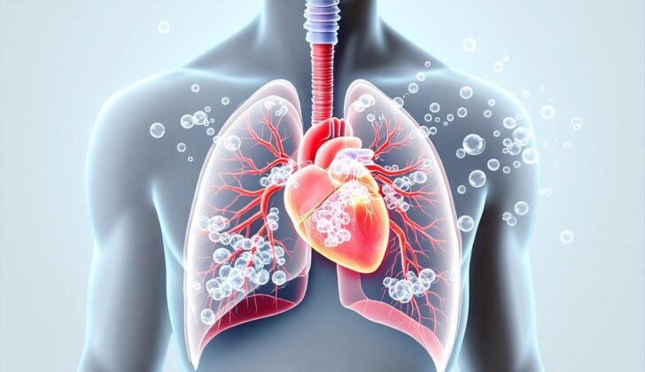
Pneumomediastinum, also known less commonly as mediastinal emphysema, refers to the presence of air in a specific part of the body called the mediastinum. The mediastinum is essentially the space in the middle of your chest. It’s nestled between your lungs, below the opening of your chest, above your diaphragm (which helps you breathe), in front of your breastbone, and behind your spine. This central chest cavity houses vital structures like your heart, windpipe and bronchial tubes (which help carry air to and from your lungs), and your esophagus (which connects your throat to your stomach).
Pneumomediastinum occurs when air leaks from your airways, lungs, or esophagus into this central chest space. This leaked air can then spread out to neighboring areas in your neck’s tissues under the skin, the cushioning layer around your spine, the sac surrounding your heart, or the abdominal cavity. Though rare and usually resolving itself, pneumomediastinum represents a medical issue that, once diagnosed and serious related conditions have been ruled out, is managed mainly by treating the symptoms.
What Causes Pneumomediastinum (Mediastinal emphysema)?
Spontaneous or primary pneumomediastinum refers to a condition that can occur in completely healthy individuals, for no apparent reason. In contrast, secondary pneumomediastinum happens when there’s a clear cause, such as an injury. Usually, pneumomediastinum is a result of rupturing of small air sacs in your lungs, more commonly referred to as alveoli.
There are risk factors for spontaneous pneumomediastinum, primarily associated with lifestyle choices. This may include smoking or using tobacco, or using certain types of drugs recreationally, such as cocaine, methamphetamine, marijuana, or ecstasy.
As for secondary pneumomediastinum, there can be a few causes, and these are often linked to pre-existing lung conditions or specific physical stressors. Some lung diseases, like asthma, chronic obstructive pulmonary disease (COPD), bronchiectasis, interstitial lung disease and lung cancer, can lead to this condition. Other factors such as something lodged in the airway, increased pressure in the chest from forceful sneezing or inhalation, childbirth, excessive vomiting or coughing, and strenuous physical activity, can also cause secondary pneumomediastinum.
There are also medical causes that can result in pneumomediastinum, which could be a side effect of a medical procedure. These procedures can range from surgeries that involve inserting a tube down into the lungs for better breathing mechanics (endoscopy or intubation), procedures involving insertion of a central line to access large veins for medication administration or monitoring, opening the chest to remove fluid or air (thoracostomy), or even abdominal surgeries.
Lastly, different types of traumatic incidents can cause pneumomediastinum, including injury from a blunt-object impact, penetrating trauma like a knife wound, or injuries as a result of an explosion or blast.
Risk Factors and Frequency for Pneumomediastinum (Mediastinal emphysema)
Pneumomediastinum, a condition characterized by the presence of air in the chest area, is extremely rare, occurring in as few as 0.002% of people. Interestingly, it often occurs in tall, thin young men (76% of cases). Experts believe it’s because the tissues in the chest region of this demographic are less hard and thickened compared to older patients, making it easier for air to escape and spread. Despite its prevalence in young adults, cases in children, excluding newborns, are not well reported.
When it comes to cases linked to severe illnesses, about 3.7% of patients with an extreme lung condition triggered by a severe infection, known as sepsis-induced acute respiratory distress syndrome, experienced a type of air leak excluding a collapsed lung. Moreover, among adults who suffered blunt chest injuries, up to 10% showed signs of pneumomediastinum.
Signs and Symptoms of Pneumomediastinum (Mediastinal emphysema)
Pneumomediastinum is a condition where air leaks into the area in the middle of the chest, between the lungs. When diagnosing this condition, doctors want to know about any risk factors or events that happened beforehand, such as:
- Smoking or using recreational drugs
- Existing airway or lung disease
- Excessive coughing or vomiting
- Recent medical procedures
- Experiencing blunt force or a cut to the chest
The most commonly reported feeling is chest pain behind the sternum (breastbone), felt by 60 to 100% of patients. This pain can move to the neck or back and might start suddenly, often after a bout of heavy coughing or vomiting. Other symptoms of pneumomediastinum include:
- Shortness of breath (in 75% of patients)
- Coughing fits (in 80% of patients)
- Neck pain (in 36% of patients)
- Pain when swallowing
- Difficulty swallowing
- Vomiting
- Abdominal pain
On a physical exam, doctors might find signs such as subcutaneous emphysema, which is when air gets into tissues under the skin (found in 70% of patients). The Hamman sign is another potential sign – this is a clicking sound in time with the heartbeat. Other signs that a doctor might look for include:
- Nasal-sounding speech
- Voice changes
- Hoarseness
- Swelling in the neck
- Rapid heart rate
- Rapid breathing
In rare cases, pneumomediastinum can become severe, blocking the major blood vessels in the heart—this could result in cardiac tamponade, a serious condition where fluid builds up around the heart.
Testing for Pneumomediastinum (Mediastinal emphysema)
Pneumomediastinum, a medical condition where air is present in the area of your chest that separates your lungs, is usually detected through a chest X-ray. The main sign that doctors look for is air that outlines the structures in your chest (occurring in 90% of cases).
Sometimes, other signs can also be seen on the chest X-ray, such as:
- Subcutaneous emphysema, which is air trapped under your skin.
- Elevation of the thymus (a small organ located in your chest) in children (otherwise known as “spinnaker sign”).
- Air surrounding your lung’s pulmonary arteries (known as the “ring sign”).
- A bright V shape seen between the descending aorta (the major artery running through your chest and abdomen) and the left side of your diaphragm.
- Seeing a double bronchial wall.
- Seeing the entire diaphragm, which is the muscle used for breathing.
- Pleural effusion, which is fluid buildup around your lungs and can suggest injury to the esophagus (the pipe connecting your mouth and stomach).
If the chest X-ray results are unclear, a computed tomography (CT) scan can be performed on your chest to confirm or rule out pneumomediastinum. CT scans are extremely sensitive and can detect even small amounts of air in the mediastinum or under your skin, and differentiate between pneumomediastinum and pneumopericardium, which can be challenging on a chest X-ray. Pneumopericardium is when air becomes trapped in the covering of your heart.
In urgent situations, ultrasound may quickly help diagnose pneumomediastinum. It could show the “air gap sign” where echoes block the sight of the heart structures beneath. Ultrasound can also spot air next to the heart or diaphragm and white spots, indicating air bubbles under the skin.
In most cases, extra tests like bronchoscopy (where a camera is passed down your throat into your lungs), esophagoscopy (where a camera is passed down your throat into your esophagus), or esophagography (an X-ray of your esophagus) are generally not required. However, these additional tests are justified according to your medical context and symptoms.
In blunt trauma, a CT scan can rule out injuries related to the aerodigestive system (the system that includes the parts taking in food and air) when pneumomediastinum is present. However, having air in the back of the mediastinum, in all medial compartments, and blood in the chest cavity (hemothorax) can result in a higher risk of death in patients with blunt trauma.
Treatment Options for Pneumomediastinum (Mediastinal emphysema)
Most people who are diagnosed with pneumomediastinum – a condition where air leaks into the area between the lungs – through imaging tests don’t have serious damage to the organs in the chest area. Instead, they usually only need treatment for their symptoms. Spontaneous pneumomediastinum typically resolves on its own and seldom happens again. These patients usually feel fine and have stable vital signs.
Treatments include resting in bed, getting oxygen if needed, taking cough suppressants, and taking pain relief medications. Unless there’s an injury to the air or food passages due to trauma or a medical procedure, antibiotics to prevent infection usually aren’t needed.
Most patients can be discharged and scheduled for a follow-up or admitted to the hospital for short-term observation. Similarly, patients who have pneumomediastinum due to blunt trauma without other complications can usually be managed with relatively non-invasive treatments.
However, hospitalization is needed for patients who are under distress, have a fever, or in cases where secondary pneumomediastinum – a similar condition but due to specific causes like trauma or disease – is a concern. This allows for observation, additional diagnostic checks, or surgery if required. Patients who are vomiting, have difficulty swallowing, are unstable, show signs of infection or pneumoperitoneum – air in the abdominal cavity – should undergo further specific examinations.
Patients with extensive subcutaneous emphysema – a condition where air gets into tissues beneath the skin – may need surgery to relieve the pressure. Those with air leakage into the space between the lungs and chest wall may need a tube inserted into their chest to remove air.
Lastly, patients presenting with malignant pneumomediastinum, a severe form of the condition, may need surgical procedures such as video-assisted thoracoscopic surgery, or thoracotomy, a large surgical cut into the chest wall.
What else can Pneumomediastinum (Mediastinal emphysema) be?
When diagnosing pneumomediastinum, which could mimic symptoms of other respiratory, cardiovascular, and gastrointestinal diseases, doctors should also look out for these conditions:
- Heart related conditions
- Acute coronary syndrome (condition brought on by sudden, reduced blood flow to the heart)
- Pericarditis (inflammation of the lining outside the heart)
- Myocarditis (inflammation of the heart muscle)
- Pneumopericardium (air in the lining outside the heart)
- Lung related conditions
- Pleuritis (inflammation of the tissues that line the lungs and chest cavity)
- Pulmonary embolism (a blood clot in the lungs)
- Pneumothorax (collapsed lung)
- Pneumonia (lung infection)
- Traumatic tracheal rupture (tearing of the windpipe caused by injury)
- Gastrointestinal conditions
- Gastroesophageal reflux disease (stomach acid frequently flows back into the tube connecting your mouth and stomach)
- Pancreatitis (inflammation of the pancreas)
- Esophageal rupture (tear in the tube that connects your throat and stomach)
- Musculoskeletal conditions
- Costochondritis (inflammation of the cartilage in the rib cage)
- Sternal and rib contusions (bruises on the breastbone and ribs)
- Rib fractures
- Other conditions
- Malignancy (cancer)
- Sickle cell crisis (a painful episode occurring in people with sickle cell anemia)
Considering these alternatives and conducting suitable tests would help arrive at a correct diagnosis.
What to expect with Pneumomediastinum (Mediastinal emphysema)
Recurrent pneumomediastinum, a condition where air gets trapped in the central area of the chest, can happen but is generally harmless. The real health risks usually come from whatever medical issue caused pneumomediastinum in the first place. In most situations, pneumomediastinum resolves on its own and only needs treatment to manage symptoms or just careful observation.
Possible Complications When Diagnosed with Pneumomediastinum (Mediastinal emphysema)
Several air-leak syndromes can further complicate an already existing pneumomediastinum. These can include conditions like pneumothorax, extensive subcutaneous emphysema, malignant pneumomediastinum, and pneumopericardium. Tension pneumomediastinum can put pressure on the body’s major blood vessels. This can disrupt the return of blood to the heart, leading to low blood pressure and low oxygen levels due to issues with the lung’s ventilation and blood flow. Certain complications could relate to the root cause of the pneumomediastinum – for example, patients with a perforated esophagus might develop mediastinitis.
Possible Complications:
- Pneumothorax or collapsed lung
- Extensive subcutaneous emphysema, trapped air in the skin
- Malignant pneumomediastinum, a rare and aggressive condition
- Pneumopericardium, air in the sac surrounding the heart
- Compression of major blood vessels
- Disrupted venous return which can lead to low blood pressure
- Low oxygen levels due to lung ventilation and blood flow issues
- Mediastinitis in case of perforated esophagus
Preventing Pneumomediastinum (Mediastinal emphysema)
In most cases, pneumomediastinum, a condition where air collects in the middle of your chest, occurs on its own. Avoiding smoking or using recreational drugs can significantly lower your chances of developing this condition. It’s also a good idea to stay away from strenuous exercises such as weight lifting and specific activities like flying, parachuting, scuba diving, or playing woodwind instruments. Keeping your asthma under control can also help.
Unfortunately, there’s not much information available about when people with pneumomediastinum can fly or participate in contact sports again. Generally speaking, it’s recommended that patients avoid these activities until they’re not experiencing any symptoms.


