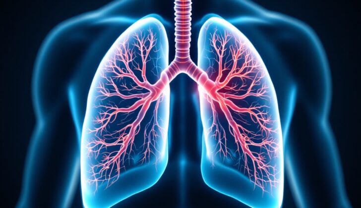
What is Pulmonary Infarction?
Pulmonary infarction is a condition where a blockage in one of the small blood vessels in the lungs happens. This can lead to lack of blood flow (ischemia) and potentially severe damage or death of lung tissue (tissue necrosis) in the area beyond the blockage. The root cause of pulmonary infarction is typically another primary disease, often a blood clot in the lungs (pulmonary embolism).
The key to understanding and managing pulmonary infarction lies in exploring the wide range of other health issues (differential diagnosis) that could be connected with it. This is important because the signs and symptoms are not usually specific to pulmonary infarction and could be attributed to other conditions. In some cases, pulmonary infarction might be the first sign of a serious underlying health problem.
What Causes Pulmonary Infarction?
Several studies have looked into how often lung tissue death (pulmonary infarction) occurs in connection with a lung clot (pulmonary embolism), which is the most common reason for such tissue death. It’s found that around 30% of people who have a lung clot experience this tissue death.
Other conditions that can lead to pulmonary infarction are infections, cancer, damage from surgery, a buildup of proteins called amyloids, sickle cell disease, inflammation of blood vessels (vasculitis), and more. Smoking increases the risk of pulmonary infarction for all causes, including those linked to lung clots.
Somewhat surprisingly, being younger (around 40 years old) and taller increases the chance of having lung tissue death from a lung clot. On the other hand, being obese seems to decrease this risk.
Earlier studies suggested that people with heart disease have the highest risk of pulmonary infarction because it was believed that poor backup blood circulation, along with a lung clot, could result in tissue death.
However, more recent studies suggest the opposite, saying that younger people without heart or lung diseases are more likely to experience pulmonary infarction due to a lung clot. Experts think this is because long-standing low oxygen levels due to chronic heart and lung diseases lead to a stronger backup supply of blood vessels in the lung airways, protecting lung tissue from dying.
Risk Factors and Frequency for Pulmonary Infarction
Pulmonary infarction is not well researched for all its causes, but its occurrence in people with lung clots, known as pulmonary embolisms, is well studied. Approximately one in every thousand individuals with a pulmonary embolism suffers a pulmonary infarction, a condition where the lung tissue dies due to lack of blood supply. This happens at a rate between 16% and 31% of the cases. The survival rate after such an episode can be around 97%, while 30-day mortality rates during one particular study were about 31%. Additionally, the mortality ratio in the first decade after developing a pulmonary embolism was 41%.
Comparing patients of pulmonary embolism with or without any visible signs of infarction, studies suggest that both groups have similar predictions about their stay in the hospital and the probability of death.
Signs and Symptoms of Pulmonary Infarction
Understanding both pulmonary embolism and pulmonary infarction is important because the former is often the cause of the latter. This means that the symptoms of these two conditions often overlap, but there are some key differences. If a person has both conditions, they might experience symptoms like:
- Difficulty breathing (between 69% and 78% of cases)
- Chest pain (49% to 70% of cases)
- Swelling or pain in one leg (27% to 31% of cases)
- Fever (5% to 11% of cases)
- Coughing up blood (4% to 19% of cases)
For those patients who solely have a pulmonary embolism, the symptoms can include:
- Difficulty breathing (72% to 75% of cases)
- Chest pain (36% to 38% of cases)
- Signs of deep vein thrombosis, like swelling, redness, or warmth in a leg (22% to 33% of cases)
- Coughing up blood (4% to 8% of cases)
Noticeably, chest pain and coughing up blood are more frequent in patients with both conditions compared to those just with pulmonary embolism. Some symptoms appear just as often in both groups, such as cough, fainting, sudden difficulty in breathing, signs of deep vein thrombosis, fever, and symptoms of right-side heart failure. Finally, some patients found to have a pulmonary infarction after a lung biopsy showed no breathing issues (65% of cases), difficulty in breathing (26% of cases), chest pain (7% of cases), and coughing up blood (5% of cases). With these statistics, it’s clear that diagnosing pulmonary infarction can be a challenging task which highlights the necessity of considering wide ranging possibilities when patients display symptoms that could potentially indicate a pulmonary infarction.
Testing for Pulmonary Infarction
Doctors use a variety of tools to detect and diagnose a condition called pulmonary infarction, which is a type of lung condition. Among these, a kind of scan known as a Computed Tomography (CT) scan is most frequently used to diagnose this condition when paired with relevant symptoms. The CT scan can show signs like a blood vessel feeding into the damaged area of the lung, a hollow or clear space in the center of the infarction, and a semi-circular shape. However, if the scan shows air-filled bronchial tubes, then it is less likely to be a pulmonary infarction.
A vessel feature and a central clear space, with no air-filled bronchial tubes on the CT scan, can make doctors 99% certain that it’s a pulmonary infarction.
An X-ray image can also provide the first hint towards diagnosing a pulmonary infarction. This image may show signs like “Hampton’s hump” (a wedge-shaped thickening on the outer part of the lung), Westermark’s sign (an area of decreased blood flow or unusually clear area in the lung), and Fleischer sign (a noticeably large pulmonary artery). However, these signs lack the ability to detect all cases of pulmonary infarction.
In fact, the “Hampton’s hump” finding was shown to detect only 22% of cases. However, when it is seen, it correctly identifies pulmonary infarction in 82% of the instances. Other signs such as a collapsed lung (known as atelectasis) or scattered thickening of the lung (termed focal consolidation) may be present, but these are not always associated with pulmonary infarction.
If doctors think a patient may have an infection of the heart lining and valves (referred to as infective endocarditis), they should also consider the possibility of a lung clot (pulmonary embolism) or pulmonary infarction, as these conditions can sometimes occur together. In fact, the occurrence of a pulmonary embolism or pulmonary infarction is considered a minor factor (in medical terms, a minor ‘Duke Criteria’) when diagnosing infective endocarditis, with reports showing that it occurs in 13% to 49% of cases.
Findings from other tests such as electrocardiograms (tests that measure the electrical activity of your heart), DD-dimers (a blood test that helps check if you might have a serious blood clot), ventilation/perfusion scans (tests that measure air and blood flow in your lungs), and echocardiograms (a type of ultrasound that lets doctors see the heart in action) also play a role in predicting whether a patient has a pulmonary infarction or a lung clot.
Treatment Options for Pulmonary Infarction
Pulmonary infarction, a potentially life-threatening condition where part of the lung dies, needs immediate attention if respiratory distress, a state of not getting enough oxygen to the body, or hemodynamic collapse, which is a sudden drop in blood flow, are present. The immediate treatment is supportive care, which is aimed at relieving symptoms and improving overall well-being.
Many patients with pulmonary infarction may develop obstructive shock, a condition with reduced blood flow in the body, due to a pulmonary embolism (a blood clot in the lungs) or due to the sudden drop in blood flow or persistent low oxygen levels. Treatment plans are based on the root cause that has led to pulmonary infarction.
Pulmonary embolism, which is a blood clot that can block the artery in the lungs, is usually treated initially with anticoagulants, which are medications that prevent blood from clotting. Hospitalized patients may be put on heparin or low-molecular-weight heparin, and transitioned to newer anticoagulation medications like apixaban or rivaroxaban, or older ones like coumadin for continued treatment after leaving the hospital.
For patients who are unstable due to moderate or high-risk pulmonary embolism, treatment options depends on the hospital’s capabilities. They could include catheter-based and systemic therapies that dissolve blood clots, or surgical procedures like embolectomies, which remove clots from the arteries in the lungs.
There is emerging evidence that patients at lower risk with pulmonary embolisms could be discharged directly from the emergency department with new direct oral anticoagulants. The treatment method for pulmonary infarctions that are not due to pulmonary embolism can vary and is dependent on the root cause.
The average time for pulmonary infarction to heal has not been thoroughly researched. However, one review discovered that, from 32 examined patients with intervals varying from 1 to 69 weeks after diagnosis, 10 patients still had signs of pulmonary infarction after an average of 10 weeks from the initial diagnosis.
Thus, there is no universal recommendation for repeat CT scans to check for resolution of pulmonary infarction. Such decisions should be guided by the individual patient’s condition. If the clinical picture aligns with the radiographic technology, biopsies, where a small tissue sample is taken from the body for examination, are rarely used to diagnose pulmonary infarction. A lung biopsy is usually saved for testing a possible pulmonary nodule, suspected mass, interstitial lung disease, or collecting a sample for culture (a test to detect bacteria or fungi).
What else can Pulmonary Infarction be?
When considering the possible diagnosis for a patient, doctors need to consider a wide range of possibilities. Here are some conditions which might be considered:
- Pulmonary embolism (a blood clot in the lungs)
- Acute chest syndrome (a condition related to sickle cell anemia)
- Infective endocarditis (an infection of the heart valve)
- Cancer
- Heatstroke
- AV malformation (an abnormal connection between arteries and veins)
- Cocaine toxicity
- Diffuse intravascular coagulation (abnormal clotting in blood vessels)
- Diffuse alveolar hemorrhage (bleeding in the lungs)
- Complications from medical procedures, such as bronchial artery embolization or IV catheter ablation
- Amyloidosis (build up of abnormal proteins)
- Pulmonary infections like pneumonia or aspergillosis (a fungal infection)
- Vasculities (inflammation of the blood vessels)


