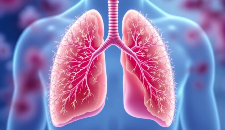
What is Pulmonary Papilloma?
Pulmonary papillomas are noncancerous growths in the lungs with a specific structure: they have a fibrovascular core, meaning they’re composed of fibers and blood vessels, and are covered by an outer layer of cells (epithelium). These growths have special outward-pointing projections and can appear in various ways.
There are three types of these growths—papillomatosis, inflammatory polyps, and solitary papilloma—and understanding these can help us comprehend how these growths affect patient care. While inflammatory polyps (swollen, round lumps) and solitary papillomas (single growths) are rare, it’s most common to see multiple papillomatosis, where there are many growths.
An overview of papillomatosis gives information on how common it is, the symptoms, the difficulties in diagnosing it, and the ways to manage it.
What Causes Pulmonary Papilloma?
Multiple papillomatosis, also known as recurring respiratory papillomatosis, is mainly caused by the human papillomavirus (HPV) infection. This infection leads to the repeated growth of non-cancerous lumps, called papillomas, in the respiratory tract, specifically in the voice box (larynx), windpipe (trachea), and airway branches (bronchi). Over 90% of these cases are caused by two subtypes of HPV, known as HPV 6 and HPV 11, which don’t usually lead to cancer. Yet, the genetic makeup of these virus types and how a person’s immune system responds can impact the course of the disease.
Other subtypes of HPV such as 16, 18, and 31 can lead to cancerous changes, especially in individuals with a type of bump called squamous cell papilloma and certain genetic changes involving a gene called TP53. Smoking, particularly among women, significantly raises the risk of these bumps turning into cancer.
People with weakened immune systems, like those with HIV or other immune-related illnesses, are more likely to develop multiple papillomatosis due to an HPV infection. This infection can be acquired either through oral-genital contact or it may be passed from an infected mother to her baby during childbirth.
Inflammatory polyps, also known as inflammatory pseudotumors, are different from other types of polyps, like adenomatous polyps, which are caused by abnormal cell growth. Inflammatory polyps are usually caused by inflammation and can appear as single or multiple lumpy lesions in persons suffering from chronic respiratory infections. Some of these cases are due to the infections themselves. An abnormal response of the body’s immune system to infections or environmental factors might lead to chronic inflammation, which in turn causes polyp formation. Genetic factors might play a part in some cases where specific genetic abnormalities are present. Exposure to environmental irritants or harmful substances can also cause chronic inflammation and promote the formation of polyps. Solitary polyps, which are rare, are often tied to infections like HPV or exposure to environmental factors such as smoking.
Risk Factors and Frequency for Pulmonary Papilloma
Recurrent respiratory papillomatosis is a condition that appears in around 4 out of 100,000 children and 2 out of 100,000 adults. Studies have shown that the frequency of this condition can be influenced by factors like how old the person is when it first starts to appear and their socioeconomic status – it tends to be more common in groups with lower socioeconomic status and education levels. However, your socioeconomic status does not have any impact on how severe the disease is. At the same time, the rate of HPV infection in women is gradually increasing, with around 26.8% of women aged 14 to 59 and 45% of women aged 20 to 24 being affected.
- Respiratory papillomatosis is common in adults, especially men (making up 84% of cases), typically between the ages of 40 and 50. There is also another surge in cases in people in their sixties.
- A specific type of tumor called a lower airway and pulmonary papilloma is pretty rare and only makes up less than 1% of all lung tumors.
- Between 8% and 9% of patients with HPV-related respiratory papillomatosis end up with lung involvement.
- Papilloma mainly shows up in children, often through childbirth from a mother who is infected with HPV-6 and HPV-11 in the genital area.
Signs and Symptoms of Pulmonary Papilloma
Pulmonary papillomas are growths in the lungs that often don’t cause specific symptoms. About a quarter of the cases are discovered by accident when doing chest scans for other reasons. Some health centers classify these growths into 3 categories—limited, moderate, or severe—based on the size and aggressiveness of the tumor. A limited type refers to a small single growth, while moderate and severe types involve multiple larger growths.
However, when there are symptoms, they often include coughing or wheezing, similar to asthma. Pulmonary papillomas can also cause a hoarse voice, trouble breathing, recurrent respiratory infections, and in some cases, can hinder airflow. These symptoms might get worse in patients with asthma, especially those using daily inhaled corticosteroids.
There are also severe symptoms such as coughing up blood and the formation of lung abscess, which might require surgical or endoscopic intervention. When a patient with bronchiectasis experiences hemoptysis or coughing up blood, they might need a bronchial artery embolization procedure, which can stop the bleeding. If a patient who smokes experiences hemoptysis, doctors must rule out lung cancer. Rarely, if there is uncontrollable bleeding, lung resection, or the removal of part of the lung, might be necessary.
Testing for Pulmonary Papilloma
Solitary papillomas, a type of lung growth, usually occur centrally in the lungs or, in rare cases, on the peripheral end. Their appearance is often like a plaque lesion.
Recurring respiratory papillomatosis, another type of lung condition, can take different forms. It may appear as nodules or growths with smooth or irregular, thin or thick walls. It can also resemble cavities or cysts, primarily located in the back regions of the lungs. If there is also swelling in certain regions near the lungs, it could indicate the possibility of a more serious, malignant condition.
Computed tomography (CT) scans, a type of X-ray scan, typically show effects such as plaques and nodules in the lungs, airway thickening, air-trapping, abscesses, foreign bodies, and bronchiectasis (an abnormal widening of the airways).
Most cases of pulmonary papilloma, about 80%, show solid and cavity-like lung nodules scattered throughout the lungs. They often have distinct patterns like calcification. However, finding isolated nodules without any airway involvement is rare on chest CT scans.
Although positron emission tomography (PET) and chest CT scans are not always accurate for detecting pulmonary papilloma, they can guide us to the right place for a biopsy. Biopsy is the procedure to confirm if the abnormal growth is malignant, especially when a severe condition like cancer is suspected.
Studies found a significant link between asthma and aggressive forms of recurring respiratory papillomatosis. Especially in patients with severe asthma who rely on inhaled steroids, they are more likely to develop this condition. Despite this association, the precise cause remains unclear.
Bronchoscopy, a procedure that lets your doctor look at your airways, is a definitive tool for confirming the presence of solitary or recurrent respiratory papillomatosis. It reveals growths within the tubes leading to the lungs or collapse of a portion of the lung. During biopsy, caution is necessary due to the risks of uncontrolled bleeding and blocked airways. If most lesions are different from each other, an excisional biopsy, one that removes an entire lesion, may be required.
Just like other lung tumors, it’s essential to get a biopsy for accurate diagnosis, followed by staging to figure out the extent of the disease. Certain procedures including endobronchial ultrasonic bronchoscopy (EBUS), which uses ultrasound during bronchoscopy, or possibly surgical staging may be required. Staging is important for developing a treatment plan with the goal of curing the condition.
Treatment Options for Pulmonary Papilloma
Treatment for pulmonary papilloma, which is a type of uncommon non-cancerous lung tumor, is dependent on factors like the tumor’s location, its detailed microscopic appearance, the patient’s lung health, and age. If it affects the lung tissue itself, as in recurrent respiratory papillomatosis, the condition tends to be more serious and may require a tracheostomy (a procedure to create an opening in the neck for breathing). Recurrent respiratory papillomatosis can also turn malignant (cancerous) and resemble a type of lung cancer known as non-small cell lung cancer. In such cases, it’s advisable to treat it similarly to lung squamous cell carcinoma, a common type of lung cancer.
Typically, pulmonary papilloma is treated with surgical removal, either by excising the affected tissue or by using an endoscope, especially in the early stages before cancer spreads. Other techniques such as laryngeal micro-debride therapy, laser treatment, antiviral drug injections directly on the lesion, photodynamic therapy, cryotherapy (using cold treatment), and interferon-alpha therapy (using a drug to stimulate the immune system) can also be used.
Some new treatments for pulmonary papilloma are currently being studied. This includes the use of anti-PD-1 therapies, which are drugs that help the immune system fight cancer, such as nivolumab and pembrolizumab. These medicines target a protein called PD-1, which can be found in higher than normal levels in these benign tumors.
Bevacizumab, a drug that targets a protein involved in blood vessel growth, is seen as a hopeful treatment, particularly for aggressive and recurrent respiratory papillomatosis. This drug, which is a type of monoclonal antibody medicine, seems to extend the time between starting treatment and needing surgical intervention. However, injecting it directly into the tumor has shown less effectiveness.
Pembrolizumab is another treatment option that is currently being evaluated in clinical trials. Celecoxib, on the other hand, a drug that blocks the production of certain chemicals in the body, has been ineffective in treating pulmonary papilloma.
Generally, solitary papillomas, when surgically removed, typically do not recur, although about 20% of patients treated with endoscopic removal may experience a recurrence. Malignant transformations of these benign tumors into cancerous ones are rare.
During laser treatments, precautions should be taken to prevent inhalation of HPV DNA particles by the medical staff. Also, the development of the HPV vaccine may help prevent respiratory papillomatosis in the future.
It’s important to monitor patients at risk of pulmonary involvement regularly. A low-dose CT scan of the chest is recommended for people who had juvenile-onset recurrent respiratory papillomatosis, starting at age 18 or earlier if risk factors like early disease onset, tracheostomy, and multiple surgeries are present.
In adult-onset recurrent respiratory papillomatosis, a low-dose CT chest may be advisable as a baseline, with subsequent scans every 5 years if there’s no evidence of lung involvement like cysts or nodules. If a nodule is found, the updated Fleischner Society guidelines for managing incidental pulmonary nodules should be followed.
What else can Pulmonary Papilloma be?
Pulmonary papillomas are a type of lump in the lungs. When a doctor finds one, they may think it might also be other conditions such as carcinoid tumors and squamous cell carcinoma. Looking at it under the microscope helps doctors figure out what kind of lump it is.
Sometimes, it’s tricky to tell the difference between these lumps and other types like inflammatory polyps and squamous cell carcinoma. Inflammatory polyps are bumpy growths, but they’re not structured like a true papillary, even if they show signs of squamous metaplasia, or cell change.
Squamous cell carcinoma is a type of tumor containing nasty bunches of cells with squamous pearls at the center. Squamous pearls are areas of dead cells that are still densely packed and they might even have cells that die off in a strange way, called dyskeratotic cells. In cases where it’s tough to see keratinization, or cell hardening, certain tests using P63 and P40 antibodies can be of significant help.
As for glandular papilloma, doctors must differentiate this from primary and metastatic adenocarcinoma, as well as other adenomas. Adenocarcinomas usually display signs indicating likelihood of spreading, such as abnormal cell changes and the invasion of what is called the basal lamina. They do not show characteristics typical to glandular papilloma, like base, cilia, and mucus cells. Additionally, adenocarcinomas often exhibit gland structures and/or mucus secretion, features that do not appear in pulmonary papillomas.
What to expect with Pulmonary Papilloma
Pulmonary papillomas, or abnormal growths in the lungs, are typically not harmful and often have a positive outcome if completely removed. Repeated cases are rare, but when the growth isn’t entirely removed, it can return. Transforming these growths into cancer is generally rare but can happen more often with certain types like squamous cell papilloma, especially when it affects the lungs. Patients who experience recurrent respiratory papillomatosis, or repeated abnormal growths in the respiratory passages, have a significantly higher risk of these growths turning into cancer.
Several factors could increase one’s chances of developing cervical cancer, including infection with certain types of high-risk HPV (human papillomavirus), smoking, previous radiotherapy, use of cytotoxic drugs (a type of cancer treatment), p53 gene mutation (a change in a gene that keeps the growth of cells in check), and high activity of a specific enzyme. Some types of HPV, considered as low risk, have shown potential for causing cancer as well. The exact way this transformation happens is still unclear, but is believed to relate to how HPV affects cell growth and change.
HPV-associated papillomas, especially some subtypes, are known for their potential to become cancerous. Laryngotracheal papillomatosis, a condition involving abnormal growths in the larynx or trachea, can spread into the lower respiratory tract in some cases.


