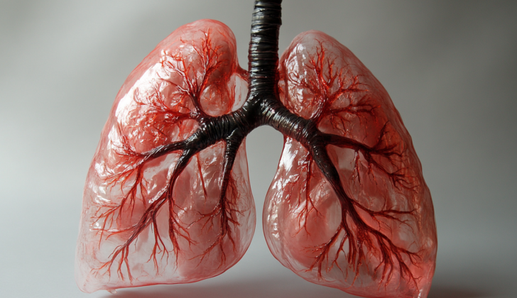
What is Restrictive Lung Disease?
Restrictive lung diseases are a varying array of lung conditions marked by difficulty in fully expanding the lungs, as indicated by lung testing. Essentially, the lungs lose elasticity, which interferes with their ability to fully expand, thus causing a decrease in overall lung capacity. This feature differentiates them from obstructive lung problems like chronic obstructive pulmonary disorder (COPD), asthma, and bronchitis that are caused by resistance to airflow due to blockages at different stretches of the airway. Statistically, restrictive lung diseases represent about 20% of lung conditions, while a larger 80% are caused by obstructive lung problems.
These restrictive lung diseases can either be due to damage to the small parts of the lungs involved in gas exchange (known as distal lung parenchyma) because of inflammation, toxins unknown, or due to issues outside these lung structures. The damage to the lung itself can be due to conditions causing inflammation changes in the structures within the lung that involve the tiny air sacs called alveoli and the surrounding parts. These conditions come under the banner of interstitial lung diseases (ILDs). ILDs contain a broad range of conditions characterized by various damage and inflammation to structures in the lung that involve changes ranging from occasional inflammation to chronic stiffening of lung tissue. Other conditions may begin within the alveoli, due to fluid buildup, for example, and spread to affect the surrounding lung structures. Whether the problem starts in the small lung structure or from the alveoli, the result is a disturbance to the normal structure of the lung, leading to functional issues.
Restrictive lung diseases can also be due to issues with neuromuscular function or movement of the chest wall that limits healthy lung function. Conditions such as neuromuscular diseases, disorders of the lining of the chest wall (pleura), extreme overweight (obesity) or bony issues with the rib joints or spine can physically restrict normal breathing. Whether caused by issues within the lung or outside it, these diseases all share the common feature of limiting lung function and can result in respiratory failure.
In this overview, we will discuss restrictive lung diseases, including how they are diagnosed, treated, and more. Keep in mind that these lung conditions are complex, and this is only a brief introduction.
What Causes Restrictive Lung Disease?
“PAINT” is a handy way to remember the causes of a disease that limits how much the lungs can expand (restrictive lung disease). It stands for Pleural, Alveolar, Interstitial, Neuromuscular, and Thoracic cage abnormalities. However, it is sometimes more useful to think of this lung disease in terms of what is causing it.
Limitations on how the lungs can stretch can be triggered by:
- Diseases that damage the delicate tissue of the lungs (known as “intrinsic” causes)
- Diseases outside the lungs (known as “extrinsic” causes)
Intrinsic causes happen within the lungs themselves, while extrinsic causes come from muscle disorders, being overweight, and other conditions outside of the lungs. Both types can lead to a decrease in lung volume because they restrict how the lungs function.
Damage to Lung Tissue (Intrinsic Causes)
An intrinsic restriction can happen because the lung tissue is inflamed from certain diseases. For example, health problems grouped under interstitial lung diseases:
- Idiopathic pulmonary fibrosis (a disease that causes scarring in your lungs)
- Non-specific interstitial pneumonia
- Cryptogenic organizing pneumonia
- Sarcoidosis
- Acute interstitial pneumonia
- Exposure to certain dusts, such as silicosis, asbestosis, talc, and others
- Exposure to organic dust, such as farmer’s lung, bird fancier’s lung, and others
- Hypersensitivity pneumonitis
- Systemic sclerosis
- Lung blood vessel inflammation
- Pulmonary Langerhans cell histiocytosis
- Some medications
- Radiation therapy
These can be grouped into diseases that make your lungs stiffer (like interstitial diseases), and diseases that fill the tiny air sacs in your lungs (like pneumonia).
Diseases Outside the Lungs (Extrinsic Causes)
Extrinsic causes can result from diseases of the chest wall, such as:
- Kyphoscoliosis (abnormal curvature of the spine)
- Conditions that affect the thin space between your lungs and chest wall
- Being overweight
- Muscular disorders like muscular dystrophy, amyotrophic lateral sclerosis, polio, and others
- Ascites (a condition in which fluid collects in your abdomen)
Extrinsic causes can be divided into those that decrease the muscle tone of the respiratory system (like myopathies and neurological deficits), deformities of the rib cage (like kyphoscoliosis), and those that take up space (like fluid between the lungs and chest wall).
In summary, many things can cause restrictive lung disease. Some are even unknown, such as in idiopathic pulmonary fibrosis. Recently, scientists are studying how genetic factors may influence the risk of this disease.
Risk Factors and Frequency for Restrictive Lung Disease
Exact numbers are hard to nail down for restrictive lung diseases because there are several variations of the disease, each potentially in a different stage. However, it’s estimated that 3-6 in every 100,000 people in the United States are affected by certain restrictive lung diseases. Sarcoidosis, another type of restrictive lung disease, has a higher prevalence in North America with 10-40 cases per 100,000 people, and even higher in Sweden at 64 cases per 100,000.
Studies have shown that certain groups of people are more prone to develop a restrictive lung disease pattern. These groups include:
- The elderly, with a sharp increase from 2.7 cases per 100,000 in the 35-44 age group, to over 175 cases per 100,000 in people older than 75. Some diseases, like sarcoidosis, affect younger patients, typically those between 20-40 years.
- African Americans, who have a higher prevalence at 35.5 cases per 100,000 compared to 10.9 cases per 100,000 in the white population.
- Women, who are more prone to sarcoidosis but also generally more likely to develop restrictive lung disease. The exception to this is a disease called IPF, which is more common in men.
- Obese individuals, as carrying extra weight can decrease lung volume and contribute to restrictive patterns.
- Smokers, particularly those with IPF, who are often current or former smokers.
Increased risk is also found among those who are frequently exposed to certain substances in their environment or workplace. These include asbestos, coal dust, and other harmful dust particles, which can lead to lung inflammation and scarring, and a restrictive pattern of lung disease. It’s worth noting though, that restrictive lung diseases are rare in pregnant people, and if they do occur, it’s usually due to neuromuscular disease or a spine condition called kyphoscoliosis.
An interesting study found that restrictive lung patterns have decreased from 7.2% to 5.4% between 1988-1994 and 2007-2010. This points to the success of improved workplace safety and a decrease in cigarette smoking rates.
Signs and Symptoms of Restrictive Lung Disease
To accurately identify and understand these complex health issues, a detailed medical history and thorough physical examination are necessary. Important information to gather includes the severity of symptoms, when and how they began, their progression rate, family health history, and any personal habits like smoking and drug use. Additionally, aspects such as exposure to certain occupational and environmental factors need to be considered. Each disease will have its own unique signs and symptoms, but most of them slowly develop into difficulties with breathing.
Common characteristics of illnesses causing external restriction include having a high Body Mass Index (BMI), abnormal spinal curvature, or a history of muscle and nerve diseases. These conditions, known as Interstitial Lung Diseases (ILDs), often lead to cough, clubbing (widening and rounding) of the nails, and abnormal lung sounds when a doctor listens to them.
The symptoms and expected disease progress can vary greatly among these diseases. For instance, Idiopathic Pulmonary Fibrosis (IPF) leads to a steady decrease in lung function, eventually culminating in respiratory failure. Unfortunately, the outlook for this condition is not good, with most patients living only 3 to 5 years following diagnosis.
- Severity of symptoms
- Time and origin of symptoms
- Rate of symptom progression
- Family health history
- Smoking and drug use
- Occupational and environmental exposure
- Progressive difficulty in breathing
- High Body Mass Index (BMI)
- Spinal curvature deviations
- History of muscle and nerve diseases
- Cough
- Clubbing of the nails
- Coarse crackling lung sounds when listened to with a stethoscope
Testing for Restrictive Lung Disease
One way to check for lung restriction is through breathing tests known as pulmonary function tests (PFTs). The first clues that can hint at a lung restriction are a decrease in total lung capacity (TLC) along with a preserved FEV1/FVC ratio above 70%. In simpler terms, this means a decreased ability to fully expand the lungs, while the rate of breathing in a second compared to the full breath (FEV1/FVC ratio) remains normal.
If these initial results indicate lung restriction, further more complete PFTs are needed. These tests will measure how much air your lungs can hold, how quickly you can move air in and out of your lungs, and how well your lungs put oxygen into and remove carbon dioxide from your blood. One of these measurements is called DLCO which is the amount of oxygen that travels from the lungs to the blood. If your DLCO is decreased, it could suggest an internal lung restriction.
People with external lung restriction may also show signs of restriction on these breathing tests, but their DLCO levels will usually be normal.
In addition to PFTs, other tests such as high-resolution computed tomography (HRCT), which is a type of detailed lung scan, inflammatory markers (signs of inflammation in the body), and specific autoantibodies (immune proteins that attack the own cells by mistake) may also be needed, especially if it’s suspected you have some type of lung restriction or damage due to a condition like IPF or idiopathic pulmonary fibrosis.
IPF is a specific kind of lung disease that falls under a larger group of conditions known as interstitial lung diseases (ILDs). It is characterized by fibrosis, which is a fancy term for tissue scarring.
The American Thoracic Society (ATS) advises that anyone over the age of 60 who has newly discovered bilateral fibrosis, a condition that causes scarring in both lungs and can be identified on chest X-rays or CT scans, should be considered for a diagnosis of IPF if there’s no known reason for this condition. They may also have bibasilar inspiratory crackles, which are specific sounds that doctors hear when they listen to your lungs with a stethoscope.
In the early stages, an HRCT scan for a patient with IPF would typically show a specific pattern with coarse crosslinking (a type of dense fibrosis) mainly in the basilar regions (areas towards the bottom of the lungs), which appears towards the back and around the edges of the pleura (the thin membrane that lines the chest cavity and surrounds the lungs). With time, you may see what’s called “honeycomb” cystic alterations (small sacs filled with air) and bronchiectasis (a lung condition that causes coughing up of mucus due to the widening of the large airways), which occur when the lung tissue becomes fibrotic or scarred.
Treatment Options for Restrictive Lung Disease
Managing restrictive lung disease can differ based on what’s causing the condition. For instance, people diagnosed with a disease called Idiopathic Pulmonary Fibrosis (IPF) traditionally receive treatments that suppress the immune system. However, doctors are now prescribing drugs like pirfenidone and nintedanib more frequently, as they have been demonstrated to slow the progression of the disease.
Patients who have autoimmune conditions that can cause lung diseases, such as systemic sclerosis, are often given drugs that suppress the immune system. These might include steroids, mycophenolate mofetil, and cyclophosphamide, and the specific drug used usually depends on how severe their disease is.
If a patient has an acute exacerbation (sudden worsening) of their condition, doctors usually prescribe steroids as a quick treatment; but the use of steroids for a long period is not recommended because they can have complications. For ongoing care, others treatments like oxygen therapy, managing other existing conditions, or rehabilitation designed specifically for lung conditions can be used. Pirfenidone and nintedanib are also used.
For obese patients, the treatment involves reducing weight through a combination of diet and physical exercise. If patients who are severely obese struggle to lose weight through traditional methods they might be evaluated for gastric bypass surgery. This procedure has been shown to be effective at helping patients significantly lose weight and improve lung function tests.
Severe scoliosis (a sideways curvature of the spine) can reduce lung function, and in such cases, surgical correction may be required to control the condition. If patients have developed a lot of lung fibrosis (thickening and scarring of the lung tissue) and chronic respiratory failure, doctors may suggest an evaluation for a potential lung transplant.
What else can Restrictive Lung Disease be?
Pulmonary restriction, once diagnosed by pulmonary function tests (PFTs), can be caused by a variety of factors. The causes are divided into two main categories: intrinsic (from within the lungs) and extrinsic (from outside the lungs).
Intrinsic causes are typically related to different types of lung diseases. These include:
- Idiopathic pulmonary fibrosis
- Non-specific interstitial pneumonia
- Cryptogenic organizing pneumonia
- Acute interstitial pneumonia
- Pneumoconiosis
- Hypersensitivity pneumonitis
- Systemic sclerosis
- Sarcoidosis
Extrinsic causes are conditions outside of the lungs that can restrict their functioning. For instance:
- Obesity
- Spinal deviation
- Neuromuscular disorders
What to expect with Restrictive Lung Disease
The outlook for patients with restrictive lung disease, a condition that makes it hard for the lungs to fully expand, can greatly vary. This variation is largely due to the cause behind the lung restriction. For example, people who have lung restriction from fluid buildup around their lungs (pleural effusion) should see their symptoms improve once the fluid is drained. Pregnant women with this condition usually get better after they give birth.
If the cause of the lung restriction is a disease called cryptogenic organizing pneumonia, which affects the air sacs in your lungs, the long-term outcomes are normally excellent with treatment.
However, patients with a disorder called idiopathic pulmonary fibrosis (IPF), a disease with unknown cause that results in lung scarring, typically live about 3 to 5 years after the diagnosis.
Acute interstitial pneumonia, a rapid and severe lung disease, has a poor outcome. More than 70% of patients with this condition die within three months.
Due to the diverse underlying causes of restrictive lung disease, it is extremely important to get a specific diagnosis once lung restriction has been confirmed.
Possible Complications When Diagnosed with Restrictive Lung Disease
Severe restrictive lung disease can lead to low oxygen levels in the blood, a condition known as hypoxemia. Only increases in breathing rate can offset this. Working harder to breathe uses more energy, which may result in loss of muscle and weight. If the body’s strategies to compensate no longer work, and the oxygen levels worsen, chronic lung failure can develop.
Patients with severe restrictive lung disease also often experience sleep disorders, such as obstructive sleep apnea (or OSA). This disorder, common in patients with lung restriction due to obesity, has been found in a significant number of patients with restrictive lung disease that is intrinsic or caused by the condition of the lung itself.
Over time, chronic respiratory failure and changes in the structure of the lung can lead to high blood pressure within the lungs (pulmonary hypertension) and right-sided heart failure (cor-pulmonale).
Common Consequences:
- Low oxygen level in the blood
- Increased breathing rate
- Loss of muscle and weight
- Chronic respiratory failure
- Sleep disorders, including obstructive sleep apnea
- Pulmonary hypertension (high blood pressure in the lungs)
- Right-sided heart failure, also known as cor-pulmonale
Preventing Restrictive Lung Disease
Once the cause of the lung restriction is identified, patients should be informed about the treatment options available to them. For patients with a condition that is getting worse, lung rehabilitation centers can help by providing advice about their breathing and oxygen needs, as well as teaching exercises that can improve lung function and manage symptoms. Sometimes, regular physical activity and weight loss can be beneficial, particularly when the major cause of the lung restriction is obesity. It’s important to remember that different treatment options work differently for everyone, so what works best will depend on the individual’s specific circumstances.


