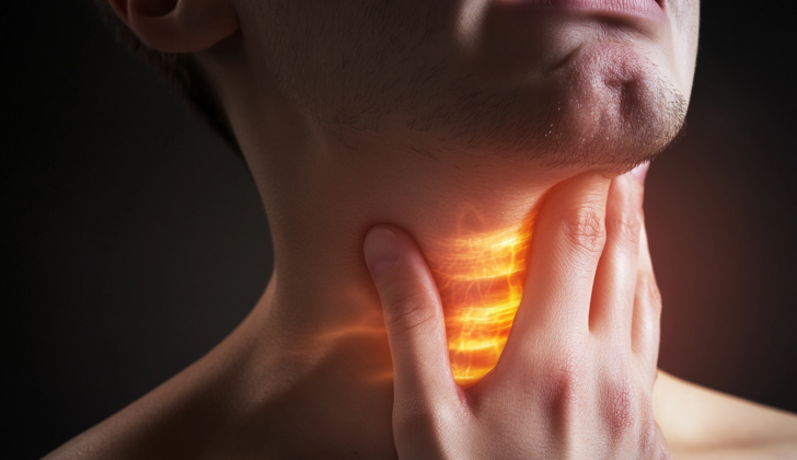
What is Tracheal Injury?
The trachea, also known as the windpipe, is similar to a tube that starts at the base of a voice box structure named cricoid cartilage and ends where the airways branch off to the lungs. It is split into two parts: one in the neck (cervical) and the other in the chest (thoracic). The trachea consists of 18 to 22 ring-like shapes that are hard in the front and sides but soft and flexible at the back. Blood reaches the neck part of the trachea from branches of the subclavian artery, a major blood vessel that stems from the heart’s largest artery, the aorta. The chest part of the trachea gets its blood supply directly from the aorta.
Several important body parts surround the trachea, including the esophagus (food pipe), different nerves, the thyroid (a gland in your neck), major blood vessels, the pulmonary trunk (major vessel carrying blood from the heart to the lungs), the azygos vein (large vein in the chest), the biggest artery in the body (aorta), and the spinal cord. Because of this, a tracheal injury could be potentially fatal before even reaching the hospital due to damage to all the neighboring vital structures.
The causes of tracheal injury often include medical procedures, direct harm to the torso, penetrating injuries (like a stab wound), breathing in harmful gases, and swallowing or breathing in liquids or objects. Injuries from direct harm to the torso happen most often within a small distance from where the windpipe branches off to the lungs. These injuries could come various forms and ranges in degree of damage to the tissue.
The key to reducing harm and death rates from tracheal injury is early detection. If the injury is suspected, the most common signs to look out for include breathing difficulty, air in the chest cavity, collapse of a lung, and air under the skin. Proper care of the airway is essential. If it’s necessary, the best approach is to insert a breathing tube while the patient is awake using a flexible telescope-like instrument, placing the tube below the site of injury. Depending on the cause, depth of injury, and any additional injuries, treatment can vary from conservative management (such as observation and medication) to surgical intervention. Nevertheless, even with early detection and suitable treatment, there may still be potential complications such as reduced lung function, infection, loss of voice due to vocal cord paralysis, and narrowing of the airway.
What Causes Tracheal Injury?
Medical procedures involving the throat and windpipe, such as inserting a breathing tube, might accidentally lead to injury. This risk increases under emergency conditions, depending upon the expertise of the person carrying out the procedure, if the stylet (a thin rod) is used incorrectly, or there’s excessive pressure on the cuff of the tube. Some people have an increased risk of such injuries, especially those aged 50 to 70, people with high body mass index, women, and those on long-term steroid use. Often, these unintended injuries occur on the back side of the windpipe.
Direct, blunt trauma majorly impacts the part of the windpipe located inside the chest. In the neck region, it usually harms the ring-like structures of the windpipe. There are three main theories about how blunt chest trauma can harm the windpipe. One, increased force like in crushing injuries could lead to more pressure on the junction of the windpipe and the lungs, causing them to separate. Two, with the vocal cords closed, increased pressure could rupture the soft tissue between the rings of the windpipe. Three, sudden deceleration, like in car accidents, could lead to tearing forces between the fixed part of the windpipe and the loosely attached lung tissue.
Although injuries from penetrating objects like knives or bullets most commonly occur in the neck region of the windpipe, they could happen anywhere along its path and can also involve nearby structures. Bullet wounds are particularly concerning due to the force of the blast.
Both breathing in and swallowing harmful substances can harm the inner lining of the windpipe, causing it to become inflamed, develop sores, and soften the ring-like structures.
Risk Factors and Frequency for Tracheal Injury
Tracheobronchial injuries, which are injuries to the airways in the chest and neck, occur in 0.5 to 2% of trauma patients with injuries in these areas, including those who didn’t survive. Around 4% of injuries that penetrate the neck and less than 1 to 2% of injuries penetrating the chest result in tracheobronchial damage. These types of injuries occur more frequently in males, and the average age of these patients is in their twenties. When it comes to medical procedures, tracheal injuries caused by endotracheal intubation – a procedure to open up the airways – happen once in every 20,000 to 75,000 routine procedures. However, this number increases to 15% for emergency procedures.
- Tracheobronchial injuries occur in 0.5 to 2% of trauma patients with chest and neck injuries.
- 4% of penetrating neck injuries lead to tracheobronchial injury.
- Less than 1 to 2% of penetrating chest injuries result in tracheobronchial damage.
- These injuries are more common in males in their twenties.
- Tracheal injuries happen once in every 20,000 to 75,000 routine intubations.
- The rate increases to 15% for emergency procedures.
Signs and Symptoms of Tracheal Injury
In a study looking back at 20 patients who had injuries to their windpipe and large airways, researchers were able to identify 45% of them based on their medical history and physical exams. The patients’ histories often included injuries to the chest and/or neck, recent procedures involving the airways, or breathing in harmful chemicals or fumes.
Some common signs and symptoms of these kinds of injuries can include:
- Respiratory failure
- Shortness of breath
- A feeling of crackling under the skin due to air escaping from the lungs (crepitus/subcutaneous emphysema)
- The bone in the front of the neck (hyoid bone) being unusually high
- Air in the space between the lungs (pneumomediastinum)
- A certain crunchy, raspy sound that can be heard along with the heartbeat when listening with a stethoscope (Hamman sign)
- A lung that keeps collapsing (persistent pneumothorax)
- A high-pitched wheezing sound when breathing (stridor)
- Coughing up blood (hemoptysis)
- A change in voice (dysphonia)
- Difficulty swallowing (dysphagia)
Some symptoms, such as a large leakage of air and the inability to re-inflate the lung after a procedure to insert a tube into the chest (tube thoracostomy), or low oxygen levels after this procedure, are especially suggestive of these types of injuries. However, many of these symptoms can also occur with other injuries to the neck and chest, so it’s always important to stay vigilant for early detection of these injuries.
Testing for Tracheal Injury
If you end up in the hospital due to a severe injury, your doctors might use different types of scans or tests to check for damage to your windpipe, which is also called the trachea. An X-ray is usually the first tool they’ll use. Signs of injury they might spot on an X-ray could include air under the skin (subcutaneous emphysema), air leaking into the mediastinum (the space in the chest between the lungs), or what’s known as a “fallen lung” sign.
This “fallen lung” sign refers to when the lung appears to be sagging or drooping and is only held in place by its blood vessels. However, it’s important to remember that X-rays aren’t always perfect and may not show evidence of windpipe injury in 10 to 20 percent of patients.
Another important tool doctors might use is CT imaging, which is effective in diagnosing windpipe injuries about 71% of the time. When aided with MPR/3D reconstructions, the accuracy of CT scans can increase to up to 100%. This technology also allows for what’s called virtual bronchoscopy reconstructions, which are so effective that they can be compared to a conventional bronchoscopy procedure. A bronchoscopy is a procedure used to view the inside of the airways and lungs, and it’s considered the best method for detecting windpipe injuries.
Treatment Options for Tracheal Injury
If someone has a tracheal (windpipe) injury, it’s important to discover it quickly, manage it well, and treat it correctly. If you have symptoms and your doctor suspects that you’ve injured your trachea, you might undergo physical examinations and imaging tests, or a procedure called bronchoscopy that lets your doctor see inside your windpipe and lungs.
After identifying a tracheal injury, your doctor must decide how to take care of your airway. The ideal way is for an endotracheal tube to be inserted while you’re awake, using a flexible bronchoscope, which is a long, thin tube with a light and camera at the end. It let the doctor see inside your body without making large incisions. Performing the procedure this way can help prevent complications like the collapse of your already damaged trachea. If your oxygen levels drop suddenly, your condition isn’t stable, or you can’t tolerate the awake procedure, they may opt for a different procedure called rigid bronchoscopy.
There are also situations where a medical procedure called a tracheostomy may be considered. This involves creating an opening in the neck to place a tube directly into the trachea, making it easier for you to breathe. This might be the case if you’ve suffered facial injuries, earlier attempts at intubation weren’t successful, or if you’ve experienced penetrating wounds in the neck region.
In some cases, the best approach is to manage the injury conservatively, especially if the lacerations are less than 2 cm, there are no additional injuries, and the patient can be stabilized without persistent symptoms. For small lacerations, a strong glue can be applied, or a stent (a device that holds open narrowed or weakened arteries or tubes) can be placed.
Stents have become a common method for treating tracheal injuries. There are two types: metallic and silicone, both can be placed using a bronchoscope. However, the use of stents can lead to complications such as migration (moving from where it’s placed), fracture (breaking), infection, obstruction (blockage), or disrupted mucus clearance. Research is ongoing for new advancements in stent technology.
Surgery can also restore lung function and reduce complications, even if a tracheal injury is discovered late. The type of surgical approach is usually determined by the injury’s location. Once the injured area is exposed, the surgeon cleans out the wound before closing it with an absorbable suture. If the injury is severe, a more complex surgical procedure could be required
After the repair, closed chest drainage and negative internal chest pressure can help the lungs to expand and seal the repaired defect. You might also have to keep your head immobilized for 1 to 2 weeks and continue using antibiotics. Other things such as cough suppressants, and medications to reduce stomach acid, could be used to ease the healing of tracheal injuries.
What else can Tracheal Injury be?
Tracheal injuries, caused by either blunt force or penetrating trauma, often affect more than just the trachea. In many cases, nearby parts of the body can also be injured. These can include:
- The larynx (voice box)
- Bronchi (airways leading to the lungs)
- Lung parenchyma (the working parts of the lungs)
- The esophagus (tube that transports food from the mouth to the stomach)
- The recurrent laryngeal nerve and vagus nerve (nerves related to speech and swallowing)
- Carotid arteries and jugular veins (large blood vessels in the neck)
- Pulmonary trunk (main blood vessel from the heart to the lungs)
- The aorta (the main artery in the body)
- Surrounding muscles
These additional injuries can have severe consequences, including conditions like pneumothorax (collapsed lung), pneumomediastinum (air in the space in the chest), dysphonia (difficulty speaking), dysphagia (difficulty swallowing), pneumonia (lung infection), sepsis (a severe infection throughout the body), bronchiectasis (damaged and widened bronchi), atelectasis (collapsed or closed air sacs in the lung), multi-organ system failure, and stroke. Any of these conditions can significantly increase the risk of death following a tracheal injury.
What to expect with Tracheal Injury
Mortality rates for tracheal injuries have significantly decreased over the years, from 36% before 1950 to just 9% in 2001. However, the prognosis largely depends on early detection of the injury, the presence of other concurrent injuries, and the specific cause of the injury.
Injuries that happened due to blunt force were often found to have better outcomes than those caused by crushing. It’s also worth noting that between 40% to 100% of fatal tracheal injuries were found to have other associated injuries that were ultimately the cause of death.
Early detection and proper treatment can make a key difference. In fact, more than 90% of patients experience satisfactory outcomes when their tracheal injuries are identified and treated promptly.
Possible Complications When Diagnosed with Tracheal Injury
A tracheal injury can lead to a series of complications, with the most immediate one being the possible collapse of the airway. However, even with the right treatment, there are other potential issues to be aware of. These range from lung infections and sepsis to failure of multiple organs, all of which can significantly increase the risk of death. Other issues that could arise include lung conditions like atelectasis and bronchiectasis, which can permanently reduce lung function.
Also, patients can experience issues like strictures, fistulas, and persistent voice changes after treatment. Due to the close alignment of the trachea and the recurrent laryngeal nerve, there’s a risk of either one-sided or two-sided nerve damage. Depending on the severity of the injury, patients could experience either temporary or permanent changes to their voice. In all instances of tracheal injury where a recurrent laryngeal nerve injury is suspected, no attempt should be made to identify the laterality or extent of the injury, as it might increase the likelihood of further harm.
Common complications include:
- Airway collapse
- Pulmonary infections
- Sepsis
- Multi-organ system failure
- Atelectasis and bronchiectasis
- Strictures and fistula
- Continued voice changes
- Potential unilateral or bilateral nerve injury
Preventing Tracheal Injury
Most tracheal injuries, which are damages to the windpipe, can be caused by medical procedures or accidents. There are several precautions both doctors and patients can take to reduce the risk of this condition. Medical practitioners can use low-pressure cuffs on breathing tubes, ensure the correct tube size is used, avoid over-inflation of the tube, and use stylets and bougies correctly to prevent injury to the windpipe. For challenging procedures and suspected windpipe injuries, the use of flexible bronchoscopes, a type of medical instrument, can help doctors see potential injuries and avoid causing further damage.
Continued education and the proper use of advanced medical procedures by healthcare professionals can also greatly contribute to lowering the death rate from tracheal injuries. Since car crashes often result in such injuries, improvements in safety features like seatbelts and airbags can help protect people from harm. Wearing neck protection while riding motorcycles or all-terrain vehicles is another way to prevent accidents that could injure the windpipe. Finally, reducing the rate of suicide can also greatly decrease the number of preventable tracheal injuries.


