What is Neurofibroma?
Neurofibromas are the most common type of benign (non-cancerous) tumors found on peripheral nerves, which are the nerves outside your brain and spinal cord. They often show up as soft, skin-colored bumps or tiny lumps under the skin. They come from the tissues within and around the nerves, and are made up of several types of cells, including Schwann cells, fibroblasts, perineural cells, and mast cells.
These tumors are caused by a mutation, or change, in a gene called NF1. There are three main kinds of neurofibromas: localized (the most common kind), diffuse, and plexiform. While most neurofibromas happen randomly and have a very low chance of becoming cancerous, the plexiform type is a hallmark of a condition called neurofibromatosis type 1 (NF 1) and has a higher chance of becoming cancerous.
The best treatment for these tumors is to completely remove them through surgery, which usually cures the condition.
What Causes Neurofibroma?
About 90% of the time, this condition occurs on its own, without any apparent cause. The other 10% of cases are linked to a condition called neurofibromatosis type 1 or 2.
Risk Factors and Frequency for Neurofibroma
Neurofibromas are tumors that commonly occur on the covering of peripheral nerves. They don’t have a preference for gender, race, or ethnicity, which means that both men and women from any racial or ethnic group can develop them. They can develop at any age but typically, isolated lesions are observed in adults between 20 and 40 years old. On the other hand, more widespread or network-like tumor types are more common in children, and the network-like type (plexiform) rarely happens after the age of 5.
Signs and Symptoms of Neurofibroma
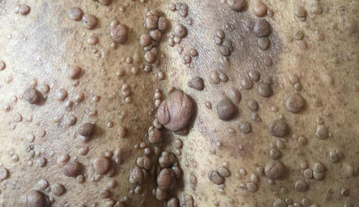
Neurofibromas are a type of lump that can form on nerves. Most of the time, they don’t cause any symptoms. However, sometimes they can result in itchiness, pain, or a tingling sensation. The symptoms can vary depending on the type of neurofibroma. Often, people seek medical help because they’re concerned about the way the lump looks.
There are different types of neurofibromas. Localized neurofibromas are often painless and may appear like a small, skin-colored or purple bump or mass beneath the skin. They can appear anywhere on the body but often show up on the trunk, head, neck, or limbs. When touched, these lumps can retract into the skin and then pop back upon release — this is called the “buttonhole sign.” They are often mistaken for moles or skin tags.
Diffuse neurofibromas usually show up on the head or neck area as ill-defined, hardened skin patches. If they’re big, they might cause a slight numbness or tingling sensation.
- Commonly appear on the head or neck
- Create hardened skin patches
- Mild numbness or tingling if they’re large
Plexiform neurofibromas often appear on the head, neck, trunk, and limbs and can grow quite large. They surround multiple nerve bundles and if on the surface, they often appear as skin-colored or darker nodular swellings. Deeper lumps coming from spinal nerve roots can become highly irregular and twisted. Pain, numbness, tingling, a sense of a mass effect, and spinal nerve compression might be symptoms if these lumps are deep.
Testing for Neurofibroma
If your doctor finds a single superficial lesion, which is simply a spot or lump on your skin, they usually will evaluate it through a physical exam or by removing a small tissue sample, called a biopsy, to look at it under the microscope. For larger spots or lumps, a biopsy and additional imaging techniques like CT scans or MRI may be required. These tools allow the doctor to see how far the spot or lump has spread in or under your skin and to plan for possible surgery to remove it.
Treatment Options for Neurofibroma
In most situations, the best treatment for skin growths known as cutaneous neurofibromas is complete surgical removal. In this case, the chance of these growths coming back is extremely low. Currently, there aren’t any other known treatments for these skin growths.
In uncommon instances, complete surgical removal might not be possible for certain types of these growths, known as diffuse or plexiform neurofibromas. In such situations, the growths are usually removed as much as possible to ease symptoms or improve appearance. Patients in these situations need to be regularly checked for quick growth or the return of these growths, as decided by the healthcare providers. A medication known as Interferon-alpha has been examined as a supplemental treatment for these specific types of neurofibromas, but the results have been inconsistent.
What else can Neurofibroma be?
Let’s break down some of the medical terms used to describe different types of tumors and their characteristics.
- Malignant peripheral nerve sheath tumor: This is an aggressive tumor that starts in the peripheral nerves. About half of these tumors are linked to a condition called NF1. They can sometimes appear in a previously benign nerve tumor, and show signs of unusual cell growth.
- Schwannoma: This is a benign (non-cancerous) tumor that’s mostly composed of Schwann cells, which insulate nerve fibers. It is linked to mutations in a specific gene, NF2. Under a microscope, these tumors have distinctive cell arrangements known as Verocay bodies and Antoni A and B areas.
- Perineuroma: This is a rare benign tumor composed of perineural cells, the cells that surround nerves. There’s no specific link to neurofibromatosis (a genetic disorder that causes tumors to form on nerve tissue).
- Dermatofibroma: This is a benign growth in the skin caused by an excess of fibroblast and histiocyte cells. It usually looks like a hard bump or papule in the skin.
- Dermatofibrosarcoma protuberans: This is a low-grade cancer that affects the dermis and subcutis, layers of the skin. It has a specific gene fusion and is known for being locally aggressive and having a high recurrence rate.
- Superficial leiomyoma: This is a benign tumor in the skin caused by abnormal growth in the smooth muscle cells. It often originates from the erector pili, the small muscles that are attached to hair follicles.
- Neurotized melanocytic nevus: This is a benign mole with melanocyte cells (which produce the pigment melanin) and loose neurotized tissue.
- Ganglioneuroma: This is a benign tumor that begins in the nerve cells and is most commonly found in the area behind the chest or in the retroperitoneum (the space in the abdominal cavity behind the abdominal lining).
- Plexiform fibrohistiocytic tumor: This tumor infiltrates the boundary between the dermis and the subcutaneous layer of the skin and is composed of fibroblasts (cells that produce collagen) and histiocytes (a type of immune cell).
- Desmoplastic melanoma: This is an invasive form of skin cancer that often resembles a skin scar. It is usually quite large at diagnosis and may have an associated in-situ melanoma (the earliest stage of melanoma).
Each of these can be identified by their characteristic features under a microscope and by using certain protein markers. This information can help doctors determine the exact nature of these tumors and suggest effective treatments.
What to expect with Neurofibroma
All neurofibromas, which are a type of benign (non-cancerous) tumor, very rarely reappear after they’ve been fully removed. Also, the risk of these tumors turning into cancer is extremely low. However, about 10% of patients with a condition called NF1 (Neurofibromatosis type 1), may experience their tumors becoming cancerous.
If someone shows up with a plexiform neurofibroma, a specific type of this tumor, they should be considered for NF1 assessment. This is because this type of neurofibroma is the most common one to turn into malignant peripheral nerve sheath tumors, which are a type of cancer.
Patients who have multiple localized neurofibromas (many benign tumors appearing in a certain area) should also be considered for further neurofibromatosis testing, which can help identify if they have NF1 or similar conditions.
Possible Complications When Diagnosed with Neurofibroma
Neurofibromas are benign nerve tissue tumors. If you have localized neurofibromas or a single tumor, the complications are usually minor and most often result from the surgical removal of the tumor. The common side effects from surgery include:
- Pain
- Bleeding
- Scarring
- Local infection
In some cases, when the neurofibromas are spread out or intertwined amongst nerves — a term we call plexiform lesions — the risks are higher. This is due to the surgery itself but also because sometimes it may not be possible to remove the entire tumor.
Amongst people with the condition known as NF1 who have persistent lesions, there is a higher chance of the harmless neurofibromas turning into harmful nerve sheath tumors. This means the tumors become cancerous, but this situation is fairly uncommon.
Preventing Neurofibroma
There are no clear risk factors for isolated neurofibromas (a type of nerve tissue tumor) that develop without an associated condition known as Neurofibromatosis Type 1 (NF1). But when a person has multiple neurofibromas, brownish patches on the skin (café au lait macules), freckling in the skin folds (intertriginous freckling), a type of brain tumor (optic glioma), certain benign growths in the eye (Lisch nodules), or bone abnormalities (skeletal dysplasias), the condition known as neurofibromatosis should be considered as a possible diagnosis.


