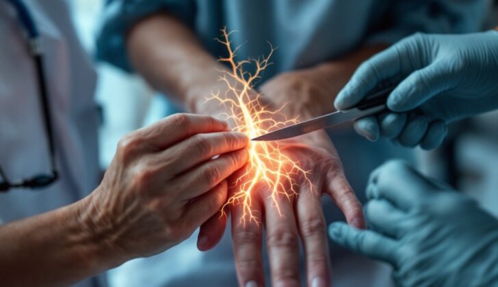
What is Neurotmesis?
Neurotmesis refers to a complete cut of a peripheral nerve, which are the nerves situated outside of your brain and spinal cord. The seriousness of this type of nerve injury can be broken down into three levels according to Seddon’s classification from 1942. These levels are neurapraxia, axonotmesis, and neurotmesis, or five different grades, rated by Sunderland’s classification done in 1951.
Neurotmesis causes complete loss of sensation and movement in the skin and muscles that the injured nerve was supplying. Without surgical treatment, the nerve function is very unlikely to recover on its own.
What Causes Neurotmesis?
Peripheral nerve injuries can occur from various causes, including high-speed accidents, cuts, broken bones, penetrating injuries, being crushed, loss of blood supply, and less commonly from heat, electric shock, radiation, hitting, and shaking. The most severe type of nerve injury, neurotmesis, involves the complete cutting of a nerve and usually occurs from sharp cuts.
During periods of wartime (such as in Iraq and Afghanistan), nerve injuries were mainly localized and caused by long-lasting blockage of nerve signal transmission or neurapraxia (45%). Other common injuries included axonotmesis, a more severe injury affecting the axon or the long part of the nerve (35%), and neurotmesis (20%). These injuries were equally common in the upper and lower limbs.
However, in the USA and Canada, during times of peace, nerve injuries most commonly occur from motor vehicle accidents.
Risk Factors and Frequency for Neurotmesis
Neurotmesis, a type of peripheral nerve injury, does not have clear statistics because it is often grouped with other similar conditions. Around 100,000 patients in the United States and Europe have peripheral nerve surgery each year. Most of these nerve injuries happen because of trauma, with roughly 350,000 cases every year.
Trauma causes 1.1 to 2.8 out of 100 cases, and most people who get these injuries are quite young, with an average age between 32 and 39. The most injury-prone age group is those aged 20-29. Depending on the type of fall, the chances of having a nerve injury vary; more forceful falls lead to higher rates whereas ground-level falls result in less.
- Women have lower rates of peripheral nerve injuries.
- 74% of those affected are men.
- In children, there’s no difference between boys and girls regarding these injuries.
When it comes to the body parts affected:
- The upper body is involved in 73.5% of cases, and 83% of these are mononeuropathies(single nerve damaged).
- In the lower body, injuries to the common peroneal nerve are quite frequent.
- Some professionals identify the sciatic nerve as the most frequently injured one, followed by the peroneal nerve.
- In the upper body, the ulnar nerve, found in your arm, tends to be the most affected, either by itself or in combination with others.
- Some experts consider the radial nerve in the arm to be the most frequently injured, followed by the ulnar and then the median nerve.
- When it comes to combined injuries, the ulnar and median nerves in your arms are most likely to be involved.
Signs and Symptoms of Neurotmesis
Understanding the cause of a nerve injury is very crucial as it helps to determine when the nerve needs to be repaired. Most full-thickness nerve injuries often happen due to sharp objects like glass, knives, or sharp metals.
A physical examination of the affected body part and the muscles controlled by the injured nerve can help identify which specific nerve is damaged. The area that the nerve provides feeling to is usually tested. If the nerve is fully damaged, there will be complete numbness in that area. Also, all the muscles controlled by that nerve will experience severe weakness. If there is a slight movement or some sensation present, it suggests that the injury is not complete. A procedure known as a Tinel sign is helpful in monitoring those with a peripheral nerve injury as it helps determine if the nerve fibers are repairing across the damaged area.
Testing for Neurotmesis
After checking your affected limb, your doctor may recommend some tests to figure out the type of damage that might be present. These tests can include EMG (a test that measures the electrical activity of muscles), an MRI scan (which uses powerful magnets and radio waves to create images of the inside of your body), or an ultrasound (which uses sound waves to produce images of structures within your body).
EMG becomes most useful after a delay of 2-3 weeks, by which time specific changes related to damage may have occurred in the affected muscles. In the case of mild nerve damage (known as neurapraxia), EMG will show no voluntary muscle action, but will also be absent of certain signals, called fibrillations, that point to severe nerve damage. If there is severe nerve damage, or neurotmesis, the EMG may start to show fibrillations and positive sharp waves, but these take time to appear and may show up 10 to 14 days later in muscles closer to the body and 3-4 weeks later in muscles further away. The appearance of certain signals called MUAPs on EMG means that the affected nerves are starting to recover. EMG can often detect this recovery earlier than a simple physical examination.
Your doctor might also recommend an MRI to help assess nerve damage. This test can show the physical changes in nerves and can track how the nerve is breaking down or healing over time. Recent advancements in MRI can provide detailed three-dimensional maps which allow doctors to have a clear view of the density and direction of nerve fibers inside your body, significantly improving the ability to assess nerve damage.
In urgent cases, your doctor may use an ultrasound to tell the difference between severe and not-so-severe nerve injuries. This is done by examining the continuity of nerves and checking if nerve segments can be seen on either side of the injury. It also allows doctors to check for the existence and location of neuromas (masses of nerve tissue that can develop after an injury), the size of any gaps, and whether the nerve is intact after procedures involving nerve grafting.
Treatment Options for Neurotmesis
The goal of nerve repair treatment is to help the nerve regenerate and reconnect with organs in the body. This process involves guiding the growth of sensory, motor, and autonomic nerve fibers carefully to avoid loss and ensure precise connectivity. The nerve ends must be correctly aligned and attached with minimal damage and ideally, as few stitches as possible.
End-to-end nerve repair techniques can involve epineural repair or fascicular repair, which treat the outer layer or internal structure of the nerve, respectively. The timing of the repair and the materials used play a crucial role. Depending on the type and severity of injury, repair could be immediate or delayed to allow damaged tissues to recover.
For injuries with sharp cuts without significant crushing, ideally, the nerve is repaired within 72 hours if the nerve bundles are still recognizable. For injuries caused by blunt trauma, repair is usually carried out after 3 to 4 weeks once the damaged area can be clearly identified. Repairing too soon could lead to failure. Gunshot wounds or stretch/avulsion injuries require a more extended waiting period, often around three months or 4 to 5 months, respectively, for recuperation before repair can proceed.
It’s vital to remember that nerves need to reconnect to their targets within 18 to 24 months; otherwise, degeneration will occur. A regrowing nerve takes approximately 1mm/day from the site of the injury or surgery. If the distance to the target is extremely long (more than 20 inches), the nerves may become nonfunctional by the time they reach their target because of ongoing muscle atrophy and degeneration.
The tension during the repair procedure is also an essential factor, as excess tension can lead to scarring, which can obstruct nerve regeneration. If there’s a significant gap to be filled in the damaged nerve, the best approach involves using a patient’s own nerve graft from a donor site in their body. While this method offers benefits, it does carry some challenges, such as the sacrifice of a donor nerve and possible mismatching.
Tissue-engineered nerve grafts can be used as a viable alternative to the patient’s own graft to bridge nerve gaps. These grafts can include scaffolds that provide physical structure and support for nerve growth or allografts, which are tissue grafts from a donor of the same species. In addition, several techniques are used to enhance the outcome, using materials like fibrin conduits or sealants, silicone conduits, or platelet-rich plasma, which is known to enhance nerve regeneration.
Based on the severity of the damage, the incorporation of stem cells along with these grafting techniques can also improve nerve regeneration. Despite the promising potential, there are concerns about the collection of such cells and potential tumor formation.
Pain management in nerve injuries can be challenging as the pain tends to be neuropathic, characterized by burning sensations and abnormal sensations. Certain specific medications are used to reduce the hyperactivity of nerves in these situations. However, common painkillers may provide short-term relief but are not the primary treatment choice.
What else can Neurotmesis be?
There are several different conditions that can affect the nerves in your body, some of which include:
- Neurapraxia, which is a temporary nerve injury
- Axonotmesis, a more severe nerve injury that can cause loss of nerve function
- Inflammatory neuritis, where your nerves become inflamed
- Infectious neuropathy, a nerve injury caused by viral infections such as HIV or bacterial infections like leprosy
- Paraneoplastic neuropathy, a nerve disorder that’s often associated with cancer
- Metabolic neuropathies, such as diabetic neuropathy caused by uncontrolled blood sugar levels
It’s crucial to identify and treat these conditions early to prevent long-term nerve damage.
What to expect with Neurotmesis
There are several factors that can improve the results after nerve injuries:
* Being younger
* The injury being closer to the end of the nerve (This is because it’s closer to the muscle, and the parts of the nerve that control movement and feeling can be better matched).
* The nerve being repaired from end to end
* The repair being done early
Younger patients usually experience better recovery times after nerve repair. Patients less than 20-25 years of age have the best recovery rates. However, as the patient’s age increases, the recovery rate can decrease significantly. In a study, children showed 95.4% partial or full recovery of both movement and feeling.
In adult patients, immediate repair resulted in improved function in 78% of cases, while delayed repair led to a 70% improvement rate. When grafts were used in nerve repair, the success rate dropped to between 63-77%.
Nerve injuries can result in long periods away from work, long-term disabilities, and in 30% of cases, it could lead to a disability pension.
Possible Complications When Diagnosed with Neurotmesis
List of Potential Issues:
- Unsuccessful tissue regrowth
- Decrease in muscle size and strength
- Tightening or hardening of muscles
- Chronic pain due to nerve damage
- Open sores that don’t heal
- Financial burdens
- Mental health problems
Preventing Neurotmesis
The best way to tackle neurotmesis, a severe nerve injury, starts with its prevention. Funding for measures to prevent, treat, and rehabilitate patients from these injuries could greatly boost patient health and improve how we manage this condition for better end results. People should understand to immediately seek medical help if they think they may have a nerve injury, as many of these cases require surgery early on.
For those who have experienced nerve injuries due to stretching, pulling, or even gunshot wounds, it could take several weeks before there is need for surgical repair. Once treated, patients may not sense any sensation or movement for a few months since nerves heal at a slow rate. Despite this, it’s important that patients continue attending their physical therapy and rehabilitation sessions to aid healing.


