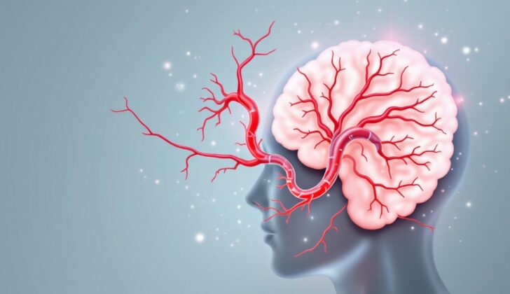
What is Basilar Artery Thrombosis?
The basilar artery is an essential blood vessel that helps circulate blood to the back part of the brain. This artery forms where the pons and medulla, two areas of the brain, meet and is created by the merging of two vertebral arteries. The basilar artery, together with the vertebral arteries, create the vertebrobasilar system, which plays a significant role in supplying blood to a part of the brain called the circle of Willis. The main job of the basilar artery is to provide oxygen-rich blood to different parts of the brain, including the cerebellum, brainstem, thalamus, and some lobes of the brain.
The basilar artery, which typically measures 3 to 4 millimeters across, produces about 20 other arteries, creating a sturdy network of blood vessels for the pons and midbrain. Spread along its length, the basilar artery distributes different types of branches. It’s situated on the underside of the pons, which is a neuron bridge linking the front of the brain to the cerebellum. Overall, the basilar artery is one of the most critical arteries in the human body.
What Causes Basilar Artery Thrombosis?
Strokes can develop from a blood clot, hardening of the arteries, or a tear in the artery wall. The exact mechanism can vary based on which part of the brain is affected. Hardened arteries mostly affect the mid-section of the basilar artery, which is at the back of your brain, as well as the point where your vertebral and basilar arteries meet.
Blood clots usually lodge in the last third of the basilar artery, especially at its top and where it connects with the vertebral arteries. A tear in the artery, also known as dissection, often occurs in the vertebral artery outside the skull and has been linked to neck injuries and neck adjustments done by chiropractors. However, dissections within the skull are extremely rare.
Risk Factors and Frequency for Basilar Artery Thrombosis
Just like other strokes, certain factors can increase your risk of posterior circulation stroke. High blood pressure is the most common risk factor, affecting about 70% of cases. Other things that can increase your risk include having diabetes, coronary artery disease, peripheral vascular disease, high fat levels in your blood (hyperlipidemia), and smoking cigarettes.
- High blood pressure is the most common risk factor and is found in about 70% of cases.
- Diabetes can also increase your risk.
- Heart-related issues, such as coronary artery disease and peripheral vascular disease, are risk factors.
- Smoking cigarettes can also increase risk.
- High cholesterol or ‘hyperlipidemia’ also adds risk.
We don’t fully know how often these strokes happen, but we do know a bit about how they work. About 20% of the blood in your brain flows through the back part of your brain, and that’s why around one out of every five strokes affect this area. Thankfully, basilar artery occlusions (blockages in a specific artery) only make up about 1% of all strokes. About 2 in 1000 people might have a blockage in this artery when they die. This kind of blockage may lead to about 27% of stopping-blood-flow strokes in the back part of your brain. Men have these strokes more often than women at a 2:1 ratio. These blockages happen more in older people, usually in their 60s and 70s, and especially if they have artery disease. This kind of blockage usually happens because something came loose and is stuck there, and it’s most common in people in their 40s.
- About 20% of the blood in your brain flows through the back part of your brain.
- One-fifth of all strokes affect the back part of your brain.
- Blockages in a specific artery called the ‘basilar artery’ only make up about 1% of all strokes.
- About 27% of stopping-blood-flow strokes happen in this part of the brain.
- Males are twice as likely to have this kind of stroke.
- People over 60, particularly those with artery disease, are more prone to these blockages.
- Blockages due to embolisms are most common in people in their 40s.
Signs and Symptoms of Basilar Artery Thrombosis
Basilar artery occlusion is a medical condition that may cause various signs related to the nervous system. Common symptoms include motor issues, partial or total paralysis of the body’s left or right side, facial muscle weakness, dizziness, and headaches. Some people may also experience difficulty in speaking, nausea, vomiting, and vision changes. It’s not uncommon for patients to have altered consciousness as well.
This medical condition may present in three main ways:
- Quickly progressing muscle related symptoms and a lower level of consciousness.
- Gradual symptoms over a few days that eventually lead to disabling muscle related symptoms and a decrease in consciousness.
- Early signs such as headache, neck pain, vision loss, double vision, difficulty to articulate words, dizziness, partial paralysis, unusual skin sensations, uncoordinated movements, and convulsive type movements.
It should be noted that the symptoms may be momentary and affect only one side of the body before turning permanent.
Mostly, an abnormal level of consciousness and focal motor weakness are typical symptoms. More than 40% of patients may also experience eye related signs, facial muscle weakness, and difficulty with speech and swallowing. Some patients may show varying degrees of partial or total paralysis in limbs. Due to the varying ways this condition may present itself, it is highly important to suspect and detect basilar artery thrombosis if patients show any of these symptoms.
Testing for Basilar Artery Thrombosis
Firstly, the main aim of any assessment is to pinpoint the exact location of a blood vessel problem and decide if immediate action is required to restore normal blood flow.
Lab tests can be useful, but they have their limits. These could comprise a complete blood count (CBC), electrolyte levels, blood urea nitrogen (BUN) and creatinine, international normalized ratio (INR), prothrombin time (PT), and activated partial thromboplastin time (aPTT). For patients who are young or who do not show signs of atherosclerosis, tests should be carried out to check if they have blood clotting conditions. These tests could include checking for protein C, protein S, and antithrombin III deficiencies, as well as for lupus anticoagulant and anticardiolipin antibodies, and homocysteine levels. An electrocardiogram can help detect irregular heart rhythms that could be a sign of a blood clot.
When it comes to imaging, a CT scan is usually conducted first. CT scans are capable of identifying large areas of oxygen-starved tissue and can highlight conditions where there is bleeding in the brain. In some cases, the CT scan may show a dense basilar artery. However, CT scanning is less reliable in identifying early oxygen starvation and is less effective in looking at the brainstem, cerebellum, and the back of the brain. As a result, high suspicion is always crucial in the right clinical context to diagnose a disease that could be easily overlooked. Sometimes, further testing with CT angiography might be discussed, with the possibility of identifying a blockage in the basilar artery. Catheter angiography is still a method of diagnosis, but with the advancement of noninvasive imaging methods like MRI and angiography (MRA), its application has transformed.
MRI/MRA is more sensitive than CT scanning in identifying early oxygen starvation and blocked blood vessels. MRI is the best option for providing images for any condition involving the lower part of the brain, including an acute ischemic infarction, which means that part of the brain tissue is dead due to lack of oxygen. DWI MRI sequence can display an acute brainstem or cerebellar infarct within seconds of the arterial occlusion. MR angiogram can non-invasively display the site of vascular occlusion. The presence of small hemorrhages in GRE-T2* or SWI imaging can also suggest an existing high blood pressure condition.
Treatment Options for Basilar Artery Thrombosis
An acute blockage of the basilar artery, a critical vessel at the back of your brain that provides oxygen and nutrients, can be life-threatening. So, when such a case is identified, patients should ideally be admitted into a specialized stroke unit, if available. Restoring normal blood flow in the blocked artery is crucial to the successful treatment of this condition, also known as basilar artery thrombosis, and can greatly improve a patient’s prognosis.
To clear this blockage, doctors can use a few methods: injecting clot-dissolving medication into the bloodstream (systemic thrombolysis or IVT), directly injecting these medications into the artery (intra-arterial thrombolysis or IAT), or physically removing the clot through a procedure called mechanical endovascular thrombectomy. More than half of the patients with basilar artery blockages who receive IAT or IVT experience flow restoration.
The timing of these treatments is important. The sooner the intervention is performed, the better the patient’s recovery. However, the exact time frame for treatment in these cases isn’t clear, as there hasn’t been a large-scale study to define it yet. But, it’s assumed to be longer than the standard 6 to 8-hour window recommended for blockages in the front part of the brain. In most cases, doctors consider treatment within 12 to 24 hours after the symptoms start.
In some cases, if a patient shows symptoms and has had a minor stroke according to an MRI brain scan, a mechanical endovascular thrombectomy could be considered up to 2-3 days later. After the blockage is cleared, the next step focuses on preventing a second stroke by addressing underlying causes and reducing risk factors.
What else can Basilar Artery Thrombosis be?
When doctors are trying to make a diagnosis, they also need to think about other conditions that may cause similar symptoms. Some conditions that might mimic the same symptoms include:
- Meningitis
- Hard to treat migraines at the base of the brain
- Bleeding in the brain that causes pressure on the brainstem
- Blockage or bleeding in a small section at the back of the brain, causing swelling
- Growths – benign or cancerous – in the back of the brain
- Non-brainstem growths that may cause severe harm by pushing on vital structures or causing herniation and pressing on the brainstem
- Other potential causes could be dangerously low blood sugar, temporary paralysis after a seizure (Todd’s Paralysis), or a mental condition known as a conversion disorder
It is crucial for physicians to consider these possibilities and perform the necessary investigations to ensure a correct diagnosis is made.
What to expect with Basilar Artery Thrombosis
The death rate for patients is often above 85%, but it can be as low as 40% if blood flow is restored. Unfortunately, only around 24 to 35% of patients treated with clot-dissolving medication have good functional outcomes. For those patients who survive but still have symptoms, they face a 10 to 15% risk of experiencing another stroke.
The two most crucial factors affecting the patient’s prognosis are how extensive the clot is and how long it has been present. As such, quickly suspecting a diagnosis and rapidly restoring blood flow provides the best chance for improved patient outcomes.
Possible Complications When Diagnosed with Basilar Artery Thrombosis
Patients with basilar artery thrombosis generally have a poor prognosis. However, new advancements in medication, mechanical thrombosis, and endovascular therapy may help decrease the likelihood of disability and death. Using methods such as catheter-directed thrombolysis and IV heparin come with their own risks, like the chance of bleeding.
Patients might encounter several complications like:
- Inhaling foreign matters such as food or liquids into the lungs (aspiration)
- Pneumonia caused by aspiration
- Disease related to blood clot formation (deep vein thrombosis and lung-related blood clots)
- Heart attack (myocardial infarction)
- Recurring stroke
Patients with severe physical impairments caused by strokes are more susceptible to muscle contractures, bedsores, and severe body-wide infections (sepsis). Many survivors of basilar artery thrombosis need ongoing physical and occupational therapy to regain and keep up their functional abilities.


