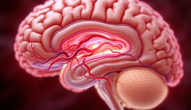
What is Duret Hemorrhages?
Brainstem hemorrhages, or bleeding in the brainstem, can be classified into two types: primary or secondary. Primary hemorrhages happen because of direct damage, high blood pressure, or issues with blood clotting. On the other hand, secondary hemorrhages might occur due to a descending shift in the brain from various causes. One form of secondary hemorrhage is known as Duret hemorrhages (DH).
The name Duret hemorrhages come from Henri Duret, a French neurologist who first explained how the blood supply is distributed in the brainstem and then in the outer layer of the brain. Dr. Duret’s research focused on brain injuries and found out that disturbances in automatic bodily functions, like breathing and heart rate, could be traced back to the brainstem. These disturbances were connected to tiny hemorrhages affecting the lower part of the brainstem and pons, which were consequences of Duret hemorrhages.
What Causes Duret Hemorrhages?
Duret hemorrhages are caused by a condition known as descending transtentorial herniation, which can happen for various reasons. This condition is a result of increased pressure inside the skull, causing different parts of the brain to shift.
Most of the time, Duret hemorrhages are associated with a boost in this brain pressure due to a variety of causes. These can range from different types of bleeding within or around the brain, injuries, brain tumors, sudden and widespread brain swelling, low sodium levels, and occasionally, certain medicines that dissolve blood clots.
In rare instances, Duret hemorrhages have been reported to result from decreased brain pressure. Typically, these hemorrhages tend to happen in certain areas within the backbone of the upper pons (which is part of the brainstem) and the midbrain.
Risk Factors and Frequency for Duret Hemorrhages
Duret hemorrhages, which are a type of bleeding in the brain, occur more frequently according to studies involving microscopic examination (30 to 60% of cases) versus those involving medical imaging (5 to 10% of cases). The reason for this difference may be that 20% of these brainstem hemorrhages are too small to be detected without a microscope. Additionally, Duret hemorrhages may not develop until after the initial CT scans have been taken. Certain factors, such as high blood pressure and older age, can increase the risk of developing a Duret hemorrhage.
- Duret hemorrhages are reportedly found in 30 to 60% of neuropathological studies, but only 5 to 10% in radiological studies.
- This difference might be because 20% of these are microscopic and can’t be easily seen in CT scans.
- Moreover, the bleeding might not start until after the initial scans.
- Having high blood pressure or being of an advanced age makes it more likely to develop a Duret hemorrhage.
Signs and Symptoms of Duret Hemorrhages
The majority of patients who experience a condition where the brain shifts within the skull often have a history of previous issues, such as a head injury, brain tumor, or abnormal growth in the brain. This can manifest as changes in awareness, from mere confusion to a state of unconsciousness, because the condition might interfere with the brain’s alert system. This can also lead to uneven pupil size due to the effect of the condition on one of the cranial nerves, and there can be weakness on the opposite side.
In some situations, they may show weakness on the same side, due to the phenomenon known as “Kernohan notch”. This happens when the protrusion of the brain presses against a structure called the tentorium cerebelli. As the condition worsens, the affected person may display unusual postures and lose their brainstem reflexes, with their breathing pattern changing from Cheyne-Stoke (an abnormal pattern of breathing) to ataxic (uncoordinated) breathing.
In cases where the shifting happens in a upward direction, especially after certain procedures to remove fluid from the brain, the patient may develop a condition known as Perinaud syndrome.
Testing for Duret Hemorrhages
If your doctor suspects abnormal growths or damage to your brain, such as tumors causing swelling, or different types of brain bleeds, they will likely order a CT scan. This imaging test can help identify these issues, plus reveal if there’s any serious compression of your brain that is erasing the normal spaces around certain structures in the brain. This condition can sometimes lead to small bleeds in specific areas of your brain or disrupt the flow of blood to the back part of your brain.
In addition to this, your doctor will need to run some blood tests to check levels of certain chemicals in your body like sodium, and to understand how effectively your body is maintaining its acid-base balance. A basic test to check the health of your kidneys, and levels of other important body substances might also be ordered.
Treatment Options for Duret Hemorrhages
When a person has a medical emergency affecting their brain, such as a brain herniation, a few key steps are taken. Firstly, doctors ensure the person can breathe and their heart is beating properly. Then, they use a type of scan called a non-contrast CT scan to check the brain’s condition. The goal is to quickly manage any increase in pressure inside the skull (intracranial hypertension) and identify the root cause to be able to treat it promptly.
One of the possible causes could be a type of bleed in the brain (epidural or subdural hemorrhage) often caused by trauma. If this is the case, the doctors will aim to remove the pool of blood (hematoma) as quickly as possible.
To handle increased pressure inside the skull, the medical team will do several things:
1. They will raise the head of the bed by 30 to 60 degrees.
2. They will control the person’s breathing to maintain a certain level of carbon dioxide in their blood.
3. They will use special fluids (hypertonic saline and mannitol) to draw excess water from the brain to decrease swelling.
4. They will closely watch the pressure inside the brain and aim to keep it at a safe level.
5. If a brain tumor causing swelling is found, they might use a medicine called dexamethasone to decrease the pressure.
6. In some cases, surgery might be needed to relieve the pressure.
Once in the intensive care unit (ICU), the goal is to keep the patient’s body in balance. This involves maintaining normal blood pressure, blood volume, sodium level, blood sugar level, and temperature. The doctors will continuously monitor the patient’s brain function and repeat the brain scans as necessary in the ICU.
What else can Duret Hemorrhages be?
Certain conditions may show similar signs as a Duret hemorrhage, a specific type of brain bleed. The comparable conditions include ‘brainstem hemorrhages’, which can be smaller and widespread (petechial) or larger and caused by high blood pressure (hypertensive).
Usually, a Duret hemorrhage shows up in brain scans forming a line; it can grow in multiple directions, and take varying shapes. It typically happens when there’s an abnormality above the tentorium cerebelli (an area in the brain), leading to pressure that causes a part of the brain to shift (called ‘transtentorial herniation’).
In cases hypertensive hemorrhages, they are generally bigger and happen in patients who had a prior history of uncontrolled high blood pressure; they do not record any abnormalities in the upper sections of the brain.
On the other hand, petechial hemorrhages, are more widespread and smaller in size. They usually occur around the mid-section of the brain (specifically around the periaqueductal and tectum regions) and are typically seen in individuals who have suffered widespread nerve damage following a traumatic brain injury.
What to expect with Duret Hemorrhages
Historically, the occurrence of a Duret hemorrhage has been seen as a bad sign for patient recovery. However, recent case reports seem to indicate that it might be possible to recover after experiencing a Duret hemorrhage. Recovery seems to depend on the root cause of the transtentorial herniation, the medical term for the brain condition that leads to the hemorrhage. Favorable outcomes have been reported for some patients with severe low sodium levels, subdural injuries, and traumatic brain injuries.
However, it’s still not known if the recovery in such cases is directly linked to the prompt correction of the underlying cause.


