What is Spinal Hemangioma?
Spinal hemangiomas, which are the most common type of tumor in the spine, are typically harmless. Like other hemangiomas throughout the body, these are blood vessel-related growths that involve an increase in regular capillary and venous structures – these are small blood vessels and veins. A study found that about 11% of people examined post-mortem had these growths. Most often, they’re found by accident during body and spine scans and X-rays, particularly of the mid to lower spine.
Only a few of these tumors cause symptoms, with back pain and neurological issues being the main complaints. Some studies suggest roughly 1% become symptomatic. The severity of the back pain can range from mild to severe, and it’s often worsened by movement. The tumors usually extend into the spinal canal or neural foramina, an opening in the spine, in cases where the pain takes on a neurological nature.
What Causes Spinal Hemangioma?
Spinal hemangiomas, which are benign growths that form on the spine, are not fully understood in terms of what causes them or how they develop. However, one thing that is well-known about these growths is that they involve the rapid growth of small blood vessels, or capillaries, a trait they share with hemangiomas that appear on other parts of the body. This rapid growth can push against bone, and in rare cases can even erode into the space around the spinal cord. Unlike hemangiomas that appear in infancy, which usually go away on their own over time, spinal hemangiomas do not disappear naturally.
Risk Factors and Frequency for Spinal Hemangioma
Spinal hemangiomas, which appear in around 11% of autopsy studies, are usually found by chance during spine-related imaging and radiographs. These types of tumors are more prevalent in women than in men. In fact, on MRI studies, the incidence of spinal hemangiomas can reach as high as 27%.
Signs and Symptoms of Spinal Hemangioma
When a patient arrives with symptoms related to a hemangioma (a noncancerous growth), the doctor’s main goal is twofold. They aim to rule out any more serious causes of the symptoms and evaluate the patient’s overall health in case treatment is needed.
It’s common for such patients to report back pain. However, it’s important not to assume this is solely due to the hemangioma, particularly if the patient has been diagnosed with it before. There are numerous, potentially serious causes of back pain that doctors should consider. These include cancer spread, infections, spinal cord tumors, and fractures caused by osteoporosis. Doctors should ask questions about the onset, intensity, nature, and pattern of the pain, as well as what makes it better or worse.
It’s useful to note that hemangiomas often occur in the middle part of the spine and usually cause pain in a single location. If a patient reports pain in multiple locations, doctors should consider the possibility that the pain may be caused by cancer spread. Other secondary symptoms, such as symptoms related to the digestive or urinary systems, are also critical in considering a diagnosis of cancer spread.
Other points in patients’ medical history that should prompt further investigation include back pain caused by a condition elsewhere in the body, a prior diagnosis of cancer, a history of trauma or osteoporosis, and pain that doesn’t improve with rest or lying down.
Physically examining the patient is also crucial. This typically involves looking at the skin over the back, checking the curvature of the spine and the patient’s walk, and assessing the spine’s range of motion. Doctors should also touch and percuss the spine, particularly at points that the patient identifies as painful.
As hemangiomas can invade the spinal canal, it’s important to check for possible nerve root compression. This check might involve performing specific tests like the straight leg raise test for the lower back or the Spurling maneuver for neck-related nerve root compression. The doctor should test reflexes and sensation, paying particular attention to the middle part of the spine.
Finally, the doctor should also examine the digestive and urinary systems as part of the physical examination.
Testing for Spinal Hemangioma
Imaging, or medical scans, play a key role in examining a hemangioma, which is a type of noncancerous growth that most commonly occurs on the skin. Through these scans, doctors can measure the size of the lesion, its location, and the extent of bone damage it has caused, particularly in the spinal canal and the small openings on each side of your spine (neuroforamina).
One common scan is a radiograph, or an X-ray, which often shows a strong pattern of small bone-like structures (trabecular pattern) with vertical lines. The affected bone (vertebral body) often appears denser, giving it a hard or sclerotic look. However, the height and size of the vertebral body should not be changed.
CT scans, another type of imaging, can provide more detailed images of the hemangioma. These scans usually show hemangiomas as having a unique ‘corduroy’ or ‘accordion’ pattern due to the coarsening or thickening of the trabeculae, which are tiny, lattice-like structures within the bone. In rare cases where the hemangioma has caused bone damage and spread into the adjacent tissues, including the spinal canal, this can be seen on the CT scan as well.
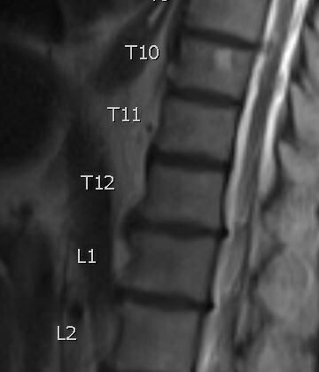
Lastly, Magnetic Resonance Imaging (MRI) scans can accurately assess if and how much the hemangioma has extended into the surrounding soft tissues. These are vital tools in ensuring the correct diagnosis and treatment plan for patients with a hemangioma.
Treatment Options for Spinal Hemangioma
Most hemangiomas, which are benign (harmless) blood vessel growths, don’t cause any symptoms. As a result, treatment usually isn’t necessary if the patient isn’t experiencing any discomfort. However, if a hemangioma does cause symptoms, there are several treatment options available.
One option is endovascular embolization, a procedure that involves blocking the blood flow to the hemangioma to prevent it from growing. This treatment has been effective in relieving pain caused by vertebral hemangiomas (hemangiomas located in the spine) and in alleviating spinal cord compression that can arise due to the hemangioma extending into the spinal canal.
Percutaneous vertebroplasty, a procedure in which a special bone cement is injected into the vertebra to strengthen it, can also be effective in relieving pain associated with hemangiomas.
Another treatment option is the injection of ethanol directly into the hemangioma, a procedure known as transpedicular ethanol injection. However, this treatment comes with certain risks and has been associated with some complications.
Radiation therapy, which uses high-energy rays to kill the cells of the hemangioma, can also be used to treat painful spinal hemangiomas. This is because these hemangiomas are generally sensitive to radiation, meaning the radiation can successfully destroy these growths.
What else can Spinal Hemangioma be?
When we are trying to identify a type of spinal lesion known as a ‘sclerotic lesion’, doctors have several possibilities to consider:
- Paget’s disease, especially if the lesion is in the body of the vertebra and looks similar to a hemangioma, a type of benign tumor. This is often accompanied by thickness in the bone’s outer layer, which shows up on X-rays as a “picture frame” sign.
- Squaring of the vertebra, a visible abnormality that can also help in diagnosis.
- Metastasis of sclerotic lesions, where cancer from another part of the body like the prostate, bone (osteosarcoma), or thyroid gland (medullary thyroid carcinoma) has spread to the spine.
- Two types of blood cancer, lymphoma and multiple myeloma.
Understanding these possibilities helps doctors determine the most likely cause of the sclerotic lesion, which can assist in establishing a more accurate treatment plan.
Possible Complications When Diagnosed with Spinal Hemangioma
Even though complications are rare, they can occasionally occur. One such complication is a pathologic burst fracture. If this happens, the highly vascular areas can bleed, leading to the formation of a hematoma and eventually to compression of the spinal cord. It’s rare, but the extension of the problem into the epidural space can also happen. This could lead to problems with neurological functions because of spinal cord compression. Nerve roots that exit the spine may also be affected, leading to neurological symptoms. Spontaneous bleeding into the epidural space can also happen, although this usually occurs as a result of a medical procedure.
Common Side Effects:
- Pathologic burst fracture
- Bleeding in highly vascular areas
- Hematoma
- Spinal cord compression
- Epidural extension
- Neurologic deficits
- Neurologic symptoms due to affected exiting nerve roots
- Spontaneous epidural hemorrhage usually as a result of a medical procedure
Preventing Spinal Hemangioma
If a patient has a confirmed hemangioma (a noncancerous growth) and isn’t experiencing any symptoms, they can be reassured that this condition is harmless. However, if the patient starts experiencing any pain or new neurological symptoms (changes in their nervous system function), they should return to the clinic. At that point, the doctors can perform a detailed examination to figure out what’s causing these new symptoms. This would also help confirm whether the hemangioma has become symptomatic (started causing symptoms) or if there’s a different underlying reason for these changes.


