What is Spinal Stenosis and Neurogenic Claudication?
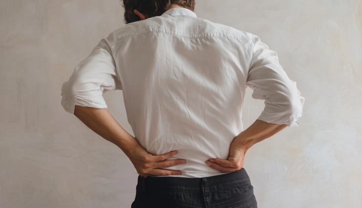
Almost everyone – around 90% of people – will experience lower back pain at some point in their life. One common reason for this pain is spinal stenosis, a health issue caused by narrowing of the central canal, side recess, or neural tunnel, which are all important pathways for nerves in your spine. This condition can cause a lot of discomfort, make it hard to carry out everyday activities, and could eventually lead to permanent disability.
As people are living longer lives, diseases of the spine like spinal stenosis, are becoming more common and can cause serious problems worldwide. It’s important to understand that spinal stenosis is often part of the natural aging process. However, it is hard to predict who will develop symptoms because there isn’t a direct link between having spinal stenosis and feeling symptoms.
While we can’t stop the aging process entirely, we can slow it down through things like regular exercise, a good diet, and other healthy lifestyle changes. Typically, spinal stenosis is most commonly found in the lower back (lumbar) and neck (cervical) regions of the spine.
What Causes Spinal Stenosis and Neurogenic Claudication?
Spinal stenosis, or the narrowing of the spaces within your spine, is most often caused by the wear-and-tear changes of osteoarthritis. This condition commonly affects the L4 to L5 level of the spine, although it can also occur at the L5 through S1 and L3 to L4 levels. Increased weight or having family members with spinal stenosis can increase your risk of developing the condition.
Several factors can contribute to spinal stenosis, such as a protrusion or bulging of the disc (this can happen as the discs in our spine degenerate with aging or after an injury), a decrease in the height of the discs, arthritis of the tiny joints on the back part of the spine, bone spur formation, or the thickening of a ligament in your spine. All of these can narrow the central canal where your spinal cord is and the openings where nerves leave the spine, and may squish and irritate these structures.
Having one vertebra (bone in the spine) misaligned, or slide forward or backward compared to the one next to it, can also narrow the spinal canal.
Other acquired causes that can lead to spinal stenosis include benign or malignant tumors or fat deposits that take up space in the spinal canal, any swelling or fibrous tissue resulting from injury or surgery or certain bone diseases like ankylosing spondylitis, rheumatoid arthritis, or Paget disease.
Certain inherited or developmental disorders can cause spinal stenosis as well. This includes conditions like a type of dwarfism called achondroplasia, Morquio syndrome, a defect with the formation of the spine like spina bifida, misalignment of the vertebrae, or a protrusion of the membranes surrounding the spinal cord through a defect in the vertebral column.
Risk Factors and Frequency for Spinal Stenosis and Neurogenic Claudication
Spinal stenosis is a condition that commonly affects people over the age of 60. In fact, it’s the most common reason why adults over 65 have spinal surgery. This ailment is typically associated with various conditions, but it’s often linked to degenerative changes in the spine.
- Spinal stenosis frequently occurs in the lower parts of the spine.
- The dorsal root of the spinal nerve tends to be larger in this area, making it more likely for spinal stenosis to occur.
- There also tends to be more spondylosis (a type of arthritis) and degenerative disc disease in these lower segments, both of which can contribute to the development of spinal stenosis.
While many people over 60 may have some form of spinal stenosis, not everyone will have symptoms. This makes it hard to gauge exactly how common this condition is.
Signs and Symptoms of Spinal Stenosis and Neurogenic Claudication
Spinal stenosis is a medical condition which narrows the spaces within your spine. This can put pressure on the nerves that travel through the spine. There are a variety of symptoms associated with this condition, and they differ from person to person. The most commonly observed symptom is neurogenic or ‘pseudo’ claudication. It’s related to the patient’s posture and most noticeable when the lower back is extended. In this condition, pain usually decreases when the patient bends forward or sits down, but can worsen while standing, walking, or doing any upright exercises. The pain often promptly resolves when the patient leans forward slightly at the waist.
Additional symptoms of spinal stenosis, likely due to spinal nerve root involvement within the lumbar spinal canal, may include general discomfort, weakness/numbness in the legs, or tingling sensations. Symptoms typically occur in both legs and can involve the entire leg, not just one part. In some patients, symptoms may not be symmetric.
While most affected individuals have a normal neurological exam, some may show evidence of weakness, loss of deep tendon reflexes, or sensory loss. A useful clinical bedside marker for lumbar canal stenosis is the wasting away of the muscle that helps move the small toes away from each other (bilateral extensor digitorum brevis).
There is also a functional test called the Five Repetitive Sit to Stand Test (5R-STS). In this test, a patient who can sit and stand five times in around 10 seconds is not considered to have significant functional impairment.
Testing for Spinal Stenosis and Neurogenic Claudication
If a person shows new symptoms or signs of conditions called radiculopathy or spinal stenosis, it’s important to do a brain and spine scan (neuroimaging). Radiculopathy happens when a nerve root in the spine is damaged or not working correctly which can cause pain, weakness, numbness, or difficulty controlling certain muscles. Spinal stenosis is a narrowing of the spaces within your spine, which can put pressure on your spinal cord and other nerves.
Magnetic Resonance Imaging (MRI) is typically the chosen method for diagnosing spinal stenosis. This is because it provides clear pictures of both the soft tissues and the parts of the nervous system within the spine, like nerves. Therefore, an MRI can confirm if the spinal canal has become too narrow or whether nerve roots are being pinched or squeezed.
Though MRI is usually preferred, a Computed Tomography (CT) scan – which uses x-rays to make a detailed image of your body – can sometimes offer a better view of bones. These scans are especially used when an MRI isn’t suitable for the patient for some reason. CT myelography, which involves injecting a contrast dye into the spinal cord before the scan, may be considered in such a case.
There are telltale signs in these scans that can reveal the presence of spinal stenosis. For instance, a ‘trefoil appearance’ (a three-leaf clover shape) detected in a CT scan and positive sedimentation signs (where certain particles settle down at the lower part of the spine) seen in an MRI scan are traditional indicators of this condition.
After the imaging tests confirm the narrowing of the spinal canal or pinching of nerve roots, further tests may be needed for a definitive diagnosis of lumbar canal stenosis (a type of spinal stenosis that affects your lower back). Tests like electrodiagnosis can detect ‘fibrillation potentials’ (irregular currents or movements in your muscles) and whether the ‘tibial H-wave’ is absent or not (related to nerve signals in your lower leg), which could further support the diagnosis.
Treatment Options for Spinal Stenosis and Neurogenic Claudication
The initial treatment for spinal stenosis, a narrowing of the spaces within your spine, often includes exercise to improve muscle strength and posture, medication to reduce inflammation and pain, and steroid injections to help reduce inflammation near the nerves. It’s usually suggested that patients avoid actions that make their symptoms worse, such as walking downhill or overextending the lower back.
Surgery is generally considered when non-surgical, or “conservative” treatments have failed to manage a patient’s symptoms. Most of the time, the goal of surgery is to relieve symptoms and improve function, rather than to prevent complications from the stenosis. Surgery might be urgently needed if a patient begins experiencing severe symptoms like rapid worsening of nerve function or problems with bladder control. This can occur in conditions like cauda equina syndrome, which occurs when all the nerve roots in the lower back are compressed. The type of surgery performed often includes removing part of the vertebra (laminectomy), with or without spinal fusion. Spinal fusion is generally reserved for patients with a condition called spondylolisthesis, where one vertebra slides over the one below it.
Patients who have surgery for their spinal stenosis often experience more substantial symptom relief compared to those treated conservatively. For patients with spinal stenosis without spondylolisthesis, a recommendation of decompression alone, which involves relieving pressure on spinal nerves, is recommended. The Spine Patient Outcomes Research Trial (SPORT) provided evidence that laminectomy and fusion can provide better results than non-surgical approaches.
Your doctor’s surgical approach will aim to balance between doing too much and doing too little. Traditional methods like a complete laminectomy can result in significant blood loss, extended hospital stays, and potential instability in the spine.
However, advancements in surgical techniques, such as laminoplasty, hemilaminectomy, and laminotomy, can help avoid some of these issues. Yet, there can still be an increased risk for neural injury. Any surgical plan should consider the stability of the spine and aim to preserve as much of the vertebra as possible to prevent instability. In certain cases, stability can be maintained by using screws to stabilize the spinal segments, but these cases often necessitate a higher rate of reoperations.
Fusing the spine can lead to a significant improvement in a patient’s quality of life. Still, it’s important to be aware that spinal fusion surgery is the most expensive type of surgery performed in U.S. hospitals. This procedure could only be justified in middle and low-income nations if significant, lasting clinical benefits were to be expected.
Recently, alternate surgical options have emerged. These include using devices to create space between the spinal processes or performing posterior fixation surgery that includes facet distraction (widening the facet joints in the spine) without decompression. These approaches have shown promise in delivering good clinical outcomes.
What else can Spinal Stenosis and Neurogenic Claudication be?
When trying to make a diagnosis, doctors will consider several possibilities. These include vascular claudication, which is pain in the legs due to inadequate blood flow, and multiple level lumbar disc protrusions, a condition where the discs in the lower spine are damaged or worn out.
What to expect with Spinal Stenosis and Neurogenic Claudication
Spinal stenosis, a condition where the spaces within the spine narrow, can have a big impact on one’s day-to-day quality of life. Over time, this condition can cause chronic pain and muscle weakness. In some situations, spinal stenosis might even lead to a serious condition called cauda equina syndrome, which involves severe pressure and swelling of the nerves at the end of the spine.
People with central spinal stenosis often find it difficult to walk and may experience abnormal movement patterns. Even though some patients may show signs of improvement with time, most see their conditions getting worse, which can eventually lead to disability.
It’s also noteworthy to mention that managing and treating spinal stenosis can be quite costly. It can often lead to high healthcare bills for patients, adding a financial burden to the physical challenges the condition brings.
Possible Complications When Diagnosed with Spinal Stenosis and Neurogenic Claudication
Not properly treating back problems can lead to a condition known as failed back syndrome. On the other hand, over-treatment might cause instability in the back.
There’s also the risk of nerve damage with minimally invasive surgeries.
Additional complications include:
- Pain radiating to the lower limbs
- Loss of muscle tissue
- Disability
- Abnormal spine shape
- Blood clot in the lung (Pulmonary embolism)
Recovery from Spinal Stenosis and Neurogenic Claudication
Differences in the shape of the facet joint and unequal size of the muscles placed either side of the spine can be helpful in checking for instability of the spine after surgery.
Interestingly, specific rehab programs have not been found to be any better than simply staying active. So, just continuing daily activities is recommended.
Preventing Spinal Stenosis and Neurogenic Claudication
“Shared decision making” is an approach where doctors and patients work together to decide on the best treatment options. This method encourages good communication and can aid in creating the most beneficial and appropriate treatment plans.


