What is Dental Caries (Cavities)?
The history of dentistry can be traced back to around 5000 B.C., when people believed that a “tooth worm” was responsible for tooth decay. The term “dental caries,” classically used for describing tooth decay, appeared first in written records around 1634. It is rooted in the Latin word “caries” which means decay, and originally, it was used to signify holes in the teeth.
Being one of the oldest and most common diseases in humans, dental caries is a widespread chronic infection. It happens when bacteria that cling to teeth metabolize sugars, creating acid in the process. Over time, this acid strips away the mineral content of the tooth, leading to decay.
What Causes Dental Caries (Cavities)?
Dental caries, or tooth decay, is a condition that is both a disease and the resultant lesion on the tooth. It takes place in the biofilm, an ever-active layer of bacteria on the teeth, with every fluctuation of pH levels. This process then leads to damage in the hard parts of the teeth.
Tooth decay happens when the natural balance of bacteria in your mouth changes. This shift usually occurs because of the frequent intake of sugars, leading to a set of bacteria that produce acid and cause decay. This problem can be invisible or can cause mineral loss in the tooth’s hard parts, causing visible damage. So, you can have the tooth decay process underway even without a noticeable cavity.
In this sense, dental caries is a diet-related disease involving harmful bacteria and regular exposure to sugars (like glucose, fructose, maltose, and sucrose) from food.
Interestingly, things like your habits, thoughts, feelings, and societal factors significantly affect this process too. It’s also well known that fluoride can prevent tooth decay, so if you’re not getting enough fluoride, it might contribute to the disease.
Risk Factors and Frequency for Dental Caries (Cavities)
Dental caries, or tooth decay, is a significant health concern that affects nearly everyone in the world, making it the most widespread disease. Despite being easily preventable, the rates of the disease have not noticeably declined in the past 30 years. Moreover, this disease is more common among people from lower socio-economic backgrounds.
Signs and Symptoms of Dental Caries (Cavities)
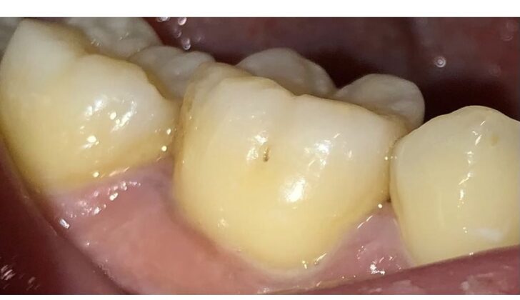
The first symptom of tooth decay is a white spot, which indicates the underneath area of the enamel is losing minerals, while the surface remains hardened. As acidic damage persists, the once smooth enamel begins to feel rough. Over time, tiny cracks and cavities may start to appear. The average tooth decay remains visible on the enamel for about three to four years, but experiences can vary a lot and sometimes reverse.
As the tooth decay progresses, the dentin, which is the tissue beneath the enamel, starts losing its mineral content and becomes invaded with bacteria. This activity causes the formation of tertiary dentin as a protective mechanism for the pulp, the central tissue of the tooth.
One can determine whether a tooth decay is active or inactive based on various factors. These factors include:
- Location: Is the decay located in an area with stagnant plaque? Is it along the gum line?
- Appearance: Is it white or brown? Does it have a dull or shiny look?
- Texture: Is it rough or smooth?
- Integrity: Is it cavitated or non-cavitated?
- Probing: Does the gum bleeding when probed?
An active tooth decay would typically be white, dull-looking, rough, cavitated, located in an area with stagnant plaque, and associated with gum bleeding upon probing. On the other hand, inactive decay generally has opposite characteristics: brown in colour, shiny, smooth, non-cavitated, unassociated with plaque, located away from the gum line, and no gum bleeding is noticed upon probing.
Root caries, which affect the root of a tooth, also begin as subsurface demineralization, similar to enamel caries. However, root caries turn soft at an earlier stage. While these lesions may seem broad, they usually do not exceed 0.5 to 1 mm in depth. Due to the slow rate of microbial invasion and tissue degradation, they allow the opportunity to take measures to control plaque and prevent further decay.
Recurrent or secondary caries are new decay forms that develop around the margins of a dental filling. They occur as a result of micro leakage from the filling, but this doesn’t cause active demineralization under the filling itself.
Testing for Dental Caries (Cavities)
When a dentist is checking for tooth decay, also known as dental caries, there are a number of techniques they might use. These could include a simple visual and touch-based examination, x-rays, shining a fiberoptic light into your mouth, or using a more advanced version of this called digital fiberoptic illumination. Interestingly, the most common method is also the simplest: a regular check-up using the dentist’s eyes and hands.
X-rays are also a practical method for spotting dental caries. These can include types like periapical x-rays, which can show the entire tooth from the crown to the root, and bitewing x-rays, which are good for viewing cavities between teeth.
A key tool that is often used to record and detect dental caries is the International Caries Detection and Assessment System. This system sorts dental caries into six categories, with higher scores indicating more severe decay. It’s very accurate and is a great way to keep track of how the caries progress over time.
Some newer ways to diagnose tooth decay involve using laser and light-based technologies. These methods give more detailed information about the cavity. For example, light-induced fluorescence can indicate how far the cavity has spread, the exact spot it’s in, and even if bacteria are currently active there. Meanwhile, laser fluorescence detection devices allow dentists to observe the cycles of mineral loss and gain in the tooth, which are key processes in caries development.
Treatment Options for Dental Caries (Cavities)
Managing tooth decay should involve:
- Identifying early stage decay
- Assessing its activity
- Evaluating the risk for further decay
- Preventing new decay from developing
- Preserving as much dental tissue as possible
- Aiming to keep natural teeth for a lifetime
Rather than removing dental tissue to treat existing tooth decay, a better first step is to use non-invasive procedures. These might include increasing mineral content in teeth, removing plaque biofilm, and sealing off teeth from decay. If there’s a cavity, it should be managed using a minimal approach, including fixing a broken filling instead of replacing it.
Non-Invasive Procedures
For tooth decay that can be reached for easy cleaning, a filling isn’t necessary. Regular teeth cleaning can disrupt the biofilm of decay causing bacteria, especially when fluoride toothpaste is used.
Decay that can be managed without filling includes:
- Early stage discoloration or damage of the teeth
- Decay seen in dental x-rays which hasn’t reached beyond the tooth’s outer layer, or enamel, or just into the layer beneath, dentin.
- Both cavity-based and non-cavity-based decay on the root surface of the tooth that can be reached with cleaning tools
- Decay recurring next to a previous dental filling that can be cleaned doesn’t need to be filled.
When should the tooth be restored?
The International Caries Consensus Collaboration in 2016 made recommendations for when to remove decay tissue and handle cavities. The decision lies on whether the decay can be accessed for cleaning. If a cavity is visibly present, the decay is considered active and beyond reach for normal cleaning.
This expert consensus agreed that:
- Cavity-based severe decay that can’t be managed with simple biofilm removal, mineral increase or sealing alone. However, in baby teeth, these lesions could potentially be converted to a manageable condition without the need to fill the cavity.
- Sometimes, even if a decay is not visibly a cavity, a dental x-ray can show that it has progressed widely into the dentin. The cavity may need to be sealed off, but it may need to be monitored regularly to ensure the seal integrity. In absence of sufficient evidence, there’s a possibility that the seal may fail and a filling would be required.
In addition to this, certain cases require invasive treatments. For instance, cavity-based decay seen on a dental x-ray reaching into dentin layer of the tooth and cavity-based decay clearly seen in the dentin layer on a dental x-ray may require invasive procedures.
What else can Dental Caries (Cavities) be?
Dental cavities, also known as caries, need to be differentiated from other conditions. These can include issues that occur as teeth develop, such as problems related to the enamel or dentine formation, insufficient minerals, or excess fluoride causing staining. It’s also important to separate them from damage caused by mechanical or chemical stress, which can wear down the teeth.
What to expect with Dental Caries (Cavities)
The prognosis for tooth decay relies on the patient’s overall health, their oral hygiene habits, and the severity and progression of the decay. If someone seeks help at the first signs of tooth decay, the damage can often be repaired with preventive measures and minor dental treatment, like remineralizing the affected area.
On the other hand, if the tooth decay has progressed to a moderate level and caused the loss of part of the tooth, then filling and rebuilding the tooth becomes necessary.
Possible Complications When Diagnosed with Dental Caries (Cavities)
Dental caries, also known as tooth decay, is generally not a life-threatening condition. However, if the infection spreads into different parts of the face, it can increase the risk of some serious complications. These can include sepsis, a potentially life-threatening condition due to the body’s extreme response to an infection; airway compromise, specifically Ludwig angina, which is a type of severe cellulitis affecting the neck and lower face; and odontogenic infections, which are infections that originate within the tooth or its close surrounding tissues. In fact, odontogenic infections were the cause for nearly half of deep neck abscess cases in a particular study.
Here are the possible consequences if dental caries or tooth decay are left untreated:
- Apical periodontitis
- Periapical abscess
- Periapical granuloma
- Periapical cyst
- Cellulitis
- Abscess
- Periostitis
- Osteomyelitis
Preventing Dental Caries (Cavities)
Preventing tooth decay can be achieved in several ways:
1. Refreshing Oral Care: Tooth decay happens because of microbes in dental plaque. The best way to prevent it is by regularly removing the plaque through brushing and flossing your teeth daily.
2. Using Fluoride: Fluoride can stop tooth decay from progressing. It does this by preventing the loss of minerals from the tooth and promoting the restoration of tooth structure. This occurs through the formation of acid-resistant crystals. Fluoride can be incorporated into your daily routine in several ways such as through drinking water that has been fluoridated, using fluoridated toothpaste or mouth rinse, and by receiving professional applications of fluoride gels and varnishes.
3. Applying Sealants: The pits and grooves of your teeth can collect food and are therefore more prone to tooth decay. Applying special sealants can create a barrier that blocks sugars from reaching these vulnerable areas, reducing the chance of decay.
4. Switching to Xylitol: Sugars, especially sucrose, play a major role in the formation of tooth decay. An alternative to sucrose is xylitol, an artificial sweetener. Xylitol is not just non-decay causing, but it also actively prevents tooth decay.


