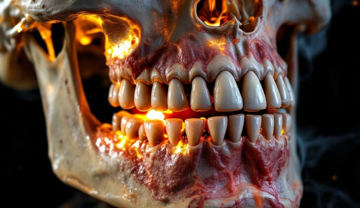
What is Mandible Osteoradionecrosis?
Osteoradionecrosis (ORN) of the lower jaw is a serious condition caused by radiation therapy used for treating oral and throat cancers. In simple terms, it’s a medical problem where the bone in the jaw loses its vitality or life, generally due to exposure to radiation therapy. This condition often occurs when the bone tissue is injured and doesn’t heal or rebuild itself properly over a period of at least three to six months.
A person can develop this wound from radiation therapy alone or from a combination of radiation therapy and physical injury. It is important to understand that ORN is different from a bone infection or persisting or returning cancer, though it can appear to look like both. While its exact cause is not universally agreed upon, it is often associated with lack of sufficient oxygen in the tissue (hypoxia) and possible damaging effects of unstable molecules known as free radicals. These factors often form the foundation of the treatment approaches for this condition.
What Causes Mandible Osteoradionecrosis?
Radiation is commonly used to treat different types of cancer. It works by damaging the DNA of cells, which prevents them from dividing and growing. The cells it targets include both cancerous and normal ones, but it strongly affects those that divide fast. That’s why cancer cells are particularly affected, although normal body cells that also divide quickly can be at risk.
Normal cells in the body that rebuild bone can be affected, which is one of the reasons why radiation can sometimes lead to a condition known as ORN, where the healing process in bones is disrupted.
The outer layer of the skin is made of squamous cells, which divide quickly and are therefore very sensitive to radiation. This sensitivity can sometimes lead to side effects such as skin inflammation and inflammation of the mucus membranes (the moist lining of some organs and body cavities). However, because these areas have a good blood supply, they often recover over time. Bones and cartilage, on the other hand, have less blood supply and might have a difficult or delayed healing process.
Radiation therapy can also harm larger blood vessels and slow down the growth of tiny blood vessels that normally help healing. This can reduce blood flow to areas that have been exposed to radiation, which can lead to slow-healing wounds. This, along with a decrease in the number of cells and limitations on the body’s repair mechanisms, can interfere with the healing of bone, leaving a long-lasting, non-healing wound.
Risk Factors and Frequency for Mandible Osteoradionecrosis
ORN, a condition caused by radiation, has remained at a steady rate of about 3% since the 1970s. Generally, ORN shows up two to four years after a patient completes their radiation treatments. However, some patients may have non-healing wounds at the site of the primary tumor (often in the jawbone and voice box) that persist immediately after radiation therapy and can eventually turn into ORN or chondroradionecrosis.
- The total radiation dose affects the chance of developing ORN – the higher the dose, the higher the risk.
- If a mechanical injury occurs after the start of radiotherapy, it can also increase the risk of ORN. This is often seen in the jawbone after treatment, mainly when implants are placed for dental rehabilitation.
- ORN can occur years after radiotherapy, and it can arise spontaneously or due to harm.
- Actions like removing teeth, placing dental implants, or wearing ill-fitting dentures can also increase the risk of developing ORN.
Signs and Symptoms of Mandible Osteoradionecrosis
There are a lot of signs indicating mouth problems that won’t improve. These problems could include a persistent oral ulcer, pain when swallowing, difficulty or discomfort in opening your mouth completely (known as trismus), changes in how your teeth come together, feeling like there’s something extra in your mouth, or spotting tiny bone fragments when you cough them up. Other complaints can be related to persistently unpleasant breath.
It is essential for the healthcare provider to know about any previous cancer experiences, such as where it was, how severe it was, when you started and stopped chemo or radiation, or if you’ve had any surgeries related to it. They also need to know about your dental background, what dental care you received before and after radiation, and any other dental procedures you’ve gone through (like getting dental implants, dentures, or special dental appliances).
An examination of your mouth might reveal a visible problem, or just show inflamed tissue. It may be necessary to use a thin, flexible viewing tube called a laryngoscope to look at the voice box and back sides of the jaw, especially if swallowing is painful.
If you have teeth, get them checked out, as loose teeth may indicate potential lingering radiation damage. Gum disease could also be a flag. Any suspicious areas or the inflamed tissue should be biotyped to make sure there’s no lingering or returning cancer before starting treatment for radiation-linked damage. Additional examination of the neck is necessary to find any lumps or swollen lymph nodes indicating recurring disease.
Testing for Mandible Osteoradionecrosis
If you’re being checked for a specific type of bone damage in your jaw called Osteoradionecrosis (ORN), there are a few steps your doctor will take as part of the evaluation.
First off, your doctor will look at your radiation therapy and dental records. They’ll also take a small tissue sample, or biopsy, of the wound area.
Imaging tests are also an important part of this process. Your doctor might order what’s called a panographic radiograph, which gives a comprehensive view of your teeth, and a CT scan of your jaw. These help the doctor see how extensive the damage is.
The CT scan can show signs like disruption of the outer layer of bone and areas of the bone that are both breaking down (lysis) and thickening (sclerosis). Pathologic fractures, which are breaks in the bone caused by disease, might also show up.
Similarly, the panographic radiograph can reveal areas of the bone that are breaking down with unclear, non-sclerotic borders – meaning, the borders aren’t hard or thick. Keep in mind, though, that standard X-rays are helpful but can sometimes understate how much the bone is affected.
Once your doctor has gathered all this information, they’ll determine the stage of the disease using the Marx Staging system. This system is also helpful in planning the treatment using hyperbaric oxygen therapy (HBO), a treatment that involves breathing pure oxygen in a pressurized room or tube.
The stages are:
Stage 1: There’s exposed jawbone that doesn’t have any pathologic fractures. The condition improves with HBO and removing small parts of diseased bone.
Stage 2: Despite 30 daily HBO treatments and removing small parts of the diseased bone, the condition doesn’t improve, or it requires major removal of the bone initially. In this case, more radical surgery is performed to remove the diseased bone followed by ten more HBO treatments.
Stage 3: Treatment has failed in stage 1 or 2, or you initially show certain signs like pathologic fractures, openings in the skin that lead to the mouth (orocutaneous fistulae), or evidence of the bottom border of the jawbone breaking down. In this stage, a large section of the diseased jawbone is removed and 30 preoperative and ten postoperative HBO treatments are done.
Treating ORN is usually a joint effort between your oral surgeon, oncologist, or ENT specialist. This team will work together to make a plan that is tailored to your specific condition.
Treatment Options for Mandible Osteoradionecrosis
Preventing ORN, or osteoradionecrosis, which is bone damage caused by radiation, is extremely crucial. There are two phases in dealing with ORN: prevention before radiation therapy, and dealing with it after it has arisen. There are several approaches including dental care, medical treatment, surgery, and the use of hyperbaric oxygen, which is breathing in pure oxygen in a pressurized room or tube.
Before radiation therapy, it is important that a dental professional with experience in head and neck cancers gives you a thorough check-up. This can include taking images of your teeth and gums, diagnosing any dental or gum diseases, and removing any teeth that might not have a good prognosis. This check-up should ideally be carried out at least two weeks before you start your radiation therapy so that you are in the best oral health possible, reducing the risk of ORN from developing. It is necessary to take good care of your teeth and gums before, during, and after radiation therapy.
During the prevention phase, medical treatment also plays a small role in reducing risk. Maintaining a good diet and rinsing the mouth with saline solution is vital to control oral mucositis, a common side effect of radiation therapy which causes inflammation and ulcers in the mouth. Antibiotics are only needed if an actual secondary infection is detected. A medication called Pentoxifylline, which is a type of anti-inflammatory, has been found to be helpful in treating ORN.
When ORN is diagnosed, surgical intervention is usually necessary. The affected, non-living bone has to be surgically removed. If you are about to undergo surgery or tooth extraction in an area that has received more than 60 Gy (the unit used to measure the amount of radiation delivered to a specific area), hyperbaric oxygen could be used to prevent further complications. This treatment helps in producing new blood vessels in the affected tissues, aiding repair. The normal course of this treatment involves multiple sessions before and after the operation, where you breathe in pure oxygen under high pressure for around 90-120 minutes.
The kind of surgery to treat ORN depends on its severity. The surgeon would generally remove the affected bone until only healthy bone remains. If the area of the operation and surrounding tissues are in good condition, local flaps, which are sections of your own tissue, can often be moved to cover the bone. If this fails or is not feasible, regional or free flaps, which are larger sections of tissue taken from another part of your body, can be used to cover and salvage the non-living bone. If these measures fail, a major operation involving the removal of a part of the lower jaw, followed by reconstruction using a vascular bone flap (a piece of bone with its blood supply) may be needed.
What else can Mandible Osteoradionecrosis be?
When examining dental conditions, professionals could also consider the following possibilities:
- Calcifying crown
- Odontoma
- Periapical abscess
- Periapical granuloma
- Rarefying and condensing osteitis


