What is Oral Leukoplakia?
Oral premalignancy is seen as a middle stage for developing oral cancer. It falls into two categories: premalignant lesions and premalignant conditions. Premalignant lesions are certain changes in the mouth tissue which have a higher chance of becoming cancerous as compared to normal tissues. An example is leukoplakia. Premalignant conditions, on the other hand, increase the overall risk of getting cancer. An instance of this is oral submucous fibrosis. The World Health Organization (WHO) recently grouped both of these under ‘Potentially Malignant Disorders’.
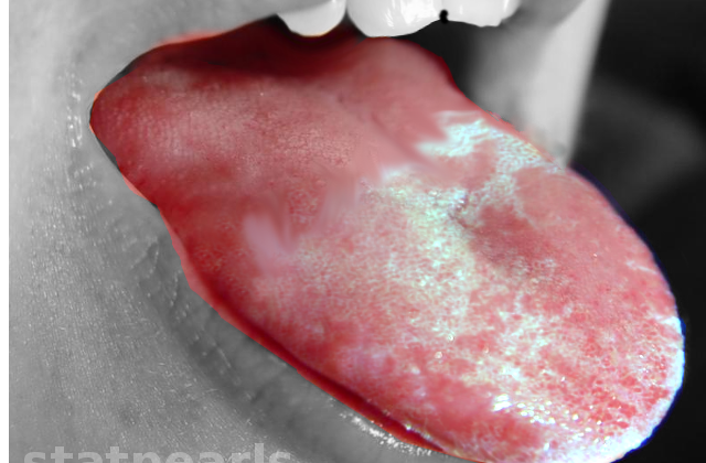
Oral leukoplakia is one such potentially dangerous disorder. It affects the lining of the mouth, appearing as a white spot or patch. This disorder is not categorized as any other specific type of lesion. Oral leukoplakia is usually linked with smoking, regardless of whether the patient uses cigars, cigarettes, beedies, or pipes.
Other factors that can increase the risk of oral leukoplakia include alcohol use, long-term irritation, fungal infections such as oral thrush, damage due to dental treatments, bacterial infections, sexually transmitted diseases like syphilis, a lack of essential nutrients, viral infections, hormonal imbalances, and exposure to ultraviolet light.
What Causes Oral Leukoplakia?
Oral leukoplakia, the appearance of white patches in the mouth, can be caused by numerous factors, and the exact reason is often unknown. A key risk factor often linked to this condition is tobacco use, in both smoked and smokeless forms.
In some regions, like the southern and southeastern parts of Asia, chewing areca (also known as betel) nuts can also increase the risk of oral leukoplakia. Smokeless forms of tobacco, like snuff, are another factor that can cause this condition.
Chronic candidiasis, a fungal infection, is also associated with the development of nonhomogeneous leukoplakia, a type of oral leukoplakia. Some types of candida fungus may produce harmful chemicals, increasing the risk.
In some cultures, holding inhaled cigarette smoke within the mouth can lead to oral lesions, including leukoplakia. People from these cultures have a 19-times higher risk of malignant transformation, where the leukoplakia becomes cancerous, compared to cultures where tobacco is used in other ways.
Risk Factors and Frequency for Oral Leukoplakia
Leukoplakia is a known potential risk factor for cancer in the mouth. The number of people affected by leukoplakia can vary, with some studies showing that around 2.6% of the global population may have it. It can evolve into cancer in 0.1% to 17.5% of these cases. In the region of India, the statistics are such that between 0.2% and 5.2% of people have leukoplakia, and it can transform into cancer in 0.13% to 10% of these cases. The high number of leukoplakia cases in India could be linked to the country’s cultural, ethnic, and geographical conditions. The estimated number of new cancer cases related to leukoplakia each year is somewhere between 6.2 and 29.1 for every 100,000 people.
- Other studies found the prevalence of leukoplakia to be within 0.4% to 0.7%, and up to 0.5% to 3.46% of the population. These studies also found that the disease could evolve into cancer in 0.7% to 2.9% of these cases.
- In another study, roughly 2% of people had leukoplakia, and about 1% of these cases converted into cancer each year.
- In one recent study, leukoplakia was found in about 1.59% of the study sample.
It’s important to note that leukoplakia is most common in middle-aged and elderly males. Generally, the older the person, the more likely they are to have leukoplakia.
Signs and Symptoms of Oral Leukoplakia
Oral leukoplakia, or simply leukoplakia, is a term that medical professionals use to describe white patches in the mouth that can’t easily be identified as another specific condition. This term has been used in different ways throughout history, often in connection with tobacco use.
Historically, other terms such as leukoma, smokers patch, leukokeratosis, and ichthyosis were used as synonyms for leukoplakia. This changed over time as awareness about the condition increased, and connections were made to smoking and oral cancer. The World Health Organization, for example, has given several definitions over the years. Now, the consensus is that leukoplakia refers to white patches in the mouth that may increase one’s risk of oral cancer, though this risk is debated and not all patches might lead to cancer.
In addition to this primary definition, different types of leukoplakia have been identified. These include:
- Homogeneous leukoplakia: This refers to white patches that are uniform in colour. These patches don’t usually cause any symptoms and are less likely to become cancerous.
- Non-homogeneous leukoplakia: This form of leukoplakia is characterized by irregular, flat, nodular, or exophytic patches of mixed red and white colour. These patches have a higher risk of becoming cancerous.
- Proliferative verrucous leukoplakia: This rare variant of leukoplakia is more widespread and commonly found in multiple areas of the mouth. It is more prevalent in older women and carries the greatest risk of turning into cancer.
Medical professionals continue to work on further refining the understanding and definitions of leukoplakia to better diagnose and treat this condition.
Testing for Oral Leukoplakia
Recent advancements in oral cancer research have led to the discovery and implementation of new diagnostic techniques to detect leukoplakia. Leukoplakia is a condition where thick, white patches form on the inside of your mouth.
Usually, the best way to diagnose leukoplakia is by taking a small sample of the affected tissue from your mouth, which is a process known as a biopsy. However, this can be seen as invasive, potentially painful, costly, and time-consuming. For smaller affected areas, the entire lesion may be removed. For larger cases, a part of the lesion and the surrounding healthy tissue can be taken for further laboratory analysis. This analysis, or histopathology, can identify several changes in the tissue, which may indicate leukoplakia:
* Formation of hard and thick layers on the skin on the inside of your mouth
* Increase in the thickness of the inner skin layer
* Abnormal thickening of the skin
* The base layer of your skin becoming thinner
* Inflammation in the connective tissues
* Changes in the cell layers
* Changes in the ratio of the nucleus to the cytoplasm in cells
* Incidences of more pigmented and enlarged nuclei
* Unusually high cell proliferation
* Abnormal cell divisions
* Increased cell divisions
* Changes in the shape and size of nuclei
* Excessive growth at the base layer of cells
* Cells shaped like a drop infiltrating deeper layers of the skin
* Loss of normal cellular arrangement
Apart from biopsies and histopathology, other diagnostic tools used for the early detection of leukoplakia include the use of a blue dye called toluidine, brush biopsy kits for oral applications, saliva testing, and optical imaging systems. Moreover, a handful of new, easy-to-use light sources and tools are being introduced to dentists to help with diagnosis right at their offices.
Treatment Options for Oral Leukoplakia
First and foremost, it is crucial to deal with anything contributing to your condition. If you have moderate to severe dysplasia (which means abnormal cell growth), the suggested treatment is typically surgical removal or laser surgery. This is especially recommended when the affected areas are on the underside and sides of the tongue, the base of the mouth, the soft part of the roof of your mouth, or the part of your throat that connects your mouth to your esophagus, also known as the oropharynx.
It is important to frequently monitor and schedule follow-up visits for lesions found in other places of the body. Surgical removal is the top recommended treatment for conditions like erythroleukoplakia (red and white patches in the mouth) and proliferative verrucous leukoplakia, which is a slow-growing, long-lasting form of white patches in the mouth that may become cancerous.
What else can Oral Leukoplakia be?
When it comes to the matter of diagnosing certain health conditions, medical professionals may come across a variety of possibilities. These include but are not limited to conditions such as:
- Candidosis (a fungal infection)
- Chemical burn
- Leukoedema (a white or grey, sometimes shiny oral lesion typically found in the oral mucosa)
- Lichen planus (a skin rash triggered by the immune system)
- Lupus erythematosus (an autoimmune disease that attacks healthy tissues)
- Morsciato buccarum (typically seen as white patches or spots inside the mouth)
- Psoriasis (a skin disease that causes itchy or sore patches of thick, red skin with silvery scales)
- White sponge nevus (a rare genetic disorder characterized by white spongy plaques in the mouth)
The process of getting to the right diagnosis involves careful consideration of these possible conditions by the doctor.
What to expect with Oral Leukoplakia
Oral leukoplakia, a condition that causes white patches in the mouth, can sometimes turn cancerous. About 1% to 9% of people with oral leukoplakia could potentially develop invasive cancer or actual malignancy in the affected area. Even if the invasive lesions are removed through surgery, it’s not unusual for the condition to come back.
Oral leukoplakia can disappear on its own sometimes. This makes it harder to track how well the treatment is working. It’s important to note that when changes occur in the cells (known as dysplastic changes), the patient’s prospects become uncertain and will require close monitoring.


