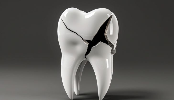
What is Tooth Fracture?
Tooth fractures, or broken teeth, are especially common in children and young adults, making up 5% of all dental injuries. Properly dealing with a tooth fracture involves correctly identifying the problem, figuring out a treatment plan, and then regular check-ups.
Fractures are often seen in the front teeth in the upper jaw, due to their positioning in the mouth. They’re most commonly caused by sports activities, car accidents, and incidents of physical violence. Depending on how severe the incident is, the tooth could be chipped, partially or fully dislodged, or even entirely knocked out of the mouth. It’s important to treat tooth fractures quickly to make sure the tooth can function properly again and to maintain the appearance of the tooth.
What Causes Tooth Fracture?
Dental injuries often happen when something hits the mouth directly or indirectly. The amount of damage depends on how much energy the hit has, where it hits, what it’s shaped like, and how the tissues around the tooth respond. Most of the time, injuries to the teeth are caused by falls, which make up to 65% of all cases. Other common causes include sports accidents, bicycle crashes, car accidents and physical violence.
The chance of getting dental injuries from sports or violence tends to increase with age. In fact, sports-related dental injuries are often seen in teenagers, and those because of violence tend to happen in people between 21 to 25 years old. But on the other hand, falls and collisions are the most common causes of dental injuries in young children’s primary teeth.
It’s important to know that unhealthy teeth or cavities can make a tooth more prone to breaking, even from a minor injury. Also, people with teeth that stick out more than normal or those who can’t close their lips completely are usually at a higher risk of getting injuries to their upper front teeth.
Risk Factors and Frequency for Tooth Fracture
Injuries to the mouth make up 5% of all injuries across all age groups, but this increases to 17% in children. These types of injuries are more commonly seen in males. The majority of dental fractures – more than 75% – happen in the upper jaw. The teeth most often affected are the central incisors, then the lateral incisors and canines, largely due to their position in the mouth.
- Fractures to a single tooth are more common than fractures to multiple teeth.
- When multiple teeth are fractured, it’s usually because of sports injuries, car accidents, or physical violence.
Permanent teeth are more likely to be damaged by trauma than baby teeth. The rate of tooth fractures ranges from 9.4% to 41.6% in baby teeth, and 6.1% to 58.6% in permanent teeth.
Signs and Symptoms of Tooth Fracture
There are different types of dental fractures, classified based on the part of the tooth that’s affected and whether or not the tooth’s pulp (inside part of the tooth) is involved:
- Enamel infractions: These are tiny cracks in the tooth’s enamel (outer layer) that don’t cause any loss of tooth structure and typically don’t cause symptoms. They can be seen with a special light and don’t cause any sensitivity or tooth mobility. X-rays don’t usually reveal any noticeable changes.
- Enamel fractures: These occur when the enamel is fractured but dentin (next layer inside the tooth) or pulp is not exposed. Such fractures are often found on the side or the top edge of frontal teeth. Normal tooth mobility and pulp sensitivity indicate an enamel fracture. The extent of enamel loss can be confirmed with radiographs.
- Enamel-dentin fractures: This type of fracture involves both enamel and dentin of the tooth but without exposing the pulp. Here, you will have a healthy tooth with no sensitivity or mobility issues.
- Enamel-dentin fractures with pulp exposure: In this kind of fracture, both enamel and dentin are lost, and the tooth’s pulp is exposed. These teeth are usually sensitive to air, temperature, and pressure. Pulp tests generally come back positive unless there is an associated injury.
- Crown-root fractures: These fractures extend from the top of the tooth down to the area below the gum line and may or may not involve the pulp. These fractures can be diagnosed clinically as well as with radiographic examination. They may cause sensitivity to percussion and pressure. Radiographic examination allows for a thorough analysis of the fracture and treatment options.
- Root fractures: This type of fracture affects the pulp, dentin, and cementum (the part of the tooth under the gum line). It can be horizontal, slanted, or both. Symptoms may include bleeding from the gum, tenderness on touch, and tooth mobility. The pulp may initially test negative due to a temporary or permanent nerve injury. Detailed x-rays or a cone-beam CT scan can confirm the diagnosis and plan the treatment.
It’s important to mention that if a piece of tooth goes missing and the patient has soft tissue injuries, additional x-rays of the lips and cheeks are advised to find the missing piece.
Testing for Tooth Fracture
Your dentist will have to decide if taking an x-ray, and with it a small exposure to radiation, will be beneficial for managing your tooth fracture. There are many different types of x-ray pictures they could take, and they will need to use their experience and expertise to choose the most suitable option for you.
Usually, the first type of x-ray that a dentist might take is a parallel periapical radiograph. However, depending on the case, they may also need to use a different type of x-ray, like an occlusal projection. For more complicated fractures, dentists might choose to use a type of scan known as a cone-beam CT scan. This scan can give a very detailed picture of the fracture, including how far it extends, where it is located, and its direction. This can greatly help the dentist in determining the most effective course of treatment.
Treatment Options for Tooth Fracture
Tooth fractures can often be accompanied by swelling, blood clot formation (hematoma), and cuts (laceration). Applying a cold pack to the affected area can help reduce pain and swelling before the dentist starts the actual treatment.
Enamel Infraction
This is a small fracture in the tooth enamel which usually doesn’t require treatment. However, if it’s a larger crack, the dentist may need to etch (roughen) and seal it with a special bonding agent to prevent bacteria from entering and discoloring the tooth.
Enamel Fracture
When the fracture only involves the crown (the visible part of the tooth), the treatments can include reattaching the broken piece, filling it with a special material (composite resin), or smoothing the tooth edges. After this, follow-up dental visits are scheduled after two months and then after a year to check for any complications like infection, inflammation at the tip of the root, or incomplete root growth in young teeth.
Enamel-Dentin Fracture
For more severe fractures involving both the enamel and the underlying layer (dentin), the treatment method involves protecting the exposed dentin. This can be done by using a bonding agent and a composite resin or glass ionomer. If the exposed dentin is close to the tooth pulp (the tooth’s sensitive core), it may appear pink but without bleeding. A product called calcium hydroxide can be used to layer the dentin, followed by capping it with a material like glass ionomer.
Restoration options can include reattachment of the broken piece, composite resin restoration, or ceramic fillings.
Fracture with Pulp Exposure
For fractures that extend to the pulp, the dentist has to be careful about how to manage the pulp exposure and tooth restoration. In these cases, a more conservative approach favors healing.
Doubts may arise between pulp capping or partial pulpotomy. The choice depends on things like how long and how wide the exposure is, the pulp’s condition before injury, the tooth’s age and root development stage, and associated injuries.
Pulp capping works in cases where the pulp exposure is brief and the exposed area is small (not larger than 1.5 mm). The chances of successful pulp healing rise if the pulp was healthy before the trauma, the tooth is young (with open apices or tips), and there are no associated injuries.
Partial pulpotomy, that involves removal of part of the pulp, might be more appropriate when pulp capping isn’t possible. For example, when a lot of time has passed since the trauma, or the exposure area is too large. The dentist may then decide to keep the tooth vital for as long as possible before eventually performing a root canal treatment.
Crown-Root Fractures
These fractures involve both the tooth’s crown and root. The treatment aims to expose the fracture margins to control the moisture level and allow better cleaning by the patient. The doctor may retrieve the tooth fragment for possible reattachment later. If the pulp isn’t exposed, the remaining dentin can be covered with a restorative material.
Other options include making a gum incision (gingivectomy), moving the tooth slowly via orthodontic treatment, using surgical procedures, or tooth extraction.
Root Fractures
With root fractures, the initial step involves moving the displaced crown fragment back into position and confirming the adjustment with an x-ray. The unstable part needs to be held in place (splinted) for up to four months, depending on the location of the fracture. The healing of the fracture needs to be closely watched for at least five years after the treatment.
What else can Tooth Fracture be?
Thoroughly examining a tooth fracture through clinical check-up and x-rays often provides an accurate identification of the problem. In baby teeth, it’s necessary to distinguish between natural tooth root disappearance and actual tooth fractures. When the tooth is tender, it may mean that the root of the tooth is fractured or the tooth has been dislocated.
What to expect with Tooth Fracture
What happens after a tooth fracture depends on several things: the type of injury, how quickly you received treatment, and the quality of the treatment. When a fractured tooth heals well, the pulp (inner part of the tooth) and the tissues around the tooth recover normally.
The healing process for a fractured tooth normally takes between 1 to 2 weeks. Smaller fractures that only involve the tooth’s outer layer, or enamel, usually have a good outcome. But if a deeper fracture is not treated, it may lead to an infection or an abscess (a pocket of pus).
Possible Complications When Diagnosed with Tooth Fracture
When a tooth gets fractured, it can lead to various problems. These include the death of the tooth’s nerve or pulp, changes in tooth color, an abscess at the root of the tooth, the closing up of the tooth’s pulp (an area in the center of the tooth), the development of an abnormal connection between two body parts (known as a fistula), and the gradual wearing away or destruction of the part of the tooth that is below the gum tissue (commonly referred to as root resorption).
The most common problem that arises from fractured teeth is pulp necrosis, which is when the nerve or pulp of the tooth dies.
Possible complications of a tooth fracture include:
- Pulp necrosis (death of the tooth’s pulp)
- Tooth discoloration
- Abscess at the root of the tooth
- Closure of the tooth’s pulp
- Fistulas (abnormal connections between two body parts)
- Internal and external root resorption (wearing away of the part of the tooth below the gum tissue)
Preventing Tooth Fracture
Preventing dental injuries can be challenging, but wearing custom-made mouthguards can protect against harms associated with contact sports. It’s crucial for parents and school teachers to learn about basic life skills and first aid techniques specifically for dental injuries. This knowledge can make a significant difference when quick response is needed.


