What is ECG T Wave?
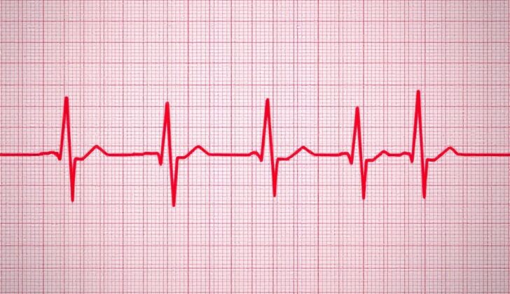
The T wave is a specific part of the heart rhythm that you see on a heart tracing (also known as an electrocardiogram or ECG). Usually, the T wave shows when the lower chambers of the heart, the ventricles, are resetting electrically after a heartbeat. But, sometimes the T wave can look different, and these changes can be perfectly normal or could indicate that there’s an issue with the heart.
Understanding the different causes of T wave changes helps to manage heart problems safely and effectively. This article will discuss about the T wave, how to define and measure it, and how it varies.
What Causes ECG T Wave?
The T wave is a part of our heart’s electrical waves that we see on an EKG (a test that measures your heart’s electrical activity). It appears as a small hump following the bigger spikes, which happens when the muscle cells in the heart’s chambers reset their electric charge to pump again.
Things can get complicated when the T wave doesn’t appear as it should. There are several reasons why this might happen. Maybe it’s just a normal variation in your heart’s electrical activity. Or it might hint towards a serious heart problem.
When T waves are too tall or “hyper-acute”, this could be an early sign of a heart attack. This is because the T wave might change within half an hour of a complete blockage in one of the heart’s arteries. If you see this on an EKG, it’s necessary to compare it with your previous scans. If you’re young or an athlete, tall T waves might be completely normal.
Just like peaked T waves, flat or “inverted” T waves can also signal a heart problem. They often appear when the heart muscle isn’t getting enough blood, a condition known as ischemia. However, flat T waves don’t always mean you’re going to have a bad outcome. They usually suggest an issue if you’ve got other signs of heart disease, like symptoms of poor blood flow to your heart. One serious condition, Wellens syndrome, shows a classic pattern of flat T waves and suggests a blocked artery in the heart. The problem is, flat T waves can also appear in lung diseases, so doctors need to consider all related symptoms and conditions.
Another important cause of peaked T waves is high potassium levels in the bloodstream or “Hyperkalemia”. Changes in the EKG due to high potassium levels begin with narrow, sharp, and tall T waves and may eventually become wavy.
Certain medications can also cause abnormalities in the T wave, like rhythm-controlling drugs, digoxin (a medication to strengthen your heart), and diuretics (drugs that help your body get rid of salt and water). These drugs can cause imbalances in your body’s electrolytes, which can alter T wave appearance. One way to differentiate whether these changes mean a heart problem versus an electrolyte imbalance is by seeing how the changes are distributed on the EKG.
Specific changes in T waves can sometimes be seen in severe brain injuries or diseases; these are known as ‘cerebral T waves’. Conditions like stroke, brain bleeding and traumatic brain injury could be associated with ‘cerebral T waves’. Similarly, conditions like inflammation of the heart sac (pericarditis) and significant lung blood clot (massive pulmonary embolism), can change the look of the T wave on the EKG.
Risk Factors and Frequency for ECG T Wave
Around 15.5 million Americans over the age of 20 suffer from coronary heart disease, as stated in 2016 by the American Heart Association (AHA). Furthermore, it’s estimated that a heart attack takes place nearly every 42 seconds in the United States.
The frequency of hyperkalemia, a condition that often affects the patterns of T-wave (a part of your heart’s electrical signal), was studied by Sanchis-Gomar and his colleagues. This investigation included about 2.2 million people. They found that nearly 1.55%, or about 3.7 million Americans, have hyperkalemia. These rates were higher amongst people with chronic kidney disease, heart failure, diabetes, and high blood pressure. Around 6% of people with chronic kidney disease and heart failure were found to have hyperkalemia during the study.
Signs and Symptoms of ECG T Wave
T-wave abnormalities in the heart can result from various causes and can often present in different ways. Understanding the patient’s medical history and doing a detailed review of their medications can give valuable insights regarding the cause of these abnormalities. For instance, if a patient reports having multiple instances of chest pain, which have worsened recently, this could suggest a heart-related issue like ischemia. On the other hand, if they’ve recently started a medication like digoxin, it could point towards drug intoxication. The timeline of these symptoms is also crucial in making the diagnosis. For example, if a patient suddenly starts experiencing shortness of breath and a fast heartbeat following recent surgery, it may indicate a possible lung clot. Despite some of these presentations having clear symptoms, we should bear in mind that T-wave abnormalities often show no symptoms. Several causes leading to T-wave changes might not be harmful, such as normal variations or incorrect placement of the lead wires during heart monitoring.
Testing for ECG T Wave
An electrocardiogram (ECG), a test that measures the electrical activity of your heart, is one part of evaluating a patient’s health status. However, it really needs to be just one part of a full health check-up to get a complete understanding of what might be going wrong.
If the doctors suspect issues like heart disease or an imbalance in the body’s electrolytes (salts and minerals that help your nerves, muscles, and other body parts work properly), they may also need to do a metabolic panel (a blood test that checks your sugar level, electrolyte and fluid balance, and kidney function) and run tests for myocardial ischemia biomarkers. Myocardial ischemia is a condition where your heart muscle isn’t getting enough blood and the biomarkers help doctors spot this.
In certain situations, if abnormal ECG results called ‘cerebral T waves’ are found, which might suggest something is wrong with brain activity, the doctors might order a CT scan. This is an imaging test that uses X-rays and a computer to create detailed images of your body, and in this case, it would be used to check for any sudden bleeding or injury to your brain and nervous system. This type of CT scan does not use a contrast dye.
In cases where a pulmonary embolism (a sudden blockage in a lung artery, usually due to a blood clot) is suspected, a special kind of CT scan known as a CT angiography may be done. This test combines a CT scan with a dye-containing substance injected into a vein to produce pictures of blood vessels within the lungs.
Treatment Options for ECG T Wave
The treatment for abnormal T-wave patterns, seen on an electrocardiogram (ECG), varies depending on what’s causing these changes. It’s important to note that some alterations in the T-wave don’t need any treatment. However, certain causes can be life-threatening without immediate medical attention.
If the changes in your T-wave are due to Ischemia or Infarction, which is insufficient blood flow to the heart, doctors are likely to prioritize restoring that flow and treating what is called an acute coronary syndrome, a group of conditions linked to sudden, reduced blood flow to the heart.
Hyperkalemia, an excessive amount of potassium in your blood, could also lead to observable changes in your T-wave. When T-wave changes occur along with severe hyperkalemia, calcium gluconate can be given rapidly to stabilize the heart and prevent irregular heart rhythm. In case of mild hyperkalemia without T-wave abnormalities, the condition can be managed with medication such as polystyrene sulfonate (a drug that lowers potassium levels), insulin (which moves potassium into your cells), or furosemide (a medication promoting the elimination of potassium through urine).
Pulmonary Embolism (PE), a condition where a blood clot blocks an artery in your lungs, is another common cause of T-wave changes. Treating PE depends on its size and severity. For critical cases of PE, doctors may use a drug known as tPA, delivered directly through a catheter, to dissolve the clot. Smaller PEs that are not causing obvious symptoms can often be managed with blood-thinning medication. Hospitals usually have a team of specialists, including a heart doctor (cardiologist) and lung doctor (pulmonologist), to promptly evaluate and determine the appropriate treatment for new cases of PE.
Pericarditis, an inflammation of the thin sac around your heart, can also result in T-wave changes. This condition can typically be managed with nonsteroidal anti-inflammatory drugs like Ibuprofen and a medication called colchicine, often for a period of at least three months. For repeated instances of this condition, you might need to take these medications for a longer time.
If a drug interaction or an overdose is causing abnormal T-wave patterns, doctors may check the levels of the suspected drug in your blood. Stopping the implicated drug is usually advised during these circumstances while further tests are ongoing. Certain medications have specific antidotes. For instance, dig immune Fab is used for treating digoxin toxicity. This consists of antibody fragments that bind with digoxin, a heart medication, and neutralize it.
What else can ECG T Wave be?
For a heart test known as an ECG, changes in the wave patterns, specifically the T waves, could indicate several possible issues. These can be split into two main categories: T-wave Inversion, and Peaked T-waves.
When considering T-wave Inversion, these may point to:
- A normal variant, or no issue
- A lack of proper blood flow to the heart (Myocardial ischemia)
- Too much pressure on the ventricles, the main pumping chambers of the heart (Ventricular strain)
- Injury to the brain (Cerebrovascular injury)
- An enlarged heart due to genetic causes (Hypertrophic cardiomyopathy)
- Unknown cause (Idiopathic)
- A blockage of electric signals on the left side of the heart (Left bundle branch block)
- A blockage of electric signals on the right side of the heart (Right bundle branch block)
- Extra heartbeats that start in one of the ventricles (Ventricular beats)
On the other hand, Peaked T-waves could suggest:
- The very early stage of a heart attack (The hyperacute phase of myocardial infarction)
- Chest pain caused by reduced blood flow to the heart (Prinzmetal angina)
- A normal variant, or no problem
- High levels of potassium in the blood (Hyperkalemia)
- Thickening of the wall of the left ventricle, the heart’s main pump (Left ventricular hypertrophy)
- The same left-side electrical blockage (Left bundle branch block)
- Inflammation of the thin layer around the heart (Acute pericarditis)
What to expect with ECG T Wave
The outlook of a patient mainly depends on the root cause of their medical condition. Abnormal T-wave patterns seen on an EKG (a test that measures your heart’s electrical activity) can either be harmless or indicate serious, potentially life-threatening health issues. An EKG, along with a detailed patient history and physical examination, can offer crucial insights into what might be causing a patient’s health problems and what their recovery may look like.
Possible Complications When Diagnosed with ECG T Wave
The most critical issue related to T-wave abnormalities is the chance of misdiagnosing serious T-wave issues or delaying necessary treatment. Other possible complications include heart diseases such as cardiomyopathy, lack of blood supply to the heart or heart attack, irregular heartbeats, tamponade (pressure on the heart due to fluid), heart failure, and even death.
Potential Complications:
- Misdiagnosis of serious T-wave issues
- Delay in necessary treatment
- Cardiomyopathy (heart muscle disease)
- Myocardial ischemia (lack of blood supply to the heart)
- Infarction (heart attack)
- Arrhythmia (irregular heartbeats)
- Tamponade (pressure on the heart caused by fluid)
- Heart failure
- Potential death
Preventing ECG T Wave
Patients should be well-informed about the signs and symptoms of heart disease caused by reduced blood supply to the heart, known as ischemic heart disease. Patients should seek immediate medical attention if they experience these symptoms. Organizations like the American Heart Association and the American College of Cardiology are dedicated to spreading awareness about heart disease. Every year, they spend millions of dollars to educate the public about this medical condition. But it’s important that all healthcare professionals regularly inform their patients about the different ways heart disease can appear, so patients can quickly seek the right help when needed.
According to a 2018 report, kidney disease is becoming more common in the United States. Chronic kidney disease, a long-term condition where the kidneys don’t work as well as they should, can often lead to electrolyte imbalances, which may in turn affect the shape of well-known part of an electrocardiogram (heart’s electrical activity) known as the T-wave. Therefore, it is critical to enlighten our patients about the benefits of controlling key health factors such as diabetes and high blood pressure, reducing the likelihood of kidney disease. Preventing kidney disease in the first place is crucial. Those who have already developed kidney disease should be closely monitored for any abnormalities in their electrolyte levels.