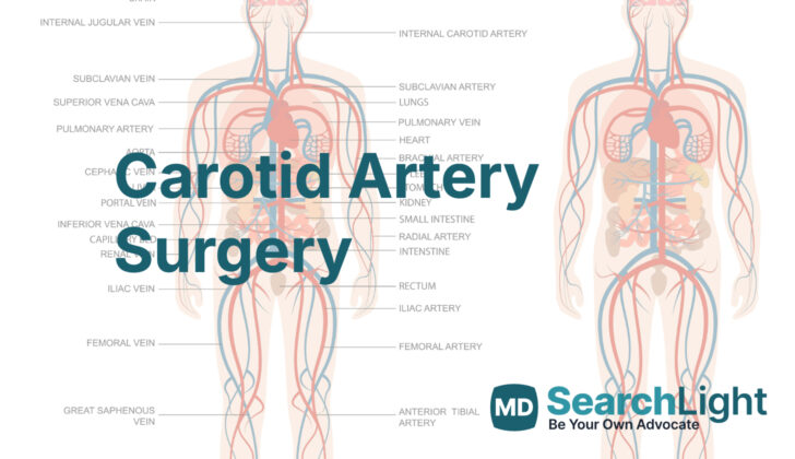
Overview of Carotid Artery Surgery
The symptoms of a specific artery disease condition often stem from what is known as embolization or the blockage caused by some form of blood clot or air bubble in the artery. In Europe and North America, this condition is responsible for nearly a quarter of incidents of stroke. The usual source of the problem is fatty tissue build-up, or atherosclerosis, within an artery located in the neck. This blockage is most commonly found at the spot where the artery branches off, the area called the carotid artery bifurcation.
Using special ultrasound technology (Transcranial Doppler), doctors have found that almost 20% of patients with moderate blockage at the bifurcation, and even higher percentage of patients with more than 70% blockage, are likely to develop emboli or blood clots. This risk increases in patients who have recently shown symptoms.
Small clots, or microemboli, can trigger sudden or temporary neurological symptoms that might include loss of sensation or movement on one side of the body, difficulty finding words (aphasia), trouble speaking due to muscle issues (dysarthria). This condition is usually brief in duration, and often less than 24 hours, and is known as Transient Ischemic Attack (TIA). When these symptoms continue beyond this period, it is then classified as a stroke, or cerebrovascular accident (CVA). A clot that reaches the first branch of the internal artery, the ophthalmic artery, can result in temporary or permanent loss of vision in one eye, a condition called amaurosis fugax.
Atherosclerosis lesions, or fatty tissue build-up, in the internal carotid artery often develop along the artery wall opposite to the branch point of the external carotid artery. A widening of the artery just beyond this branch point can result in slower blood flow and changes in the direction of the flow. This allows more interaction between the fatty tissue particles and the artery walls, which can contribute to the localized fatty build-up or plaque at the carotid bifurcation.
Fortunately, this localized atherosclerosis accumulation can be effectively removed, and the risk of stroke reduced. Without treatment, 26% of patients with TIAs and more than 70% with blockage of the carotid artery are likely to develop permanent neurological damage from continued clot formation at two years. This risk can be lowered to 9% by removing the fatty build-up and is usually even lower for patients with temporary or permanent loss of vision.
Why do People Need Carotid Artery Surgery
In simple terms, carotid stenosis refers to the narrowing of carotid arteries. These are the blood vessels that supply your brain with much-needed blood. This issue can be detected in two major ways: by using an angiography, which is a type of X-ray, or by duplex ultrasound, which uses sound waves to create images of your blood vessels. If your carotid arteries are narrowed by more than 60% according to angiography, or 70% according to duplex ultrasound, even though you have no symptoms, you may need treatment.
For individuals who do have symptoms like a sudden weakness or numbness often occurring on one side of the body, difficulty speaking, loss of vision, dizziness, or severe headache, these could be signs of two things: a stroke (also known as a cerebrovascular accident), a small stroke that improves within 24 hours (transient ischemic attack), or sudden, brief vision loss (amaurosis fugax). If any of these occur and a health professional confirms you have over 50% carotid stenosis, you may need treatment.
People with other medical conditions like severe chronic obstructive pulmonary disease (a type of lung disease), heart disease, or other complex health problems might need a procedure called a carotid endarterectomy. This is a surgical procedure that helps unblock the carotid arteries. The procedure can be performed safely using regional anesthesia, meaning you would be conscious but unable to feel pain.
However, if you have a history of neck radiation treatment, a type of neck surgery called a modified radical neck dissection, or if you have needed a carotid endarterectomy in the past, you might be considered for a less invasive treatment. This is known as carotid stenting, where a small, metal mesh tube is inserted to keep the carotid artery open.
If you have symptoms of both carotid stenosis and heart disease, doctors may consider combining two treatments: carotid endarterectomy and coronary artery bypass grafting. This is a type of heart surgery that aims to improve blood flow to the heart. However, this combined treatment option is only considered under certain circumstances.
When a Person Should Avoid Carotid Artery Surgery
Sometimes, there are certain conditions that can prevent an individual from undergoing specific medical procedures. In cases involving the distal internal artery, if there is a blockage (referred to as occlusion), it may serve as a reason to avoid certain treatments. This consideration is essential to ensure the safety and well-being of the patient.
Who is needed to perform Carotid Artery Surgery?
A vascular surgeon, general surgeon, or neurosurgeon specialized in cerebrovascular arterial disease performs your operation. These are doctors specifically educated to operate on the brain’s blood vessels. They work alongside assistants, who could be other doctors in training, or a physician assistant (a healthcare professional who performs many of the same duties as a doctor).
An anesthesiologist will also be present during your operation. This type of doctor is responsible for providing anesthesia, which means they will ensure you don’t feel pain during the surgery.
Nurses also play an important role- usually there’s a scrub nurse technician (a specially trained nurse who assists in surgical operations by preparing the operating area) and a circulator nurse (a non-sterile nurse who manages the overall nursing care in the operating room). These medical professionals all work together to make sure your operation is safe and successful.
Preparing for Carotid Artery Surgery
Before carrying out certain medical procedures, doctors use a special kind of ultrasound called a ‘duplex ultrasound’. This type of ultrasound is excellent at detecting problems or diseases in the body. For most patients, this is the only imaging they will need before the surgery.
If the patient has had radiation treatment, neck surgery, unusual ultrasound results, or narrowing of their arteries again after treatment, they might need a more detailed scan. This is known as an ‘arteriography’, and can be done traditionally or with a CT scan.
Preparing for the procedure can involve setting up in a specific way to ensure the patient’s comfort and safety. The patient will be positioned in a chair slightly leaned back, with a small cushion at the back of their shoulders. This posture helps gently tilt and rotate their head to the opposite side.
The arm on the same side as the procedure is relaxed and padded around the elbow and wrist to keep it protected and comfortable.
Doctors must be careful with the head position. Excessive turning or nodding above the midline level can potentially lead to restrictions in blood flow through the arteries in the neck and on the side opposite to the procedure’s location.
Doctors will also take into account and consider various physical features on the patient’s body, like the earlobe, the angle of the jaw, the bone behind the ear, the dip between the neck and the chest, and the collarbone. These are key body landmarks and must be considered when preparing the patient for the procedure.
How is Carotid Artery Surgery performed
Carotid endarterectomy is a surgery performed to clear the carotid arteries, which carry blood to our brain. This procedure can be done under different forms of anesthesia, depending on various factors. Some doctors use anesthesia that lets the patient stay awake during surgery, while others prefer general anesthesia, where the patient is completely asleep.
The procedure itself starts with the surgeon making an oblique incision, or cut, along the anterior border of the sternocleidomastoid muscle, a noting muscle in our neck. They’ll use a tool called electrocautery to divide the platysma, a superficial neck muscle, and begin a careful dissection to identify important structures, like the intern internal jugular vein, which carries blood from the head back to the heart.
Certain veins are tied off and divided to allow for better visibility of the surgical area. It’s important for the surgeon to carefully identify and avoid various cranial nerves, which include nerves vital for functions like speech and swallowing. They dissect around the common carotid arteries, external carotid arteries, and internal carotid arteries, and then isolate them for the procedure.
If the disease is located high up inside the carotid artery or spans a long area, the surgeon may need to make some additional maneuvers to access the affected area. Once the surgeon has great access, they then insert a medical device called a shunt if needed to temporarily maintain blood flow to the brain during the procedure. The plaque causing the problem is then carefully removed. Keeping the arteries clean not only allows proper blood flow to the brain but also prevents clots or plaques from breaking off and causing a stroke.
When the plaque has been removed, the surgeon thoroughly rinses the area with a saltwater solution. Then, they place a patch over the area where the artery wall had been opened. This patch can be made of different materials, and it’s tailored to match the size and shape of the artery it’s being used to repair. Finally, they remove the shunt, allow for any remaining blood to drain out, and restore the full blood flow into the carotid arteries.
In some cases, the surgeon might perform a slight variant of the operation called an eversion endarterectomy. The decision to perform this variant, which slightly changes the way the surgery is performed and how the artery is repaired, will depend on where most of the plaque is located.
After the surgery, overnight monitoring is done to keep an eye on your recovery. This includes keeping a close eye on your blood pressure to minimize the risk of any complications. Some patients might be able to go home the same day if everything is going well and their blood pressure is in check.
Possible Complications of Carotid Artery Surgery
After a person undergoes an operation, different issues might happen that relate to the brain and heart. These include:
1. Cranial nerve injury: Harm to one of the twelve nerves that come from the brain. These nerves are in charge of things like your sense of smell, eyesight, or ability to chew and swallow food. An injury like this might disrupt those abilities.
2. Stroke: This is when blood supply to part of your brain gets interrupted or reduced. This means the brain can’t get the oxygen it needs, which can lead to damage in the brain cells.
3. Myocardial infarction: You might commonly know this as a heart attack. This happens when blood flow to a part of your heart is blocked, usually by a blood clot.
4. Carotid restenosis: This situation refers to the narrowing of the carotid arteries (major vessels that carry blood from the heart to the brain), after they’ve already been opened up by a procedure. This can reduce the flow of blood to your brain and could lead to stroke.
What Else Should I Know About Carotid Artery Surgery?
Carotid endarterectomy (CEA) is a surgical procedure performed to prevent stroke. This surgery is important for people with a significant narrowing (stenosis) of the main blood vessels supplying blood to the brain, known as the carotid arteries, regardless of whether they have symptoms or not. The effectiveness of this operation relies on it being carried out safely and effectively, with minimal complications.
A significant study carried out in 1991, the North American Symptomatic Carotid Endarterectomy Trial (NASCET), compared the outcomes of patients undergoing CEA with those receiving medical treatment alone. All the patients in the study had either experienced a minor stroke or had visual disturbances due to inadequate blood flow to the eye, within the four months leading up to the study. They also had significant narrowing in their carotid arteries. The study found that the risk of stroke was significantly lower in the group that had surgery compared to those who just received medication. Specifically, 9% of the surgical group had a stroke within two years compared to 26% in the medical group.
Similar findings were reported in the Veterans Affairs Cooperative Study and the European Carotid Surgery Trial. All of these studies highlight the significant benefit of CEA in reducing the risk of stroke in patients with moderate to severe narrowing of their carotid arteries.
It’s important to note, though, that while the risk of stroke and death due to CEA is generally low (around 2% to 4%), it can be slightly higher in patients who have already had symptoms like a minor stroke or visual disturbances. Despite this, the strong evidence from these studies shows that CEA can substantially reduce the risk of stroke in these patients and is considered a core part of their treatment.


