Overview of Chest Tube Insertion in the Neonate
Pediatric chest tube placement, a type of procedure known as thoracostomy, is commonly used for different purposes in children. These purposes include supporting breathing after heart surgery, managing emergency airway issues, and treating diseases affecting the chest cavity. This procedure mainly involves the use of a chest tube and is especially important for treating a condition known as pneumothorax.
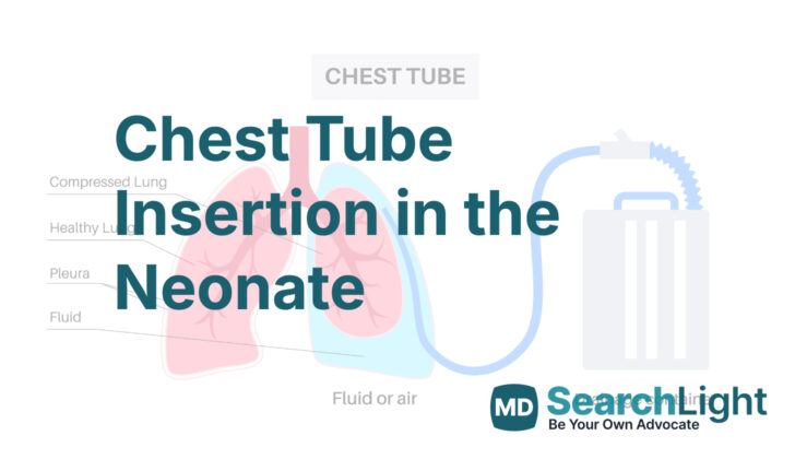
Pneumothorax is a condition where air gets into the space between the lung and the chest wall, which can cause the lung to collapse. The chest tube helps to take this air out, allowing the lung to re-expand. While other devices like pigtail and venous catheters are sometimes used, the chest tube remains the most common tool for treating this condition.
However, placing and maintaining chest tubes in children can pose special challenges. This is because children’s chest anatomy and functioning are different from those of adults. Therefore, it’s very important for doctors to understand these differences. This knowledge can help ensure the chest tube is placed properly and managed effectively, while also reducing the risk of complications.
Anatomy and Physiology of Chest Tube Insertion in the Neonate
Newborn babies have a different chest structure compared to adults. Their chest wall is very flexible and their lung tissues are extremely delicate. The organs around the middle part of the body, or ‘mediastinum’, are close to the chest in babies. The lungs are lined with a thin, fine tissue known as the ‘visceral pleura’, which aids in avoiding friction between the lungs and the chest wall. Another protective layer, the ‘parietal pleura’, lines the chest wall and the muscle that helps you breathe known as the ‘diaphragm’. The space between these two protective layers is known as the ‘pleural cavity’.
Any build-up of substances like air, fluids or blood in the pleural cavity may lead to health problems. One common method to solve this is by a procedure called ‘thoracostomy’, which drains these substances and reduces discomfort. This procedure lets the lungs expand fully, pressing directly against the chest wall, which helps in better breathing and removes any leftover space in the pleural cavity.
The chest wall consists of the ribcage and muscles that are located between the ribs. Every rib has a bundle of nerves, arteries, and veins in between a set of these muscles. The empty space between each rib is called the ‘intercostal space’. Careful handling of these nervous structures is critical, hence a chest tube is always inserted under a rib.
Before inserting a chest tube, the medical practitioner should understand how the structure of a newborn’s chest changes during breathing. The ‘phrenic nerves’ control the movement of the diaphragm, which is the major muscle involved in breathing. Its contraction creates space in the chest for the lungs to expand during taking in air, and comes back to its place on breathing out. It’s important to note that the structure of the insides of the chest changes in 3 dimensions during breathing in and out.
If a chest tube is inserted too low, it might enter into both the pleural cavity and the abdominal spaces depending on where it is put through a rib. Several medical conditions could cause the diaphragm to be positioned higher than usual, like paralysis of the diaphragm, partial loss of its movement (paresis), a condition where abdominal organs push through the diaphragm into the chest (hernia), and if there is an abnormal enlargement of the abdomen. Extreme caution is required in these cases because contents from the abdomen might enter the chest and cause problems. Also, patients who have previously gone through certain procedures like chest tube placement, procedures that bind the layers of the pleura together (pleurodesis), infections in the pleura, trauma, and heart or lung surgeries should be operated by a surgeon because, in these cases, the lung tissues might stick to the chest wall and get rid of the pleural space. Sometimes, the doctor might suggest more invasive procedures like camera-assisted surgeries or opening up the chest cavity (thoracotomy), especially in complicated cases.
Why do People Need Chest Tube Insertion in the Neonate
A chest tube is a device that doctors might need to place in newborns for different reasons. It’s a bit like a medical version of a drain. When inserted into the chest cavity, it can help remove air or fluid that has built up around the lungs. By getting rid of this excess air or fluid, the chest tube helps the lungs to expand properly, which is crucial for the baby to breathe comfortably. In some rare cases, doctors might use a chest tube to put fluids or medications directly into the space surrounding the lungs.
One scenario where a chest tube can be lifesaving is in a medical situation known as tension pneumothorax. This occurs when a significant amount of air has entered the chest cavity, causing high pressure that can restrict blood flow, leading to low blood pressure and possible collapse of the body’s vital functions. In this case, doctors first use a needle to quickly release the air (a procedure known as needle decompression), followed by inserting a chest tube to keep the pressure in the chest stable.
Other situations where a chest tube might be needed include removing different types of fluids from around the lungs. For example, a chest tube could be used in babies who are receiving a treatment called extracorporeal membrane oxygenation. This is a treatment where a machine takes over the work of the lungs, but can sometimes cause fluid to leak into the chest. Similarly, after heart surgery or injury, blood can build up around the lungs (a condition called hemothorax), blocking them from expanding properly. A chest tube can help drain this excess fluid.
Some babies might develop a condition called pleural effusion, where there’s an abnormal amount of fluid in the chest, or chylothorax, where a specific type of fluid known as lymph accumulates in the chest. Both of these conditions can be caused by different factors including heart or vein problems, birth defects or heart surgery, and a chest tube can help resolve them.
Lastly, a chest tube might be needed if there’s an infection around the lungs that causes pus to build up (a condition known as empyema). The chest tube can help drain the pus and control the infection.
Just like every medical procedure, inserting a chest tube comes with risks, and doctors weigh these against the potential benefits in each unique situation. For instance, chest tubes can occasionally lead to significant bleeding and other complications. However, when needed, a chest tube can be a crucial tool in helping newborns bounce back to health!
When a Person Should Avoid Chest Tube Insertion in the Neonate
Up till now, there are no definite reasons which completely stop a chest tube placement (a procedure where a tube is inserted into the chest to drain fluids or air) from being done in newborn babies. But, there are some conditions that might make it a risky procedure. These include the existence of diaphragmatic hernias (a birth defect where there’s a hole in the diaphragm, the muscle that helps you breathe), or when the baby’s blood coagulation (clotting) system is medically suppressed especially if they are getting extracorporeal membrane oxygenation (a treatment that uses a pump to circulate blood through an artificial lung back into the bloodstream).
In babies who are under such blood-thinning treatments, putting a chest tube could potentially lead to serious negative effects, including heavy bleeding complications and, in worst cases, even death. Special care needs to be taken for newborn babies who have gone through heart or chest surgery because children’s body structure is unique and different from adults.
Equipment used for Chest Tube Insertion in the Neonate
Doctors should be knowledgeable about what medical equipment and supplies are included in the kits used to place chest tubes at their hospital, because the contents may vary. Doctors’ personal preferences might also affect which tools are available. Here are the necessary items required before starting the procedure:
* Chest tube: A tube that helps remove air, fluid, or pus from the space around your lungs. For babies weighing more than 1500 grams, a 10F to 12F tube is used. For babies weighing less than 1500 grams, an 8F to 10F tube is used.
* Atrium, sealed, or Pleur-evac® drainage system: These are different systems that help to safely drain the removed substances from around your lungs into a collection container.
* Sterile attire: This includes gowns, gloves, and hats that are completely clean and germ-free, to prevent any risk of infection during the procedure.
* Sterile prep and drapes: Clean and sanitized materials used to prepare the area for surgery and to maintain a sterile environment.
* Scalpel: This is a small, sharp knife used for surgeries. The options often used for this procedure are either a 15-blade or 11-blade scalpel.
* Clamp: A tool used to carefully create an opening into the space surrounding the lung. The choices include either a curved Kelly clamp or a mosquito hemostat.
* 4-0 (or 3-0) silk suture on a cutting needle: This is a thread-like material used to close wounds or surgical incisions. It’s attached to a sharp needle that cuts through tissue.
* Occlusive dressing: This is a type of bandage that covers and seals the wound tightly (like Tegaderm, xeroform, or 4 x 4 gauze).
Who is needed to perform Chest Tube Insertion in the Neonate?
Only a professional who has been trained properly on how to use a chest tube should be the one to do this procedure. Sometimes, there might be an assistant who helps the doctor. But only one person really needs to do it. Depending on how old and how big the patient is, another doctor might need to give the patient medicine to help them relax and take away any pain. This helps the patient stay still and feel less discomfort while the chest tube is being placed.
When it comes to babies and children, another doctor or specialist might need to be there to make sure their airway is open and their vital signs like heartbeat and breathing are watched closely during the procedure. For very small babies and infants, they might need to be put to sleep completely with a tube down their throat (this is called general anesthesia with endotracheal intubation) for everyone’s safety.
There could also be other staff in the room to help keep an eye on the patient, give medications, get any supplies that are needed, and keep track of the patient’s vital signs during the procedure.
Preparing for Chest Tube Insertion in the Neonate
Before any procedure, doctors meticulously check all prior imaging results such as ultrasound, x-ray, CT scans, and MRI results. When preparing to insert a chest tube, doctors administer pain relief and sedation through the vein. This eases the discomfort during the procedure. However, they do this with extreme caution to ensure the patient remains stable, particularly if the patient is already showing symptoms due to fluid or air in the space around the lungs.
As an extra measure to reduce discomfort, a local anesthetic may be used. This numbing medication is carefully measured within safe limits and is injected directly under the skin and along the path of the planned deep incision.
Doctors use sterile tools during the procedure, including sterile towels, gloves, hats, masks, gowns, and equipment. The skin where the incision is planned is thoroughly cleaned to prevent infection.
Throughout the procedure, the patient’s vital signs are closely watched, making sure pulse and blood pressure stay within normal range. There are dedicated staff to ensure that all equipment works properly and that backup equipment is ready in case something stops working.
Before the procedure begins, the health care provider confirms the patient’s identity and double checks the side of the body that needs treatment. There are cases, however, when this final check isn’t possible, like when a chest tube is needed immediately in emergency situations.
How is Chest Tube Insertion in the Neonate performed
A chest tube, also known as a thoracostomy tube, can be used to treat various conditions in a newborn baby. It is a flexible plastic tube that is inserted through the skin and muscle on one side of the chest to remove any air or fluid from the space between the lungs and the chest wall. The preferred location to place the chest tube is in the 4th or 5th rib space. It’s important to choose the right spot to avoid injuring important organs such as the liver, spleen, the thymus gland, major blood vessels, or the heart. The direction of the tube will vary depending on whether it is to remove fluid or air from the chest cavity.
To place the chest tube, a cut is made in the skin over the 4th or 5th rib space. A clamp is used to delicately separate the underlying tissue with care to avoid hurting the nerves or blood vessels that run along the lower edge of each rib. Once the chest tube is in place, it is then secured to the skin, typically by a stitch that does not get absorbed by the body, like silk or nylon. Then, the tube is then connected to a drainage system and air is sucked out. A chest x-ray is obtained immediately to make sure the chest tube is in the right place.
Additionally, a type of chest tube called a pigtail catheter can also be used and is commonly placed using a technique known as the Seldinger technique. This process inserts a needle into the chest cavity, followed by a wire, which guides the insertion of the pigtail catheter.
After the chest tube is secured, it should be covered with a dressing according to standard guidelines. The dressing should remain until the chest tube can be taken out, unless it gets soaked. If that happens, a healthcare professional should change the dressing. It’s also important to take steps to prevent infection after the procedure, though taking antibiotics may not be necessary. This would be up to the discretion of the doctor.
The chest tube is linked to a drain system that collects the fluid or air removed. To begin the draining process, sterile water is added into the drain until it reaches a certain level. After the drain is attached to the patient, the tube is connected to a suctioning system. This system will need to be monitored and the output recorded, at the minimum, every eight hours.
The chest tube will be removed when it is either blocked or when it is no longer needed based on improvement in the patient’s condition. Before removing the chest tube, it might be clamped shut to see how the patient does without it for a bit. If the doctor finds that the patient is doing well, the tube may be removed.
Possible Complications of Chest Tube Insertion in the Neonate
Inserting a chest tube involves some risks, especially for newborn babies. This is due to their thin chest walls which increases chances of piercing the layer around the heart. No differences in death rates have been found between using a needle to take out air or a chest tube in treating air leakage in the lungs of newborns. Other possible issues include holes in the lung, the muscle that separates the chest and the belly, or the middle part of the chest. It’s also worth noting that symptoms of these complications might not be immediately obvious. Additionally, chest tubes placed in a certain part of the right lung, and in contact with a certain nerve, hold a higher risk for a paralysis of the diaphragm, the muscle that aids in breathing.
After a chest tube gets inserted, it’s important to properly manage and monitor it. Removing it too soon might lead to the build-up of fluid between the lungs and the chest wall, which could require reinsertion of the tube, thus prolonging the hospital stay. Also, a collection of air in the chest cavity might not always alter a patient’s condition, especially after heart surgery, hence the need to closely watch these patients.
In case a chest tube gets dislodged or disconnected, the primary healthcare team should be immediately notified. While observing the patient’s vital signs, the tube should be quickly cleaned with an alcohol wipe before reattaching it to the drainage system. If the patient’s condition is deteriorating fast and supplies aren’t readily available, the tube should be reconnected to the drainage system as soon as possible and the attending doctor informed. Afterwards, an X-ray of the chest should be taken to check on the patient’s condition.
What Else Should I Know About Chest Tube Insertion in the Neonate?
Some conditions in the chest can be dangerous for newborn babies and may cause severe health problems. If these are not addressed in time, they can lead to a dangerous state where the pressure in the chest increases and can even cause the heart and lungs to stop working. For medical professionals, it’s crucial to understand how to perform a procedure called a thoracostomy in newborns. They should also be familiar with the unique chest structure in this group of patients to be efficient in treating diseases affecting the space around the lungs in newborns.


