Overview of Lung Decortication
Every year in the United States, around 1 million people are hospitalized due to pneumonia. Among them, 20-40% will develop a condition called parapneumonic effusion, which is when there’s excess fluid around the lungs. This can further progress into a more serious condition called empyema in about 5-10% cases, affecting approximately 32,000 patients each year in the United States. Empyema is a collection of pus in the space between the lung and the inner surface of the chest wall. Unfortunately, about 15% of these patients pass away, and 30% need surgery on their chests to get rid of the infection.
The surgical procedure typically used in these cases is called lung decortication, which was first carried out to treat empyema by Delorme in 1895. This procedure is mainly used in cases of chronic empyema, a condition caused by a severe, extended phase of the disease, or when there’s hemothorax, which is blood within the space between the lung and chest wall, and pleural thickening, or increase in the thickness of the lung coverings.
The procedure involves the removal of a thick layer known as a fibrinous peel, covering the lung, chest wall, and diaphragm. This layer forms due to the growth of fibroblasts, a type of cell that contributes to the formation of connective tissues, during the advanced stages of empyema.
Not only has decortication been useful in treating advanced stages of empyema, but it has also been found to have positive results when used as the first-line treatment, as shown by a study by Shin and colleagues.
The main goals of this surgical process are to allow the lung to expand again, to get rid of the source of the infection, and to prevent deformity resulting from fibrothorax, a condition where the lung covering becomes thick and hard due to scar tissue.
Anatomy and Physiology of Lung Decortication
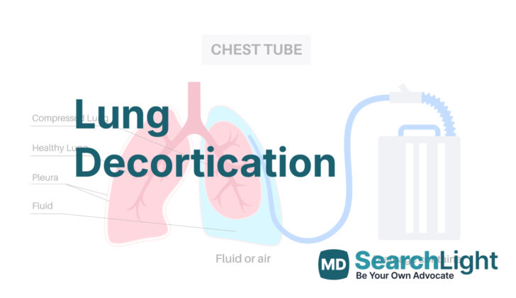
The area around your lungs is called the pleural space, which is wrapped by two layers known as the parietal and visceral pleura. Empyema is a condition where pus accumulates in this space, usually due to lung infections. When empyema progresses, fibrous tissue (fibrin) starts to accumulate around the outer surface of the lungs. This can trap the lung and stop it from expanding properly.
When the lung cannot fully expand, it collapses partially or completely. This results in a mismatch between the amount of air going in and out of your lung (ventilation) and the blood flow in the tiny vessels of your lung (perfusion). This can make it hard for you to breathe, and it requires prompt medical attention.
Why do People Need Lung Decortication
Lung decortication is a medical procedure often recommended for patients with pleural empyema, which is an infection in the area around the lungs. This infection can be chronic, meaning long-lasting, and may be caused by several different bacteria like Streptococcus pneumoniae, Staphylococcus aureus, or Klebsiella pneumonia. The buildup of pus from the infection causes the layers of tissue surrounding the lung (the pleura) to stick together, and decortication is a surgery that removes this unhealthy tissue.
Lung decortication can also be needed for other health conditions. These include hemothorax, which is when blood collects between the chest wall and the lung, and pleural thickening which can happen because of inflammatory conditions like Rheumatoid arthritis. It can also be done if there is a growth or tumor like malignant mesothelioma (a type of cancer) around the lungs.
There’s no universally accepted rule about when it’s the best time to do lung decortication. But, one suggestion is that if the capacity of a patient’s lungs to hold air drops to 70% or less of what’s usually expected, that can be a good sign that decortication is needed.
When a Person Should Avoid Lung Decortication
Sometimes a person can’t undergo a decortication procedure due to several reasons. This procedure is usually performed on someone with a collapsed or trapped lung. However, if the lung is severely damaged due to disease, it might not expand again after surgery. Even after surgery, these patients might not get better, and as an alternative, they could be suggested to remove the lung completely, either immediately or later during follow-up.
Another roadblock for performing a decortication procedure is bronchial stenosis. This is a condition where the person’s bronchial tube has become narrow. In this case, apart from the decortication, the patient needs the narrowed segment to be removed, and the passage needs to be reconnected.
Furthermore, the procedure might not be suitable for patients with unstable blood pressure and heart rate (hemodynamic instability), problems with blood clotting, failure of multiple organs, or generally poor health status. These conditions can make it too difficult for the patient to handle the risks and recovery of a major surgery. So typically, these patients won’t be recommended for a decortication procedure.
Equipment used for Lung Decortication
Here’s a simple breakdown of the tools and materials a surgeon needs for a procedure called lung decortication, which is a surgery to remove a layer of tissue over the lung:
– A solution for cleaning the skin, made up of either 10% povidone-iodine or a mix of 2% chlorhexidine gluconate and 70% isopropyl alcohol.
– Protective wear for the surgeon and medical team, like gowns, masks, goggles, and sterile gloves.
– A scalpel, which is a small, sharp knife used in surgeries.
– Electrocautery and bipolar forceps, which are tools that use electric current to cut or control bleeding in tissues.
– A rib spreader (known as Finochietto’s rib retractor), used to separate the ribs and create a bigger space for the surgeon to work.
– Bone instruments, such as a periosteal elevator, rib raspatory, bone cutter, and bone nibbler, used if it’s necessary to remove a portion of a rib.
– Lung grasping forceps (specifically Duval lung grasping forceps), which are used to hold and manipulate the lung.
– Sponge holding forceps, used to hold a surgical sponge.
– Hemostats, either curved or right-angled – these are tools that help control bleeding by clamping onto blood vessels.
– Sutures, the medical term for stitches.
– Intercostal drains, which are tubes inserted between the ribs to remove fluid or air from the chest cavity.
– Dressings to cover and protect the wound after the operation.
Who is needed to perform Lung Decortication?
Decortication, a surgical procedure on the lungs, must be performed by a specially trained heart and lung surgeon, known as a thoracic surgeon. A supporting team of medical professionals — including an anesthesiologist (the doctor who puts you to sleep for surgery), surgical assistants, a technical assistant, and nurses — helps out during the surgery.
It’s also very important to have experienced lung doctors, known as pulmonologists, and radiologists, doctors who can interpret X-rays and other images. These specialists are greatly involved in taking care of patients before and after the surgery, helping to make key decisions.
Patients who have this operation because of chronic empyema, a long-term lung infection often filled with pus, may need to be watched very closely using advanced monitoring technology in the intensive care unit (ICU) right after the surgery. This helps to ensure the best outcome possible.
Preparing for Lung Decortication
Before you have any surgery, your doctor has to make sure you are healthy enough and that the surgery is the best solution for your medical issue. If you’re scheduled for a surgery that involves your chest, specific tests have to be done beforehand to help plan the operation. These tests might involve X-rays and an advanced type of scan called a contrast-enhanced computed tomogram (CECT). This helps doctors check the condition of your lungs, see if anything is out of place, and decide the best treatment plan.
Sometimes, a procedure called bronchoscopy is also performed. This involves a small camera that is used to look into your lungs. Blood tests are usually done as well. All these are important to ensure everything goes well during and after the surgery.
During surgery, it’s important that doctors have the necessary equipment and materials ready in case of high blood loss. This is because some procedures may involve carefully peeling off a layer from around your lung and that could bleed quite a bit.
When the day of the procedure comes, you will be gently positioned on your side with the affected side facing up. You will have supports like a folded towel or a roll placed under you for comfort. One leg will be bent, with a pillow for support between your legs. Doctors ensure any pressure points of your body are properly cushioned to avoid discomfort.
For some surgeries, a tube is sometimes inserted into your esophagus, the tube that connects your mouth to your stomach, to make sure the doctor can see it during the operation. This helps prevent any accidental injuries. Finally, the skin on your chest will be cleaned thoroughly before the surgery starts, to prevent any infections.
How is Lung Decortication performed
A posterolateral thoracotomy is a complicated surgical operation performed on the chest area. Here are the main steps involved:
1. Cutting the skin: The surgeon will make an incision starting halfway between your spine and the edge of your shoulder blade, moving downwards and forwards. This cut is about two inches below the tip of your shoulder blade. The doctor will then use a special device that uses heat to make this area deeper. They do this by cutting through two of your muscles, the latissimus dorsi and the serratus anterior. The surgeon will then count your ribs to know exactly where they are operating.
2. Getting to the chest cavity: This is achieved through the space between either the 5th or 6th ribs. You need to be careful during this step because the surgeon must avoid injuring a bundle of nerves and blood vessels while they are cutting through the muscles between your ribs. Sometimes, they might need to remove a piece of rib if the ribs are too close together.
After the surgeon has accessed the chest cavity, they would move on to a region called the extrapleural space. The surgeon would take special care so as not to enter into what is known as the empyema cavity. They also aim to prevent injury to any of the important structures surrounding that region, such as the lung tip, esophagus (food tube), main veins, and the diaphragm (a muscle that helps with breathing). In this step, the surgeon would also remove any abnormal growth on the outer surface of your lungs.
After this, the surgeon will ask the anesthesiologist (a specialist who helps with pain control during surgery) to fill up your lungs with air. This helps the surgeon find and repair any leaks in your lungs. The surgeon also needs to ensure that there is no bleeding. Once they ensure these, they will put a drain in the space between your ribs. This drain helps prevent fluid build-up after the operation. Some surgeons might place two drains – one high up and another one low in your chest. These tubes stay in place until there is sure that your lung has expanded fully. At the end, the surgeon closes the cut layer by layer.
With advancements in medical technology, a minimally invasive surgical procedure called Video-assisted Thoracoscopic Surgery (VATS) is sometimes used. With this technique, the surgeon makes small cuts and uses a small camera to guide their work. Research has proven that this method is generally safer and more effective for early stages of empyema, which is pus in the chest cavity surrounding the lungs.
Finally, after the operation, you will still get antibiotics, pain control, and enough water and food. If you are very sick, you might be placed on a mechanical ventilator (a machine to help you breathe) and would need intensive monitoring. In addition to taking regular Chest X-rays, the doctors might also check the oxygen and carbon dioxide levels in your blood.
Possible Complications of Lung Decortication
Lung decortication is a surgical procedure to remove a layer of fibrous tissue that prevents the lungs from expanding properly. This surgery can sometimes have complications, some of which include:
1. Hemorrhage: This means that there might be significant bleeding from the part of the lung that was operated on. After surgery, your doctor will check your blood levels to check if you need a transfusion.
2. Persistent air leak and bronchopleural fistula: Sometimes, small leaks in the air can occur during this surgery. They usually heal on their own after several days. But the larger leaks need to be sewed up by the surgeon to prevent a bronchopleural fistula, which is an unnatural connection between your bronchial tubes and the space between your lungs.
3. Persistent lung collapse: In some cases, the lung tissue might not expand as it should after surgery. In situations like this, breathing exercises and chest physiotherapy can help the lung to expand again. However, some patients might not respond to these measures if their lungs are too damaged.
Another complication is injury to vital structures in the chest during the surgery. This can include injuries to the blood vessels under the collarbone, the diaphragm (a muscle that helps you breathe), the esophagus (the tube which connects the mouth with the stomach), and the sac around the heart.
If all the pus from an infection isn’t thoroughly removed during surgery, it can cause further infection and make you very sick. This is why it’s really important that the surgeon does a good job cleaning during the surgery.
The surgery can also cause severe pain afterwards. Good pain relief is really important and sometimes a combination of different pain relief methods may be needed. For example, you may need to take medication through a needle in your vein (intravenous analgesia), and you may also need medication delivered to your spinal area (epidural analgesia).
The surgery can also sometimes cause changes to the shape of the chest wall and cause an abnormal curvature of the spine (scoliosis).
What Else Should I Know About Lung Decortication?
The results of decortication surgery, a procedure to remove a layer of tissue covering the lung, depend largely on the existing condition of your lungs. Interestingly, the length of time you’ve had fibrothorax, a condition where the outer layer of your lung becomes thick and fibrous, doesn’t necessarily impact the surgery’s outcome.
If your lungs are severely affected by a disease affecting the lung tissue, the surgery might not improve your lung capacity (the largest amount of air that you can hold in your lungs). In fact, your lung capacity could even decrease after the operation.
That’s why doctors must carefully choose who should undergo this procedure. It’s usually recommended for patients with significant fibrosis (scarring or thickening) of the pleura (the thin lining that covers the lungs) and a relatively healthy underlying lung. It’s often beneficial for those whose daily lives are impacted by breathing difficulties when exerting themselves.


