Overview of Mitral Commissurotomy
Mitral valve commissurotomy is a type of heart surgery performed to help alleviate a condition called mitral stenosis. Mitral stenosis typically results from rheumatic heart disease, where the mitral valve, which allows blood to flow from one part of the heart (the left atrium) to another (the left ventricle), becomes narrowed. The narrowing obstructs the flow of blood and results in higher pressure in the atrium and potential high blood pressure in the lungs (pulmonary hypertension).
Mitral valve commissurotomy helps to fix this problem by separating or dividing the stuck parts (fused commissures) of the mitral valve. The technique of doing this operation has evolved since the early 20th century, moving from techniques that require larger incisions to those that are less invasive, such as balloon procedures that use small tubes inserted through the skin (percutaneously).
This procedure is particularly helpful for patients who have symptoms due to mitral stenosis but can’t undergo a valve replacement surgery, especially in areas where healthcare resources are limited and rheumatic heart disease is common. Depending on the patient’s health and the shape of their valve, the operation can be done using either a conventional surgical approach (an “open” operation) or through the less invasive percutaneous approach. Both methods have their own unique benefits.
Anatomy and Physiology of Mitral Commissurotomy
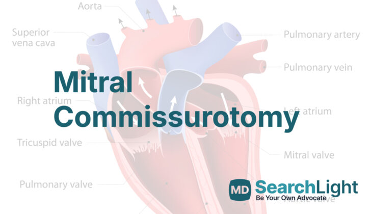
The mitral valve is an essential part of our heart. It’s made up of two flaps, or “leaflets”, that control blood flow between the left upper (atrium) and lower chamber (ventricle) of the heart. The larger flap is closest to the back of the heart, and the smaller one lies near the front, close to the aortic valve (the door between the left ventricle and the main artery, the aorta). These flaps are held in place by string-like structures (chordae tendineae) that attach to small muscles in the heart. This system keeps the valve from flipping backward when the heart squeezes to pump blood.
During heart surgery, care must be taken not to harm these structures. Understanding this anatomy is essential to avoid complications.
Mitral stenosis (MS) happens when the mitral valve becomes narrower than it should be. The most common cause is an inflammatory heart condition called rheumatic heart disease. This disease makes the valve leaflets stick together over time, restricting blood flow through the valve. This typically happens years after the initial infection that caused rheumatic heart disease.
When the valve becomes too narrow, it can cause an increase in pressure in the left atrium. This high pressure stretches the left atrium and could lead to a heart rhythm problem called atrial fibrillation. It can also lead to blood clots forming in the left atrium. Over time, this high-pressure situation can affect the lungs, causing pulmonary hypertension (high blood pressure in the blood vessels of the lungs). In advanced stages, this can affect the right side of the heart, leading to heart failure and other complications.
There are other, less common, causes of MS, including congenital defects (defects you are born with), infections of the heart valves, autoimmune diseases, and radiation therapy to the chest. Certain conditions can cause symptoms similar to MS.
Pregnancy can sometimes reveal MS because it increases the amount of blood the heart needs to pump. This can lead to heart failure symptoms in pregnant individuals. Therefore, a procedure to widen the valve (percutaneous mitral valve valvuloplasty) can be used to improve the condition of these patients.
Doctors assess the severity of MS using different scoring systems. These systems grade the valve based on its thickness, movement, and other factors. These scores guide treatment decisions, including the need for surgery.
Doctors diagnose MS through a thorough physical examination and the use of advanced imaging techniques. These techniques are used to confirm the presence and severity of MS, helping to identify symptoms, changes in the heart’s pumping function, and structural changes of the mitral valve.
Patients with MS might feel tired or experience shortness of breath due to reduced blood flow and fluid build-up in the lungs. An enlarged left atrium can cause a fast, irregular heartbeat (atrial fibrillation), which may be felt as heart palpitations. Long-term MS can lead to symptoms related to high pressure in the lungs and right heart failure.
A physical examination may reveal changes in the pulse and the sound of blood flow through the heart. An electrocardiogram, a test that measures the electrical activity of the heart, may show changes related to enlargement of the left atrium and atrial fibrillation.
Imaging tests are essential for diagnosing MS. For instance, a chest x-ray can show if the left atrium is enlarged. However, an echocardiogram (an ultrasound of the heart) is the best way to assess the size and function of the heart’s chambers and the structure and function of its valves.
Why do People Need Mitral Commissurotomy
Mitral valve commissurotomy is a procedure where doctors operate on or insert a small device into the heart to fix a problem with the mitral valve. The mitral valve is responsible for the flow of blood in one direction from one chamber of your heart (the left atrium) to the next (the left ventricle). This process needs to function properly for the heart to pump blood effectively. However, sometimes the flaps of the mitral valve can stick together (a condition known as mitral stenosis) and prevent normal blood flow. The goal of the commissurotomy procedure is to separate these flaps and restore normal valve function.
This intervention is usually performed on patients who have moderate to severe symptoms of mitral stenosis, often stemming from rheumatic heart disease. The following are key factors considered before deciding to perform the procedure:
– Symptoms of Mitral Stenosis: If a patient exhibits symptoms like shortness of breath, fatigue, and difficulty breathing when lying flat (orthopnea), doctors may suggest a commissurotomy. These symptoms line up with class II to IV of New York Heart Association’s classifications of heart disease severity. Often this means that the area of the mitral valve is less than 1.5 cm², causing significant obstruction to blood flow.
– Pulmonary Hypertension: Mitral stenosis can cause pulmonary hypertension, which means high blood pressure in the arteries to the lungs. In severe cases (when the pulmonary artery systolic pressure is over 50 mm Hg), doctors may recommend this procedure even if the patient only has minimal symptoms. Treating mitral stenosis in this way can lower the pressure in these arteries and prevent further complications in the right side of the heart.
– Atrial Fibrillation: This is a condition that causes the heart to beat irregularly. If a patient’s mitral stenosis results in new or recurring atrial fibrillation, particularly if the patient also has issues with medication to prevent blood clots or abnormally fast heartbeats, the commissurotomy could help improve the situation and reduce the size of the left atrium.
Doctors may opt for a less invasive version of the procedure, called percutaneous mitral balloon commissurotomy (PMBC). This method is often chosen for patients who would be at high risk for open-heart surgery. PMBC has the advantage of less invasiveness, quicker recovery, and lower risk during the process.
When a Person Should Avoid Mitral Commissurotomy
Mitral commissurotomy, a medical procedure to treat mitral stenosis (narrowing of the valve that controls blood flow from the lung to the heart), can be performed in two methods: open and percutaneous (non-surgical). However, not everybody is eligible for the procedure. Which one to choose depends on one’s physical condition, diseases they’re currently dealing with, and any factors that might make the surgery harmful or impossible for them.
Generally, certain conditions foreclose options to consider mitral commissurotomy, whether performed via open surgery or percutaneous method:
If a person has severe mitral regurgitation – a condition in which the heart’s mitral valve doesn’t close tightly, allowing blood to flow backward in the heart – these procedures might worsen their condition. Both aim to reduce stenosis by dividing the fused parts of the heart valve, but may also intensify the regurgitation due to potential harm to valve flaps or altered valve structure. In these cases, repairing or replacing the valve might be a better option.
Commissurotomy, either open or percutaneous method, is performed when the heart’s mitral valve area is less than 1.5 cm². This surgery aims to alleviate the pressure that builds up in the left chamber of the heart due to reduced blood flow, which subsequently causes heart failure symptoms. If a person’s mitral valve area is equal or more than 1.5 cm², commissurotomy might not be recommended.
You may not be a potential candidate for the procedures if you have an extensively hard and thickened valve. The inflexibility of the valve makes it difficult to separate the tissue adequately during commissurotomy. If a patient has a high Wilkins score, indicating a hardened, thickened, and less movable heart valve, valve replacement might be the preferred choice.
If a blood clot exists in the left atrium of the heart, particularly in the left atrial appendage, it may prevent open and percutaneous commissurotomy. Messing with the valve during the procedure might cause the blood clot to break loose, increasing the chances for stroke. Before surgery, a transesophageal echocardiogram (use of sound waves to create detailed images of your heart) can help to exclude the existence of any blood clot.
If you have an ongoing heart infection (endocarditis), it may get worse with valve treatment and increase the chances for embolic complications (obstruction of an artery). The infection also weakens the tissue, which might cause damage or rupture to the valve. If you have irreversible severe high blood pressure in the arteries of your lungs, brought about by longstanding mitral stenosis, relieving the mitral obstruction might not improve your condition. Both open and percutaneous methods may not be recommended as the benefit may be insignificant.
If you have a severe issue with your aortic valve (largest valve in the heart), you may not be considered a suitable candidate for commissurotomy since you might require broader valve surgery. Double valve replacement might be the suitable option for you. If you have inborn abnormalities in your heart, some may limit the success of commissurotomy, such as a parachute mitral valve (where all heart’s tendons connect to only one papillary muscle). If you have such congenital issues, surgical valve replacement might be necessary rather than commissurotomy.
There are certain specific conditions which might prevent a person from having a percutaneous mitral commissurotomy:
If your heart valve is heavily hardened, or the space under the valve is extensively calcified, percutaneous mitral commissurotomy might not be a good option for you. In such conditions, the risk of complications such as mitral regurgitation (backward flow of blood) is high. If there is an extensive hardening around the mitral valve, this could hamper adequate balloon expansion during the procedure, causing failure of stenosis relief or rupturing of the valve. In such scenarios, surgical options might be more suitable.
If you suffer from serious chordal thickening or fusion in the structures under the valve, percutaneous commissurotomy may not be effective. This procedure may not adequately reduce the stenosis, or worse still, pose a risk of damaging the structures below the valve, leading to significant mitral regurgitation (backward flow of blood). Treatment options in such instances could be open commissurotomy or valve replacement.
Percutaneous commissurotomy creates a septal defect to access the left atrium. Therefore, preexisting septal defects may increase the risk of paradoxical embolism (blood clot that travels from the venous side to the arterial side) or complicate the procedure. In such cases, open surgery might pose a safer option.
If coronary artery disease requires a bypass grafting, open surgery might be a better option for mitral commissurotomy than a percutaneous method.
Just like there are conditions barring a percutaneous method, there are also certain situations where an open surgery might not be a good fit:
If you have severe concomitant disorders such as advanced heart failure, long-lasting obstructive pulmonary disease, or significant kidney issues, the risk of open surgery might outweigh the benefits, and less invasive options might be better suited. If you’ve experienced prior heart surgery, particularly mitral valve surgery or other complex procedures, you may have adhesions or scarring that make it difficult to repeat open-heart surgery. In such cases, less invasive alternatives such as percutaneous methods or valve replacement might be considered.
Equipment used for Mitral Commissurotomy
The equipment needed for your doctor to carry out the procedure depends primarily on the technique being used.
If they use a percutaneous technique, which means they will be making a small incision and then inserting a thin, hollow needle into your body, they will need the equipment listed below:
– Various sizes of wires and sheaths. These are used to help guide the tools through your body.
– A special room fitted with a cath laboratory and hybrid fluoroscopy. Fluoroscopy is a type of medical imaging that allows your doctor to view internal structures in real time.
– Machines for Transthoracic Echocardiography (TTE) and Transesophageal Echocardiography (TEE). These tests use sound waves to create pictures of your heart and let your doctor see how it is working.
– The mitral valve repair device. This is a tool your doctor will use to fix the valve in your heart.
– A Code cart. This is a special cart that contains extra medical supplies and medications in case of an emergency.
– Sterile gown and gloves, sterile drape. These are used to ensure that everything remains clean and germ-free during the procedure.
– Anesthesia is a medication used to numb you or make you sleep during the procedure to ensure you don’t feel any pain.
– An operating cardiac surgical team and open surgical trays. These are the medical staff and tools that will be present during the procedure.
– X-ray lead is a type of shielding material used to protect your body from unnecessary x-ray exposure.
If they use an open technique, which means they will be making a larger incision and using traditional surgical tools, they will need the following:
– The operating cardiac surgical team
– Open surgical instruments
– Machines for TTE and TEE tests
– Code cart
– Anesthesia
– Sterile gown and gloves
– Ink and saline injectors. These are used to help your doctor see more clearly during the procedure.
Who is needed to perform Mitral Commissurotomy?
A successful heart repair, whether performed through a small skin puncture (percutaneous technique) or a more traditional open technique, requires a team of highly trained professionals. Here’s who might be involved:
A cardiac surgeon, who is a doctor specially trained to perform surgeries on the heart. An interventional cardiologist, who uses small tubes, inserted through the skin, to treat heart problems (this is the percutaneous technique).
A perfusionist, who operates the heart-lung machine that keeps blood flowing through your body during surgery. An anesthesia team, who will make sure you’re asleep and pain-free during the surgery.
Operation room personnel which includes nurses and scrub techs who help the doctors during the surgery. And in many cases, a skilled ultrasonographer, who uses sound waves to create images of your heart. Sometimes, this can be a member of the anesthesia team using a tool called a transesophageal echocardiogram (TEE).
All these professionals work together to ensure your heart repair is successful and safe.
Preparing for Mitral Commissurotomy
Before a patient undergoes a surgery known as a mitral commissurotomy (a procedure where doctors open up a narrowed heart valve), doctors gather a lot of information about the patient’s current health. This involves not only asking about the patient’s health history and performing a physical check-up, but also running various tests.
These tests can tell the doctor if the patient has a higher risk of complications such as heart failure, irregular heart rhythm (atrial fibrillation), or blood clots that could travel to the brain or lungs (a condition known as thromboembolism). Certain conditions might require the patient to take blood thinners.
One important tool doctors use is echocardiography, which involves taking ultrasound images of the heart. This allows the doctors to study the specific structure of the mitral valve, which separates the upper and lower chambers on the left side of the heart, checking for any issues like calcification that might affect how the valve moves.
Sometimes, in patients with signs of heart disease, doctors will also perform a procedure called coronary angiography, where they take an X-ray of the blood vessels in the heart. The goal here is to rule out any significant narrowing of the arteries, which may require additional surgery to encourage blood flow.
Doctors also check the pressure in the patient’s lungs, as elevated pressure can impact the approach to surgery. If the patient has irregular heart rhythm or history of blood clots, the medical team will use a special type of echocardiography to make sure there’s no clot in a small pocket of the upper left chamber of the heart.
Prior to the surgery, doctors work on balancing the patient’s fluids, stabilizing any symptoms related to heart failure, and ensuring the heart rate remains regular. The anesthesiologist also checks if the patient is fit enough for the surgical procedure and plan how they will monitor the patient during the operation.
Lastly, it’s crucial to keep the operating room as clean as possible to prevent infections. That’s why the surgical procedures are performed after taking all cleanliness measures.
How is Mitral Commissurotomy performed
In simple terms, an Open Mitral Valve Commissurotomy is a surgical procedure that is performed to fix a problem in your heart’s mitral valve. The mitral valve is like a door that controls the flow of blood between two rooms of the heart. When this doorway gets narrow or stenotic, a surgical procedure could be performed to widen this door, allowing the blood to flow more freely. The procedure, known as Open Mitral Valve Commissurotomy, has been the preferred choice of surgery for many years due to its excellent results.
The procedure happens like this. Firstly, the surgeon creates a big opening in the middle of the chest to access the heart. Sometimes if needed, a side opening also might be used. Once this opening is made, the surgeon connects the patient to a heart-lung machine that takes over the functions of the heart and lungs during surgery. The surgeon then opens a portion of the heart to expose the defective mitral valve. To get a clear view of the valve, a special thread or hooks are used to pull back the ‘door leaves’ of the mitral valve.
The surgeon now examines the parts of the mitral valve and where they are sticking together due to disease. With the help of tools that function in right angles, the surgeon carefully cuts through the glued parts of the mitral valve without damaging its healthy parts. The efficiency of the surgery is then tested utilizing the heart-lung machine before, and after the cutting operation.
During the surgery, tests similar to liquid checks are done to ensure the valves close properly without any leaks. Moreover, an ink test is also done which allows the surgeons to mark the points of surgery on the valve leaves creating a reference point for optimal door closing. Any leaks, if present, are corrected using appropriate methods.
Once the heart-lung machine is turned off, another diagnostic test called transesophageal echocardiography (TEE) is done. This test is considered reliable to see the repaired valve and measure issues like stiffness or leaking in the valve.
There is also another method called the Closed Mitral Valve Commissurotomy, which was largely used in the past and is still used in developing countries because it is less complicated and cheaper than open surgery. Nevertheless, this technique does not give a direct view of the defective valve and the repairing process.
In the closed method, the surgeon accesses the defective part of the heart through a smaller incision on the side of the chest. With careful manipulation, the surgeon feels the defective valve and assesses conditions like hardening and narrowing of the valve. After ensuring there is no clot present, a smaller cut is made on the left lower heart chamber, known as the left ventricle. Now the surgeon uses a specialized instrument that gradually widens the narrow valve, improving the blood flow.
In essence, both these techniques aim at improving blood flow by repairing the defective valve in the heart. The surgeon decides which approach to use based on the patient’s health condition and the technical requirements of the procedure.
Possible Complications of Mitral Commissurotomy
There are risks that come alongside any repair of the mitral valve, regardless of whether it’s done by open surgery or a less invasive procedure known as a transcatheter percutaneous repair. Studies recently conducted demonstrate no recorded deaths in relation to these repairs and a less than 3% rate of complications. Here are some of the main complications that could occur:
* Problems related to the catheter used to reach the area of repair. These could be things like a hematoma (a clump of blood that forms outside of a blood vessel), arteriotomy (a cut in an artery), dissection (a tear in an artery) or arteriovenous fistula (an abnormal connection between an artery and a vein)
* Stroke
* Heart attack due to damage or irritation to the left coronary artery (a major blood supply to the heart)
* Excessive bleeding during or after the operation and complications related to blood transfusions
* Mitral regurgitation (where the mitral valve doesn’t close properly and some blood flows back into the heart chamber)
* Pericardial tamponade (where fluid builds up around the heart and impacts its ability to pump blood)
* Heart rhythm disorders and heart ‘blocks’ (interrupted signals between the heart chambers)
* Heart failure
* Adverse reactions to drugs
* Infections at the site of surgery
* Sepsis (a severe infection that spreads in the bloodstream)
* Lung-related complications, such as a collapsed lung (pneumothorax), lung infection (pneumonia), or embolisms (a sudden blockage in a blood vessel)
In addition to these, there’s a risk of kidney damage due to the use of a contrast dye during the procedure.
What Else Should I Know About Mitral Commissurotomy?
Mitral commissurotomy is an important treatment for a condition known as mitral stenosis (MS). This condition makes it harder for your heart’s mitral valve to open, which can cause symptoms like breathlessness and fatigue. This operation can improve the mitral valve’s function and improve these symptoms, enhancing your overall quality of life.
During the procedure, the size of the opening in the mitral valve is increased. The reduction in pressure in the left half of your heart helps to lessen symptoms. The procedure can also prevent complications related to MS, like an irregular heartbeat (atrial fibrillation) and blood clots (thromboembolism), while also reducing high blood pressure in the lungs and improving the heart’s ability to pump blood. By treating these problems, the procedure can often delay or even eliminate the need for a valve replacement operation, which can be especially important for younger patients.
There’s also a less invasive option called percutaneous mitral balloon commissurotomy (PMBC). This involves a quicker recovery and less risk of complications, making it a great choice for patients who are at high risk for the surgical procedures or prefer a less invasive method. Both methods of mitral commissurotomy, whether performed using traditional surgery or the percutaneous technique, can lead to excellent long-term results. The choice of procedure depends greatly on the patient’s condition, and both have been proven to significantly improve both heart performance and overall quality of life.


