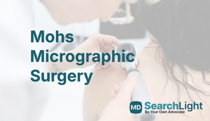
Overview of Mohs Micrographic Surgery
Mohs micrographic surgery is a specialized way of operating on skin cancer which was developed by a surgeon named Frederick Mohs. This surgery is a great option for treating skin cancers such as basal cell carcinomas (BCC) and squamous cell carcinomas (SCC). The benefits of this surgery are that it can help get rid of the entire cancer while saving as much healthy skin as possible.
This technique was created by Dr. Mohs back in the 1930s. Originally, it was called “chemosurgery,” because it involved using a chemical solution (zinc chloride) directly on the tumor in the skin. After this solution had been on the tumor for about a day, the tumor was cut out and looked at under a microscope. If the tumor was not completely gone, the process would be repeated.
Over time, the surgery has evolved. Now, instead of using zinc chloride, the tissue is cut out fresh and quickly frozen. Doing so allows this to be done more quickly than before (15 to 30 minutes), it’s less uncomfortable for the patient, and even better at saving healthy skin.
This surgery is usually suggested for skin cancers that are likely to come back and when it’s important to keep as much healthy skin as possible. During the operation, a thin layer of skin is cut out around the tumor. The tissue is then frozen and cut into sections quickly (about 15 to 30 minutes). Looking at the tissue this way lets the surgeon see almost 100% of the tissue’s edge under the microscope. The surgeon keeps cutting out tissue and looking at it under the microscope until no more cancer cells can be found.
Anatomy and Physiology of Mohs Micrographic Surgery
Mohs micrographic surgery is a type of procedure that helps preserve as much healthy tissue as possible. It’s especially beneficial when applied to areas where both functionality and appearance matter a lot. These areas include the face and neck, the area around the genitals and anus, hands, and feet.
Why do People Need Mohs Micrographic Surgery
Mohs surgery is a procedure used to treat certain types of skin cancer, particularly those at high risk of coming back, or when it’s important to save as much healthy tissue as possible. There is a set of guidelines, known as the Mohs Appropriate Use Criteria (AUC), which help doctors identify whether a specific skin tumor would be best treated with Mohs surgery. These guidelines can also be accessed through an app available for mobile devices.
This method is often used for skin cancers in specific areas of the body, including the central face (eyelids, eyebrows, nose, lips, chin, ears), genitalia, hands, feet, ankles, the areas around the nails, and nipples. It’s also recommended for patients considered high risk, such as those with compromised immune systems, certain genetic syndromes, a history of radiotherapy to the skin, or a history of high-risk tumors.
The type of tumor also influences the decision to use Mohs surgery. If a tumor previously removed still had cancer cells at the edge of the removed tissue (a positive margin), Mohs surgery may be used. A tumor showing aggressive features that make it more likely to recur might also make Mohs surgery the better option.
For basal cell carcinomas (a type of skin cancer), these features may include a certain microscopic pattern, cancer cells growing into nerves (perineural involvement), or metatypical/keratotic features. For squamous cell carcinomas (another type of skin cancer), aggressive features include undifferentiated appearance under the microscope, involvement of nerves or blood vessels, being of a spindle cell type, having a depth of more than 2 mm, or being a Clark level IV or greater.
Although the Mohs AUC offers guidance, this does not mean Mohs surgery is the only valid option. Other treatments, such as curettage (scraping away of the tumor), electrodesiccation & curettage (scraping and using an electric needle) or excision (cutting it out), can still be used successfully to treat the lesion.
When a Person Should Avoid Mohs Micrographic Surgery
Basically, if a doctor thinks that someone is healthy enough for surgery in general, there aren’t any specific reasons why they couldn’t have Mohs surgery. Mohs surgery is a precise surgical technique used to remove skin cancer.
Equipment used for Mohs Micrographic Surgery
Mohs micrographic surgery, a procedure to treat skin cancer, needs specific tools for both the surgery room and the laboratory. The surgery room needs bright lights and a flexible table to make sure the doctor can clearly see and reach the tumor. The tools used for the surgery are pretty simple. They include a sharp, clean knife (scalpel), precision tweezers (fine forceps), scissors, medical absorbent fabric (gauze), and a machine that uses electric current to stop bleeding (electrosurgical device).
If the surgeon needs to repair an area (reconstruction), they might also need a tool tray with holders for the stitching needle, cutting shears, delicate pliers, skin lifting hooks, and a surgical knife.
The laboratory that examines the tissue samples – the Mohs histology lab – uses a machine called a microtome to freeze the tissue and cut it into super thin slices. These slices are then placed on glass slides. The tissue samples are then slapped with a color (stained), either by a machine or by hand, to make the cells easier to see. This might require a special hood to make sure people aren’t exposed to the chemicals used in the coloring process.
The colored slides are then examined by the surgeon under a microscope to check if there’s any cancer left in the tissue. Some Mohs labs also have special coloring agents to allow more specific kind of staining of the tissue, called immunohistochemical staining.
Who is needed to perform Mohs Micrographic Surgery?
The operation involves the main doctor, also known as the surgeon, and at least one helper doctor in the surgery room. Also, there needs to be at least one lab technician, known as a histotechnician, in the laboratory doing the Mohs technique. This person helps with examining the tissues that are removed during surgery.
How is Mohs Micrographic Surgery performed
Mohs surgery is a special procedure used to treat skin cancer. The process of Mohs surgery is divided into several steps:
1. The doctor first outlines the area where the tumor is present. Afterwards, they use a local anesthetic to numb this area. Once numb, the visible tumor is delicately scraped off or “debulked” with a small scraping tool, flexible knife, or scalpel.
2. Before removing the tumor, tiny superficial etch marks are made around the tissue layer and the skin’s surface with a scalpel. These markings usually mirror the positions of a clock face: 3, 6, 9, and 12 o’clock.
3. Then, a thin ring of tissue is cut from around and underneath the debulked tumor. This “layer” of tissue is cut out at an angle of about 45 degrees to aid in the next stages of the process.
Once the tissue layer is removed, it is often cut into halves or quadrants. It is then marked with colored dyes to help map the exact location of the tumor. Usually, the tissue is pressed flat so that the outer layer and the deeper tissues lie on the same plane.
This “beveled” edge makes this flattening easier. The tissue is then cut horizontally to assess virtually every part of the area under the microscope. This provides a complete view of the tumor’s boundary.
If the tumor remains under the microscope, the corresponding tissue is precisely removed from the part of the patient’s skin that still contains the tumor. This process repeats until all the tumor is removed, with utmost care to preserve healthy skin. Once the tumor is completely taken out, the wound is closed using a variety of methods such as stitches, skin grafts, or natural healing. More complex cases may require doing the process repeatedly.
In Mohs surgery, tissue stains like hematoxylin and eosin (H&E) and toluidine blue are commonly used. These special dyes help the surgeon to see the tumor more clearly. Although most surgeons prefer H&E, some favor toluidine blue for treating a specific type of skin cancer known as basal cell carcinoma. This is because toluidine blue highlights specific substances associated with basal cell carcinoma with a magenta color.
What makes Mohs surgery effective is that most skin cancers grow continuously without skipping any areas. Thus, Mohs surgery allows maximum removal of the skin cancer and minimum removal of healthy tissue.
What Else Should I Know About Mohs Micrographic Surgery?
Mohs surgery is a specialized skin cancer treatment that has proven to be exceptionally successful. This procedure has notably high 5-year cure rates for two non-melanoma skin cancers: basal cell carcinoma (cells that line the bottom of the skin’s outer layer) and squamous cell carcinoma (cancer that occurs in the squamous cells, a type of skin cell). The statistics show: primary basal cell cancer has a cure rate of 99%, while it’s 94.4% for its recurrent cases. Primary squamous cell cancer has a cure rate between 92-99%, and its recurrent cases have a 90% cure rate.
Apart from these, Mohs surgery can also effectively treat a number of less common tumors. Some examples are dermatofibrosarcoma protuberans (a rare skin cancer that begins as a small patch of rough skin), microcystic adnexal carcinoma (a rare skin cancer that typically affects the face), extramammary Paget disease (a rare skin cancer that typically starts in the breast area), Merkel cell carcinoma (a very rare and aggressive skin cancer), and sebaceous carcinoma (a rare and aggressive type of cancer that begins in the oil glands in the skin).
More recently, with the availability of better testing methods, this surgical procedure has shown promising results in treating some types of malignant melanoma (the most dangerous type of skin cancer), including lentigo maligna (a type of melanoma that slowly progresses over many years), lentigo maligna melanoma (a common type of melanoma in older people), and thin melanomas (a type of skin cancer that appears as a thin layer on the skin).


