Overview of Oophorectomy
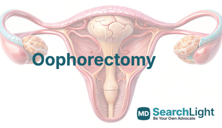
An oophorectomy is a medical procedure that involves the removal of one or both ovaries, which are the organs that produce a woman’s eggs. It’s often done along with a hysterectomy, a procedure that removes the uterus, making it one of the more common gynecological surgeries. However, the removal of the ovaries due to problems specific to these organs is quite common among women of all ages.
If the surgery involves removing only one ovary, it doesn’t majorly affect a woman’s hormone levels. However, if both ovaries are removed, it will result in infertility and menopause. This means the woman will be unable to have biological children and will experience changes in the body and potential health risks generally associated with menopause.
Because of the serious implications, it is crucial to have thoughtful discussions with the doctor before deciding to undergo this surgery.
Anatomy and Physiology of Oophorectomy
The ovaries are vital organs in the female reproductive system. They are responsible for producing the majority of the female sex hormones and for releasing eggs (ovulation). When a girl is very young, her ovaries are inside the abdomen, about the level of the bellybutton, and are quite small, measuring less than 2 cubic centimeters. As she grows and goes through puberty, the ovaries move down into the pelvic area and get bigger, growing to about 10 cubic centimeters on average. In women who have gone through menopause, the ovaries stay in the same position they were in adulthood, but they become similar in appearance to the ovaries of a girl who hasn’t started menstruating.
The ovaries get their blood from an artery (a large blood vessel) known as the ovarian artery. This artery runs through a structure known as the infundibulopelvic ligament. However, blood can also reach the ovaries from another artery – the uterine artery – via the utero-ovarian ligament. The ovarian artery is a branch of the largest artery in the body, the aorta. The veins that carry blood away from the ovaries also have a unique pathway: the right ovarian vein drains directly into the large vein called the inferior vena cava, but the left ovarian vein drains into the left renal vein (which is attached to the kidney).
There are three main paths that the lymphatic fluid (which white blood cells use to fight infection and disease) can take away from the ovary. The most common paths are through the infundibulopelvic ligament to the lymph nodes near the aorta, and through the ovarian ligament to the lymph nodes in the pelvic area. A less common path is through the round ligament to the lymph nodes in the groin area.
Looking at the ovary under a microscope, you would see three main components: the medulla, cortex, and mesothelium. The innermost part is the medulla, which consists of a mixture of different cell types including fibroblasts, cells that produce collagen, smooth muscle cells, and others. The cortex contains cells that respond to hormones and a store of undeveloped eggs known as primordial follicles. These follicles are formed before a girl is born and over time, they either disappear or grow and develop into a mature egg ready for ovulation. The outermost layer of the ovary is the mesothelium, comprised of a thin layer of cells. Unfortunately, the most common type of ovarian cancer in women over 20 can start in this epithelial layer, although tumours can develop from any cells within the ovary.
Why do People Need Oophorectomy
The way doctors manage adnexal masses, or abnormal growths in the area around a woman’s reproductive organs, relies on several factors. These include the woman’s age, how the mass has changed, what the mass looks like on a scan, whether any cancer-related substances (tumor markers) are present, what disease the patient has, and what symptoms they are experiencing. If a woman may have ovarian cancer, doctors specializing in cancer affecting women’s reproductive organs, or gynecological oncologists, should be consulted.
In children and teenagers, any suspected ovarian cancers are managed by a team of doctors from different specialities. Regardless of the patient’s age, if the ovary is removed due to suspected ovarian cancer, it should be done carefully to avoid rupturing and spilling the mass.
Simple cysts, which are growths filled with fluid, can be managed by monitoring them to see if they cause symptoms or grow larger. This is especially true if they are less than 10 cm in size. But they may twist, which could cause pain or block blood supply, requiring surgery. Complex masses, such as suspected teratomas or endometriomas, can be managed the same way, although they may eventually need surgical removal.
Surgery to remove the ovary or ovarian cyst can be performed during pregnancy if there are concerns about cancer, high risk of twisting, or if the patient is experiencing significant symptoms. During surgery, there’s always a risk of having to remove the potentially affected ovary if it’s severely damaged, if the mass can’t be fully cut out, or if there’s excessive bleeding.
If it is thought that the ovary might be twisting, which is a condition known as ovarian torsion, quick surgical intervention may be required to untwist it and possibly remove the cyst. This helps to keep the ovary working and preserve the woman’s ability to have children. Removing the ovary should only be considered if the ovary’s tissue is seriously damaged and fragile.
In conditions such as pelvic inflammatory disease where women develop abscesses, or pockets of pus in the tube connecting the ovary and uterus (tubo-ovarian abscesses), removing the ovary is rarely necessary. Instead, antibiotics are the first line of treatment. If the abscesses do not respond to antibiotics and remain large, draining them might be considered.
In certain cases, a surgeon may decide to remove both the fallopian tubes and ovaries without a specific reason but, instead, because it makes a hysterectomy procedure (removing the womb) easier. This is known as elective bilateral salpingo-oophorectomy. The decision should be made after discussing it with the patient before the operation, considering the risks of onset of menopause. If performed on women under 65 with no serious health condition, it is associated with an increased risk of dying due to any cause.
There is a procedure where both fallopian tubes and ovaries are removed to reduce the risk of further surgeries or cancer. Patients should be made aware of the risks of menopause compared to the risks of needing another surgery, especially in situations such as history of pelvic inflammation, abscesses in the tubes and ovaries, endometriosis, scar tissue in the pelvic cavity, or severe menstrual cramps. This procedure could also be a good option for those with a significantly increased lifetime risk of ovarian cancer or hormone-sensitive breast cancers due to inherited genetic conditions, such as BRCA mutations.
Finally, removing and freezing an ovary for future use, a procedure known as ovarian tissue cryopreservation, is still experimental. However, it can be an option for preserving fertility and has resulted in over 100 live births since 2004.
When a Person Should Avoid Oophorectomy
An oophorectomy is a surgery to remove one or both of your ovaries. There’s no definitive reason why you shouldn’t have this surgery, but it’s important to consider that your ovaries produce beneficial hormones and also play a role in fertility. If both ovaries are removed, especially at the same time (known as a bilateral oophorectomy), this can have some risks. It’s essential that you talk through these with your doctor before making a decision. The hormones produced by the ovaries, known as estrogens, have wide-ranging effects on your heart health, bone health, and mental well-being. However, we’re still learning more about how these hormones work in our bodies.
According to a large study called the Nurses’ Health Study, having a bilateral oophorectomy at the same time as a hysterectomy (removal of the uterus) for non-cancerous reasons can reduce your risk of getting breast and ovarian cancer. However, this surgery can also increase your risk of dying from any cause. It also ups the risk of heart disease and lung cancer, but it doesn’t make you likely to live longer.
For younger patients with non-cancerous tumors in their ovaries, doctors usually try to keep as much of the ovaries as possible. If a younger patient’s ovary twists around (a condition known as torsion), the first step is usually trying to untwist the ovary and maybe remove the cyst causing the twist. Removing the twisted ovary is usually only done as a last resort.
Equipment used for Oophorectomy
The process of laparoscopic surgery involves several specialized tools:
– A laparoscopic monitor is a special screen which helps the doctors to see inside your body without making large incisions.
– The laparoscope is a tiny camera (5 or 10 mm in size, with the ability to move 0 or 30 degrees) that ensures the area doctors work on is visible.
– The carbon dioxide source, along with tubing for insufflation, is used to create space within your abdomen for the surgery, making it safer.
– Trocars (two 5 mm ones and one larger, between 5 to 12 mm) are special tubes used as pathways for the surgical instruments to be inserted.
– Atraumatic graspers, electrocautery, or ligation device, are tools used to handle tissues and blood vessels without causing harm.
– An endoscopic retrieval bag is used to collect any tissues or fluids that need to be removed.
– A scalpel (like a very sharp, precise knife) may be needed, usually with an 11 or 15 blade.
– Forceps are a type of tweezers used to hold or move tissues around.
– A needle driver is a device used to guide sutures (stitches made with absorbable thread) used to close any incisions.
– Finally, the doctor will apply a dressing of your choice to cover the small wounds on your tummy where they did the surgery.
Who is needed to perform Oophorectomy?
The doctor who will be doing the surgery is called an operating surgeon. This is a doctor who is specially trained in performing operations. Another medical professional, referred to as a surgical assist, will be there to help the surgeon during the operation.
Furthermore, there’s also a scrub tech or scrub nurse on the medical team. This person helps by preparing and managing the surgical tools that the surgeon will be using in the operation.
Preparing for Oophorectomy
Before the operation, doctors ensure that the patient is thoroughly prepared. There’s usually no need to give antibiotics before surgery, unless there are other procedures being performed or if the patient is suspected to have an infection.
In the operating room, a grounding pad is used. This helps to prevent any accidental electrical shocks during surgery. Doctors may also discuss with the patient about inserting a Foley catheter. This is a tube that goes into the bladder to help with urine flow and could be required depending on the specific patient’s needs.
The patient’s proper positioning is crucial in preventing nerve injury. They are placed in a way that pressure points are cushioned. An anti-slip pad may be placed under the patient for additional safety. This pad can help to keep the patient in place during the procedure.
They might also use compression devices on the lower legs. These devices gently squeeze the legs to help blood flow and prevent clots in the deep veins of the lower extremities, a condition known as deep venous thrombosis. Again, this is decided on a case-by-case basis and depends on the medical center’s rules and policies.
Lastly, to reduce the risk of infections, the surgical area is cleaned and prepared using aseptic techniques, creating a germ-free surgical field on the body for the operation.
How is Oophorectomy performed
If a woman needs an oophorectomy, which is a surgery to remove one or both of her ovaries, a laparoscopic approach is usually the best way to go. This is a type of surgery that makes small incisions and uses a camera to guide the surgery. However, sometimes a traditional open surgery might be needed, especially if there’s a risk of ovarian cancer.
During the surgery, the patient might be in a certain position to allow for the surgical team to access the perineum or vagina, if needed. This is called a modified lithotomy position. Care is taken to prevent any injury during the positioning. If access to the perineum or vagina isn’t needed, the patient can simply lie on her back (in a supine position). Sometimes, a tube might be placed in the bladder to help it drain, depending on how long the surgery is expected to take, the patient’s age, and the size of the ovarian mass.
The surgeon will usually create the first incision at the belly button area. However, if there are complications like scarring from previous surgeries, abnormal anatomy, or a large ovarian mass, the first incision might be made at a different place, such as Palmer’s point. Other small cuts (trocars) will be made as needed. The trocars placement is a meticulous process to avoid any potential injury and make the surgery process smoother.
After making the incisions, the patient is often tilted slightly to give the doctors a better view of the pelvic region.
The oophorectomy procedure involves identifying certain anatomical structures like the Infundibulopelvic ligament (IP ligament) and the ureter (the tube that carries urine from the kidneys to the bladder). The IP ligament, which includes the ovarian artery, is then separated from the ureter and cut (ligated). Next, the surgeon cuts the mesovarium, which are the tissues holding the ovary, until they reach the uterus. Then, the connection (ligament) between the uterus and ovary is cut, and the ovary can be removed. The procedures vary slightly depending on the individual’s anatomy, but these are the basic steps.
In cases where the surgery is being done as a preventive measure (risk-reducing oophorectomy), the IP ligament is cut at a safe distance from the ovary to make sure all potentially harmful tissue is removed. The cut-off tissues are then carefully examined to make sure no malignancy is missed. If the left ovary is to be removed, the doctor may need to move the sigmoid colon (part of the large intestine) a bit. This ensures they have a clear view of all the anatomical structures.
When a benign (non-cancerous) ovarian mass is being removed, it is sealed in a bag and removed through one of the incisions. The integrity of the bag is kept intact to prevent spillage and possible spread of cells.
Possible Complications of Oophorectomy
While getting laparoscopic surgery in the field of gynecology, which involves procedures on the female reproductive system, there’s a small chance (0.1% to 0.64%) of causing harm to the large vessels in the body. But don’t worry, this doesn’t happen very often, and no particular surgical methods have been found to increase or reduce this risk.
In 1% to 2% of gynecologic surgeries, there can be injuries to the genitourinary tract, which is the system of the body involving the reproductive and urinary organs. This rate can be higher if certain risk factors are present. Unfortunately, many times, these injuries are not recognized during the surgery. So, it’s vital to be watchful for any symptoms after the surgery.
About 2% of major gynecologic surgeries may cause neuropathies or damage to the nerves. This is more common in patients who are overweight or underweight, have mobility issues before surgery, have a longer-than-usual surgery time, or need to be moved around a lot during surgery. With good positioning before surgery, the risk of these injuries can be reduced. Don’t stress too much – most of these injuries get better or completely resolve over time.
What Else Should I Know About Oophorectomy?
Oophorectomy, or the removal of one or both ovaries, is usually a safe procedure that can typically be done as an outpatient surgery, meaning you can go home the same day. However, it’s a decision that has important long-term effects, especially because it can lead to early menopause if both ovaries are removed.
When dealing with ovarian growths, tests and scans ahead of the surgery are useful, but they’re not 100% accurate at telling us what the growths are exactly. Therefore, in some cases, a final diagnosis can only be made once the growth has been surgically removed and examined under a microscope.
Similarly, imaging tests can be useful in assessing possible twists in the ovary (ovarian torsion). However, this condition can only be firmly diagnosed in surgery and notably, it’s only confirmed this way about half of the time. This suggests that our methods for diagnosing ovarian torsion could be better.
As for preserving ovarian tissue through freezing (ovarian tissue cryopreservation), it’s an area that’s still in the research stages. The goal of this procedure would be to preserve fertility (the ability to get pregnant) and it is not yet a standard treatment option.


