Overview of Parotidectomy
A parotidectomy is a surgical procedure where a part or all of the parotid gland is removed. This gland is one of our salivary glands, which helps to keep our mouth moist. This surgery may be needed due to different reasons such as inflammation, infections, birth abnormalities, or benign (non-cancerous) or malignant (cancerous) tumors.
The operation needs to be done very carefully and should be carried out by experienced surgeons. This is because the parotid gland is close to the facial nerve, a crucial nerve that controls the muscles in our face. To make sure this nerve is not damaged during the surgery, it is located and preserved as one of the most important steps of the operation. This is second only to making sure that all of the cancer is removed, if the surgery is being done because of a cancerous tumor.
It’s important to note that before the surgery, it’s not always possible to accurately predict exactly where the tumor and the facial nerve will be in relation to each other, unless the patient already has facial paralysis before the surgery. That’s why identifying the nerve during the operation is such a crucial step.
Anatomy and Physiology of Parotidectomy
The parotid gland is the largest salivary gland in the body. It’s located near several important facial structures such as the masseter muscle (the muscle that helps us chew) on the front, the zygomatic arch (or cheek bone) on top, the sternocleidomastoid muscle (the muscle that helps us turn and nod our head) on the bottom, and the external auditory canal (or ear canal) at the back. The facial nerve, which controls our expressions, runs through the gland and divides it into two parts: the superficial lobe and deep lobe.
The gland is surrounded by a type of protective tissue layer known as deep cervical fascia. The parotid gland produces a watery type of saliva, different from the thicker saliva produced by the submandibular gland, located below the jaw. This distinction explains why thick saliva stones form more often in the submandibular gland.
The saliva produced by the parotid gland exits through a duct, called the parotid duct, which runs parallel to the cheek bone and enters the mouth near the second upper molar tooth. There is important anatomy related to the facial nerve that runs through the parotid gland. After leaving a small hole at the bottom of the skull called the stylomastoid foramen, the main trunk of the facial nerve splits into two divisions, each giving rise to branches that control various facial movements.
Also running through the parotid gland is a vein called the retromandibular vein, which can serve as a useful point of reference for identifying the different lobes of the gland during medical imaging procedures.
There’s another nerve traveling alongside the parotid gland called the greater auricular nerve, which gives feeling to the skin at the angle of the jaw, the side of the upper neck, and the lower half of the outer ear. If this nerve is damaged, these areas can lose sensation. Sometimes this nerve may need to be used in nerve grafting procedures, which is when damaged nerves are repaired using sections of healthy nerves.
Finally, within the parotid gland are small structures called lymph nodes. These are distributed mostly in the superficial lobe and less in the deep lobe. The lymph nodes here play a role in draining fluid from various head and neck regions, and in the event of conditions such as melanoma (a type of skin cancer) or other epithelial (skin or mucous membrane related) tumors in these areas, sections of the parotid gland may need to be removed to ensure the complete removal of these lymph nodes.
Why do People Need Parotidectomy
A parotidectomy is a surgical procedure to remove the parotid gland, which is a type of salivary gland. Most of the time, this surgery is performed because of a tumor in the gland. The majority of these tumors, about 75% to 80%, are benign, meaning they aren’t cancerous. Pleomorphic adenoma and Warthin tumors are the most common types of benign tumors in the parotid gland. The most common malignant, or cancerous, tumors in this gland are mucoepidermoid and adenoid cystic carcinoma. Sometimes, cancer from the skin can also spread to the parotid gland.
A parotidectomy may also be performed for conditions such as chronic inflammation of the parotid gland or repeated infections that don’t get better with medication. Other reasons for the surgery could include tuberculosis, a type of infectious disease, or the presence of abnormal growths or cysts.
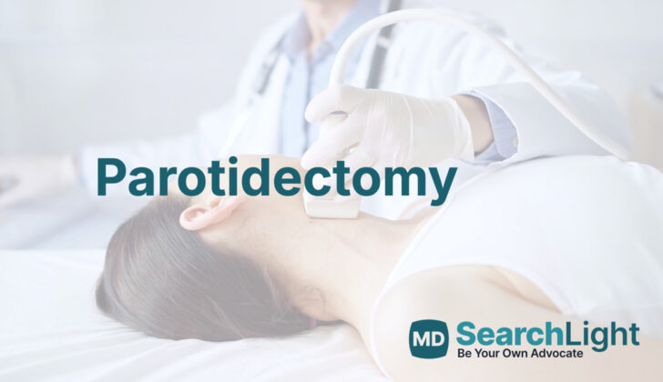
Before the surgery, the doctor will conduct a thorough examination of the patient, looking for any signs of a tumor or other abnormalities. This includes an evaluation of the function of the facial nerve, which is close to the parotid gland and could potentially be affected by a tumor. An ultrasound-guided biopsy may be done to distinguish between benign and malignant tumors.
Additional tests such as computed tomography (CT) scans and magnetic resonance imaging (MRI) can help provide detailed pictures for a more accurate diagnosis. Advanced imaging techniques can help differentiate between different types of benign and malignant lesions or tumors with a high degree of accuracy, which aids in planning the appropriate treatment.
About the treatment, the decision to remove lymph nodes in the neck as part of the surgery will depend on various factors such as the grade of the tumor, its size, and the patient’s age. Following surgery, radiation therapy may be recommended, especially for advanced tumors or if there is a risk of local recurrence.
The exact surgical approach will depend on the specific case. Different procedures include superficial parotidectomy, total parotidectomy, or a more radical form that involves removing the entire gland along with the facial nerve. The choice between these options depends on the location, size, and type of tumor or other abnormalities.
When a Person Should Avoid Parotidectomy
A parotidectomy is a surgical procedure that can generally be done for any patient who is healthy enough to go under general anesthesia, which is a medicine that causes you to sleep during surgery. There are some cases where the surgery is done under local anesthesia (medicine that numbs a specific area of the body), but these are rare and not commonly recommended. Sometimes, putting a tube in the patient’s throat to help them breathe during surgery can be difficult. This can happen if the tumor is in a challenging location, like deep within the parotid gland or spreading to the joint of the jaw, which can cause locking of the jaw.
In such cases, doctors may have to help the patient breathe either with a special procedure using a thin, flexible tube with a light at its end (awake nasal fiberoptic intubation), or by creating a hole in the front of the neck (tracheostomy).
It’s also worth noting that a surgical removal of the parotid gland might not always be the best solution. While most growths in the gland are usually treated with surgery, not all lumps or masses are cancerous. For instance, if a patient with HIV has swelling in the parotid gland, it’s more likely that they have fluid-filled sacs (lymphoepithelial cysts) inside the gland. These cases need careful examination with imaging tests and/or microscopic examination of cells (cytology).
Surgery is considered a last option because of the disease’s pattern, which is expected to affect both glands and worsen over time, and also the increased chances of injuring the facial nerve during the procedure. Therefore, doctors usually prefer other methods such as monitoring the condition, draining the fluid from the cysts repeatedly, administering antiretroviral medicine (drugs that treat HIV), sclerosing therapy (treatment involving injection of a substance to shrink the cysts), or radiation therapy.
Equipment used for Parotidectomy
Parotid surgery, a procedure performed on salivary glands located near the ears, can be performed using basic tools typically used in surgeries involving the head and neck. A device called bipolar is used to stop bleeding. It’s possible to monitor nerve activity by having someone observe the patient’s face for muscle twitches during the procedure. However, many hospitals commonly use something called continuous electromyography nerve monitoring for increased accuracy. This method uses devices like a specialized tool for tracking nerve activity during surgery and a handheld device to identify exact nerve locations.
Evidence from medical research provides useful information about electromyographic facial nerve monitoring. This specialized monitoring can reduce the likelihood of immediate facial weakness after the surgery. It aids in identifying the location of the facial nerve. It is also considered essential in some specific types of surgery, where the facial nerve isn’t directly identified. Additionally, it can provide valuable predictions in terms of how the patient’s facial nerve function will recover after the surgery.
How is Parotidectomy performed
The procedure is performed while you are under general anesthesia, which means you will be completely unconscious and won’t feel any pain. The breathing device used during the procedure, called an endotracheal tube, is secured on the opposite side of where the surgery is being performed. Facial nerve function needs to continually be monitored, so your medical team will avoid using muscle-relaxing drugs during the procedure.
Your head and neck will be positioned in a way that provides the best access to the surgeon. Part of your face will be cleaned with an antiseptic solution to help prevent infection. If a nerve integrity monitor is being used, it will be set up to accurately assess the nerves that control facial expressions throughout the procedure. The surgeon will then make a specialized incision. The exact location and method can vary, but it typically starts near a crease in front of your ear and extends downward.
The surgeon will lift an area of the skin on the front side of the surgery area. They will be very cautious during this part to avoid any injury to the facial nerves. The nerve that controls sensation on the ear’s outer part will likely be cut in the process. However, it can sometimes be preserved by carefully working around it.
During the procedure, an assistant will continuously watch your facial movements. If they see any muscle contractions, they’ll notify the surgeon right away. The surgeon needs to separate the bottom part of the salivary gland from a thick neck muscle, divide and seal off a nearby vein, and identify important muscle and nerve landmarks to guide their work.
The longer and more complex portion of the procedure involves identifying the facial nerve and tracing it along its many branches. The surgeon will be very cautious during this part to avoid any nerve injury. After this delicate process, they will be able to remove the problematic tumor or affected part of the gland.
If the surgeon needs to remove the deep lobe of your salivary gland, they will either entirely remove the superficial lobe (the top part of the gland), or lift it away to reach the deep lobe and then put it back into place after the procedure. Finally, all facial nerve branches are carefully separated from deep gland tissue, and the tumor can safely be removed.
Possible Complications of Parotidectomy
After getting a parotidectomy, which is a surgery to remove the salivary gland near your ear, some people might experience complications, including:
– Hematoma: This is when blood collects outside of the blood vessels, causing swelling, or a clump to form. This happens in about 1% of cases. To prevent this from happening, doctors make sure they stop all bleeding during the surgery, and may need to perform another surgery to control the bleeding.
– Facial paralysis: Sometimes after a parotidectomy, people may experience weakness or total loss of facial muscle movement. This is usually due to the stretching of the facial nerves during surgery. Temporary facial paralysis may occur in 16.6% to 34% of people, and it tends to get better within a month. However, in rare cases, it can last up to 18 months. To protect your eyes, doctors might provide eye drops, ointments, or suggest a visit to an eye specialist.
– Surgical site infection: As with any surgery, there is a chance of infection at the site of the operation. The best approach to preventing infection is debated, but antibiotics are often used, especially for more complex surgeries.
– Frey syndrome: This is a condition where you might start sweating and swelling on the side of your face when you eat. It happens when sweat glands become connected to the nerves cut during surgery. Doctors diagnose it based on your symptoms. If the diagnosis is unclear, a test can be done where starch, applied to the affected area, turns blue when you sweat. This used to be more common, but changes in surgical techniques have made it increasingly rare. If necessary, the condition can be treated with medication injected into the skin that blocks nerve signals for up to 36 months.
– First-bite syndrome: After surgery, some people experience a painful spasm in the jaw area when they take their first bite of food. This usually gets better as the meal goes on, but can continue to happen with each new meal. It’s thought to be caused by an imbalance in nerve signals after surgery. The treatment often includes medication, and the condition usually improves over time.
– Loss of feeling around the ear: This happens quite frequently. Most patients notice an improvement within a year. Surgeons try to preserve specific nerves to help speed up the recovery.
– Trismus: This is a temporary and mild inflammation of the muscle that controls jaw movement.
– Sialocele: This condition happens when a gland or duct that carries saliva gets connected to the skin, causing saliva to drain from the wound. It happens in 4% to 14% of cases. Doctors treat it with frequent drainage and compression dressings and sometimes recommend a reduction in oral intake. In some cases, an injection of medication could be used to stop the saliva flow.
It’s important to remember that these risks are generally low, and your doctor will take measures to minimize them. However, understanding the potential complications can help you prepare and know what to look out for after your surgery.
What Else Should I Know About Parotidectomy?
The parotid gland, a type of salivary gland near your ear, can sometimes develop health issues that medication cannot effectively treat. In these cases, surgery is often the best course of action. This surgical process, called a parotidectomy, is complex and requires careful planning and execution.
During a parotidectomy, the surgeon must consider the functionality and appearance of the area, as well as potential cancer-related factors. It is important for the doctor to have detailed knowledge of the area’s structure, preparation before the operation, techniques to spot the facial nerve during surgery, and understanding of possible complications. This ensures they can carry out the parotidectomy safely and effectively.


