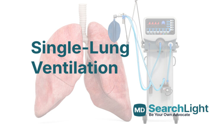
Overview of Single-Lung Ventilation
Single-lung ventilation, sometimes colloquially referred to as ‘one-lung’ ventilation, is a process where doctors temporarily restrict breathing to one lung while allowing the other one to deflate, or ‘collapse’. This is done to either make surgical procedures in the chest area easier or to avoid damage to one lung from fluids (like blood or disease-related secretions) in the other.
It’s very important to position the medical tube properly during this procedure because if the tube is misplaced, it could prevent the desired lung separation or differentiated control of each lung’s breathing. After placing the tube, doctors use a device called a bronchoscope (a thin tube with a light and camera) to confirm that it’s in the right position.
Single-lung ventilation is often used when doctors need to perform procedures on structures in the chest (referred to medically as thoracic or mediastinal structures) or to isolate a lung. This is done using specialized equipment like double-lumen tubes (tubes with two distinct passageways), bronchial blockers, and endobronchial tubes (tubes inserted into the bronchus, a passage way in the lung). Understanding how to use these tools and the way single lung ventilation works is crucial for safe administration of anesthesia.
The article touches upon the details of how single-lung ventilation works, why it is needed, the equipment used, and how to prepare for it. The discussion of traditional mechanical ventilation (the process of using a machine to assist or replace natural breathing) is separate.
Anatomy and Physiology of Single-Lung Ventilation
The tracheobronchial tree is a system of airway passages inside the body that helps us breathe. It starts with a large tube called the trachea, which is about 10 to 13 cm long and has a structure made up of 12 rings. These rings are incomplete at the back, giving them a ‘C’ shape when looked at from a cross-section. The trachea then splits at a point called the carina into two main airways called bronchi, one for each lung. This is just like how the trunk of a tree divides into branches.
The left bronchus continues onward for about 5 centimeters before it branches out to supply air to the upper and lower parts of the left lung. Meanwhile, the right bronchus is a little different. It’s shorter, wider, and more vertical than the left one. Not far down its length, it forms an offshoot that supplies air to the upper part of the right lung, then goes on as an intermediary bronchus.
The precise details of this airway system matter a lot when it comes to procedures that involve ventilating only one lung – a common practice during surgeries involving the chest. These details help doctors to correctly place a special kind of ventilation tube known as a “double-lumen” tube. Their placement is verified using an instrument called a fiberoptic bronchoscope that lets doctors see directly inside the bronchi.
Normally, when we breathe both lungs get equal amounts of air. But during single-lung ventilation, only one lung receives air, which may lead to low levels of oxygen, or hypoxia. However, the actual amount of hypoxia can depend on the patient’s position during the operation. The lung that is being ventilated can be constrained by the weight of the other lung and the elasticity (stretchability) of the lungs can increase when the patient is lying on their side. This could possibly contribute to injury related to mechanical ventilation (a machine-based method of assisting or replacing the natural process of breathing).
Why do People Need Single-Lung Ventilation
Single-lung ventilation, a medical procedure, is primarily used for two reasons: to help surgeons operate on the lung by keeping it separate from the rest of the body and to avoid infecting a healthy lung in the case of lung trauma by focusing the airflow away from the affected lung.
Single-lung ventilation is often used during certain operations which include:
- Video-assisted thoracoscopic surgery (VATS), a type of surgery where a small camera is used to see inside your chest.
- Pulmonary resections, including pneumonectomy (removal of a lung) and lobectomy (removal of a lobe of the lung).
- Surgeries on the mediastinum, the space in the chest between the lungs.
- Thoracic vascular surgery, surgeries on the blood vessels in your chest.
- Esophageal surgery, surgery on the tube that connects your mouth to your stomach.
- Spine surgery.
- Minimally invasive valve replacement surgeries, operations to replace the valves in the heart through a small incision.
Also, single-lung ventilation is used to protect one lung when the other is at risk of contamination, for example, during a massive bleeding episode in the lung, or when there is infection or draining of lung secretions. It is also utilized to manage and control airflow when there is trauma to the airway or a fistula, an unusual connection between the bronchus and your skin or pleura, the tissue that wraps around the lungs.
When a Person Should Avoid Single-Lung Ventilation
There are cases when a person may not be suitable for single-lung ventilation, a procedure where only one lung is supplied with air. The reasons might include:
If a person can’t tolerate single-lung ventilation, or they need both lungs to be ventilated, it may not be possible.
If someone has abnormal growths inside their airways (known as intraluminal airway masses), it could make it difficult to place a double-lumen tube (DLT), which is often used in lung ventilation procedures.
People with unstable blood pressure and heart rate (a condition termed as hemodynamic instability) might not be eligible for this procedure.
The presence of severe low oxygen levels in the blood (also known as severe hypoxia) could make this procedure inappropriate.
People with severe Chronic Obstructive Pulmonary Disease (COPD), a type of lung disease that blocks airflow, may not be suitable for single-lung ventilation.
Severe pulmonary hypertension, a form of high blood pressure that affects the arteries in the lungs, could make the procedure too risky.
If there are known or suspected difficulties with inserting a tube into the windpipe (a process called intubation), the procedure might not be safe.
Equipment used for Single-Lung Ventilation
The concept of breathing with just a single lung during surgery was first thought up by physiologists Eduard Pflüger and Claude Bernard, who experimented with this approach using dogs. Over time, the technique has been refined by many medical professionals and is now a crucial part of certain types of surgery.
Performing surgery with single-lung ventilation means using special devices to direct airflow to only one of the patient’s lungs. This allows the other lung to collapse safely during the operation. The particular device used during the procedure will depend on the patient’s individual body structure, the type of surgery they’re having, and the preference of the medical team.
One common tool used in this process is a double-lumen tube (DLT). A DLT is a special kind of breathing tube that has two separate pathways or ‘lumens’. These allow the medical staff to control the air flow to each lung independently. There are different types and sizes of DLTs. The type selected for use will depend on which lung is being operated on, the size of the patient, and the specifics of their surgery.
DLTs have special features to make them easier to use. For example, there are different color-coded sections for the connectors to the left and right lung pathways. This helps the medical team ensure they’re working with the right pathway during surgery. There is also a safety feature in place that allows the team to briefly stop air reaching the lung that’s being operated on.
Apart from DLTs, other devices called bronchial blockers can be used to achieve single-lung ventilation. These blockers work by sealing off the airway to one lung. They’re simple, inflatable devices fitted onto the end of a tube, but they can take longer to properly position and allow for slower lung collapse compared to a DLT.
Before a DLT is put into place, a fiber-optic scope is used to make sure it’s positioned correctly. This is crucial to ensure a good seal and to safely isolate the lung.
Endobronchial tubes are another tool that can be used for this process. They provide air supply to one lung while bypassing the other. However, they have a downside: it’s challenging to access the lung that’s not intubated (inserted with a tube). Despite the challenges, these tubes are sometimes preferred in specific circumstances, such as for patients who have had previous neck or oral surgeries and need careful lung separation.
While the usage of these devices has transformed the way certain operations are conducted, they’re not without risks. Placing a DLT could potentially cause injury and bleeding to the airway. These tubes might also be necessary for patients who already have a tracheostomy—a medical procedure that involves creating an opening in the neck to place a tube into a person’s windpipe.
Preparing for Single-Lung Ventilation
If a patient needs to have surgery on one lung, the doctor must first thoroughly check their overall health. The main disease for which the patient is being treated needs to be well-understood before the operation can take place. An echocardiography – which is a test that creates images of the heart – is particularly helpful for patients with heart and lung issues. This test provides important information about how well the heart is working.
To plan for the surgery and prepare the patient, the doctor will also assess relevant information from scans of the lung. This will help determine how to manage the anesthesia given during surgery and also prepare the patient. If the patient has illnesses such as fluid-filled cavities, blocked lungs or collapsed lungs (a condition called atelectasis), this can decrease the efficiency of their lungs and increase their risk of low oxygen levels during the surgery. This might mean that they would need to be given a higher concentration of oxygen to breathe.
Examining any large hollow spaces (called bullae) in the lung not being operated on can suggest if the patient is more likely to develop a collapsed lung during or after surgery. If the patient has tumors, they need to be assessed for any additional conditions caused by the tumor which might change how the anesthesia is managed.
Age also plays a significant role in planning the surgery, as older patients have a higher risk of complications and death from lung surgery. Additional tests may need to be conducted to gauge the risks involved apart from age. Two commonly used tests are the FEV1 (forced expiratory volume of one second) and DLCO (diffusing capacity of the lungs for carbon monoxide). The FEV1 test measures how much air a person can exhale in one second, while the DLCO measures how well your lungs transfer oxygen into your bloodstream.
Doctors consider a FEV1 and DLCO less than 60% as a risk factor for potential complications after lung surgery. However, rather than outright preventing patients with lower DLCO values from having lung surgery, doctors use these values to predict how the patient will cope after their operation.
If patients have predicted FEV1 and DLCO values more than 60% after their surgery, further tests aren’t usually needed. But if these values are less than 60% but more than 30%, patients will need additional tests, such as stair climbing or a shuttle walk test. If both values are less than 30%, patients should undergo a test measuring their maximum capacity for exercise along with their maximum oxygen consumption.
The stair climb test measures the maximum height a patient can climb, with 22 meters being the deciding height. Alternatively, a patient can walk back and forth between two points at an incrementally increasing pace – if they can complete more than 400 meters, this signifies a satisfactory maximum level of oxygen use. This information is vital for the doctor to understand the risks involved and predict the success of single lung ventilation surgery.
If the patient is at a high risk of developing acute lung injury (ALI) or worsening to intense lung distress (ARDS), a low-breath volume method, called a lung-protective strategy, is used. The volume of breath should be initially set at 4 to 6 mL/kg based on the person’s ideal body weight. This volume can be adjusted and monitored to avoid injury to the lung.
A respiratory rate (RR) of 16 breaths per minute is typically appropriate for most patients to maintain the right carbon dioxide balance in the body. A blood sample should be taken approximately 30 minutes after the ventilator is started to assess the acid-base balance and carbon dioxide levels in the patient’s blood so that the RR can be adjusted if necessary.
The positive end-expiratory pressure (PEEP) – the amount of pressure applied by the ventilator at the end of each breath – is usually set at 5 to 10 units depending on the patient’s heart performance, oxygen requirement, and if there’s any obstructive lung disease present. An oxygenation goal of 88%- to 95% is aimed for, according to ARDSnet protocol.
How is Single-Lung Ventilation performed
In medical procedures that need a lung to be isolated, a tool called a double-lumen tube (DLT) is often used. More commonly, doctors prefer to use a left-sided DLT. This is because inserting a left-sided DLT is less challenging than inserting a right-sided one. The right-side is a bit trickier to deal with due to the design of the body’s airways, increasing the chance of dislodging the DLT and causing problems with right lung ventilation.
To understand how a doctor places a left-sided DLT, it’s important to know that the patient is given general anesthesia first. After ensuring the patient’s muscles are fully relaxed, the doctor proceeds with the following steps:
- Using a specific tool known as a Macintosh blade, the doctor gets a clear view of the throat. This tool is used because DLT is large and needs more space for insertion.
- The DLT, helped by a stiff guiding wire known as a stylet, is inserted into the throat. The stylet helps the DLT navigate past the voice box (vocal cords).
- The doctor then removes the stylet.
- Next, the doctor turns the DLT 90 degrees to the left to ensure the tube lines up correctly with your airways.
The doctor then either pushes the tube in further until it hits resistance, or uses another tool known as a fibreoptic bronchoscope to help guide the tube into the correct position. Once the DLT is in place, the doctor can then start supplying air (ventilation). Using the bronchoscope, the doctor can then ensure that the DLT is in the right position for proper ventilation. To further confirm that the procedure is successful, the doctor will listen for the sound of air entering both lungs. They will then temporarily close off (clamp) parts of the DLT to confirm that each lung can be separately ventilated.
Remember that the DLT can’t be used if a doctor is unable to get a clear view of the throat. In such cases, the doctor may use a bronchial blocker, which is a different tool designed to isolate a lung for certain procedures.
Possible Complications of Single-Lung Ventilation
When we breathe, our lungs both receive the air with oxygen (ventilation) and return the oxygen to the rest of the body through the bloodstream (perfusion). In some medical procedures, only one lung is used for this process. Although that one lung isn’t receiving fresh air, it is still trying to send oxygen out into the body. This can cause a problem where the lung is using up its oxygen but not getting any more, which is called hypoxemia.
To avoid this, your body has a way to cut off blood flow to that one lung to save the oxygen for the parts that are still working. But this might not be enough. So, during a single-lung process, your anesthesiologist (the doctor who helps with pain and makes sure you’re okay during the operation) will have ways to help you get more oxygen if needed.
Making sure air and blood are working together well in your lungs (V-Q matching) is very important when you’re using just one lung. Some researchers think that you can get more oxygen when you’re lying on your side instead of flat on your back.
Usually, the doctor will give you a high mixture of oxygen during the operation. But having this much oxygen can sometimes cause your lung to collapse. So they might start with a lower amount of oxygen and add more if necessary.
If you report having hypoxemia using just one lung, the doctor will:
1. Make sure the tube that helps you breathe is still in the right place.
2. Increase the amount of oxygen you’re getting.
3. Do special actions to make sure all parts of your active lung are working, and keep you from getting hypoxemia.
Sometimes they can add oxygen to the lung that’s not being used, but this can make the operation trickier, so they only do it if nothing else is working. If none of these steps help, then doctors may have to start using both lungs again. A serious case of hypoxemia can cause problems like a collapsed lung, especially in people with lung disorders. If this happens, the operation would have to be stopped, and a chest tube would be put in to inflate the lung.
Using one lung can occasionally cause damage. Doctors avoid this using special protocols that help to protect your lungs.
What Else Should I Know About Single-Lung Ventilation?
There are two different positions that can affect our breathing: lying flat on our back (supine) and lying on our side (lateral decubitus). When you lie on your side, two things happen. First, more blood flows to the lung that’s closer to the ground (dependent lung), compared to the upper lung (nondependent lung). Second, the dependent lung inflates and deflates more efficiently when you breathe in and out. This is due to the position of the diaphragm, a muscle that helps you breathe, being more advantageous in the dependent side. The lower lung is also in a position on a compliance curve that is better for efficient breathing. The “compliance curve” measures how easy it is for your lungs to stretch and expand.
However, things change when you are under anesthesia. Anesthesia causes a decrease in the remaining space in your lungs after you breathe out (functional residual capacity). When this happens, the upper lobe of the lungs under anesthesia moves to a better position on the compliance curve while the lower lung moves to a less favorable position. Furthermore, neuromuscular blockers used during the anesthesia process allow the belly contents to push up against the dependent diaphragm, making breathing difficult. An open non-dependent lung can complicate this more, leading to mismatches in ventilation and blood flow (ventilation-perfusion or V/Q mismatch), which could result in low levels of blood oxygen (hypoxemia).
If only one lung is ventilated during a specific medical procedure, this can cause a right-to-left intrapulmonary shunt. This is a condition where blood from the right side of the heart bypasses the non-dependent lung (which is not being ventilated) and goes straight to the left side of the heart. This heightens the difference in oxygen levels in the alveoli (tiny air sacs in the lungs) and the blood vessels (alveolar-to-arterial (A-a) oxygen gradient), which can also contribute to low oxygen in the blood (hypoxemia). Certain factors, such as low oxygen levels, high pressure in the airway, taking vasoconstrictor medicine (meds that narrow blood vessels), and PEEP (an amount of air left in the lungs during the respiratory cycle to avoid collapse) can make this worse.
An interesting point is that the Carbon dioxide elimination from our bodies is usually unchanged when only one lung is ventilated, as long as we maintain adequate breathe-out capacity. But, single-lung ventilation may affect both lungs in different ways. The ventilated-dependent lung might be susceptible to damage from the high volume of air used (ventilator-induced lung injury), while the nonventilated lung could be at risk for damage from surgery trauma and insufficient blood supply injury (ischemia-reperfusion injuries). Therefore, it is crucial to consider these physiological changes during single-lung ventilation for safe anesthesia and airway handling.


