What is Cerebellopontine Angle Cancer?
The cerebellopontine angle (CPA) is a triangle-shaped area in the back of the brain’s floor. It is found between the tentorium at the top, the brainstem in the back and middle, and part of the temporal bone at the back and side. This area is significant not only because of its location in the brain, but also because it contains the CPA cistern which holds cranial nerves V, VI, VII, and VIII, as well as the anterior inferior cerebellar artery.
The CPA area’s importance comes from the wide range of issues that can occur in this region. These problems can lead to a range of vague symptoms, most commonly sensorineural hearing loss, ringing in the ears, and dizziness. The tumors that affect this area of the brain can be divided into two categories: those that start in the CPA, and those that originate from nearby areas and spread into the CPA.
What Causes Cerebellopontine Angle Cancer?
CPA tumors are often harmless, slow-growing tumors with a very low chance (about 1%) of becoming cancerous. What causes a specific type of these tumors, known as vestibular schwannoma, is still unclear. However, two main kinds of these have been identified:
* Sporadic: These are single-sided tumors often found in individuals aged between forty and sixty.
* Those linked with a condition called neurofibromatosis type 2: These typically appear as two-sided acoustic tumors in younger patients with a family history of the condition. A mutation on chromosome 22q12 leads to this kind of tumor and can increase the risk of other brain tumors as well.
Schwannomas are mainly found in certain cranial nerves which include the trigeminal, facial, glossopharyngeal, vagus, and sometimes even the accessory cranial nerve.
Meningiomas come from an overgrowth of certain cells in the meninges (the protective layers of the brain), usually from bone structures near the ear or the internal auditory canal.
Epidermoid tumors originate from misplaced skin cells which improperly located during the formation of the neural tube during embryonic development.
Arachnoid cysts are fluid-filled sacs that come about due to the splitting of embryonic arachnoid membranes (one of the layers protecting the brain and spinal cord).
Risk Factors and Frequency for Cerebellopontine Angle Cancer
Between 5 and 10% of all brain tumors are found in a specific region of the brain called the cerebellopontine angle. The most frequent types of tumors in this area are vestibular schwannoma, meningioma, and epidermoid tumors. Out of all the tumors found here:
- 75-85% are vestibular schwannoma
- 10-15% are meningiomas
- 7-8% are epidermoids
Other less common types of tumors in this area, each making up roughly 1% of cases, include a variety of different types of schwannomas, arachnoid cysts, lipoma, glomus jugulare, and metastatic lesions. There are also cases of epidermoids, dermoids, and intraventricular tumors like choroid plexus papilloma. Some very rare tumors found in this region are hamartomas, cysts, teratomas, different types of bone tumors, astrocytomas, medulloblastomas, ependymomas, brainstem gliomas, and heterotopias.
Even though they are not common, primary intracranial melanomas in this region can cause severe symptoms when they do occur, including loss of balance, hearing loss, and difficulty swallowing.
Signs and Symptoms of Cerebellopontine Angle Cancer
Lesions in the cerebellopontine angle (CPA) area of the brain can cause several symptoms. The most commonly observed ones include hearing loss, ringing in the ears (tinnitus), dizziness, swirling sensation (vertigo), headaches, and difficulty in walking (gait dysfunction). Hearing loss is usually one-sided and sensory-related, caused by damage to the cochlear nerve in the ear. Larger tumors in the CPA area can also lead to more severe effects, like other cranial nerve damage, brainstem compression symptoms, and a condition known as hydrocephalus.
- Hearing loss
- Ringing in the ears (tinnitus)
- Dizziness
- Swirling sensation (vertigo)
- Headaches
- Difficulty in walking (gait dysfunction)
- Other cranial nerve damage (with larger tumors)
- Brainstem compression symptoms (with larger tumors)
- Hydrocephalus (with larger tumors)
Testing for Cerebellopontine Angle Cancer
Diagnosing CPA (cerebellopontine angle) tumors involves several steps. It starts with taking the patient’s medical history and carrying out a physical exam. Additionally, the doctor will need to conduct audiometric and radiological tests.
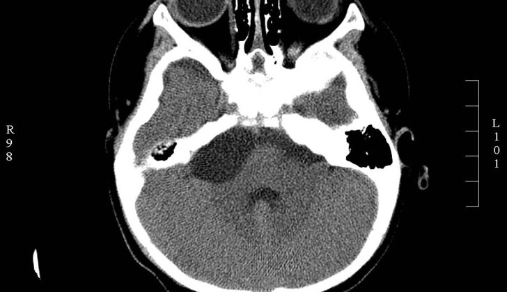
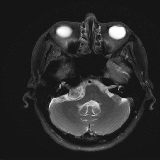
Magnetic Resonance Imaging (MRI) is the paramount tool for diagnosing these kinds of tumors. It can deliver a clear picture of the affected area and the size, shape, and exact location of the tumor. A high-resolution Computed Tomography (CT) scan can also be useful, especially for seeing if bone structures are affected.
There are various types of CPA tumors, each appearing differently on imaging tests. For instance, a type of tumor called vestibular schwannoma typically shows up as having the same density as the surrounding tissue on a CT scan – this is known as being ‘isodense’. On MRI scans, it will have a lower intensity on T1 images and a higher intensity on T2 images.
Meningiomas, another type of tumor, typically appear denser or ‘hyperdense’ on CT scans, and show a similar pattern of intensity on T1 and T2 MRI scans as vestibular schwannoma. However, some unique features can help doctors differentiate between the two. These may include the absence of damage to the internal auditory canal, a broad attachment to the protective membranes of the brain and spinal cord (dura mater), a clear separation between the tumor and brain tissue, and a thickening around the tumor. Meningiomas often come with headache symptoms, while vestibular schwannoma is usually associated with severe hearing loss.
Epidermoid tumors, on the other hand, exhibit typical CPA symptoms but can vary in their appearance on imaging tests. The most common presentation is a reduction in intensity on T1 MRI and an increase on T2 MRI, known as the ‘black lesion’.
More detailed MRI data, such as MR spectroscopy metabolite peaks and perfusion ratios, can help further differentiate between meningioma and vestibular schwannoma. Other features offering more specific information, like an ice-cream-cone shape of the tumor, changes in the bone adjacent to the tumor, the presence of calcification, or a dural tail extending into a skull base opening, can finalize the diagnosis.
Treatment Options for Cerebellopontine Angle Cancer
Cerebellopontine angle (CPA) tumors can be managed with observation, radiation therapy, or microsurgery. Observing the tumor with yearly scans is a good option for older patients or those with unstable health. However, if the tumor shows signs of growth, more aggressive treatment might be needed. One downside to this strategy is that it can make it harder to protect the function of the cranial nerves if treatment is delayed.
Radiation therapy is another treatment option, which can control the tumor well with minimal chances of it coming back. But, it can also lead to some side effects, such as worsening hearing or other nerve-related issues.
Surgery can also be tricky in treating these tumors because of the intricate and significant structures in the brain that may get in the way during surgery. For some specific types of CPA tumors like meningiomas, the preferred treatment is surgically removing the entire tumor, along with any bone and tissue around it. However, for other tumors like vestibular schwannomas, surgeons might opt for a more careful approach, removing most but not all of the tumor, to try and protect nerve function.
Microsurgery is a precise surgical approach that aims to remove the entire tumor while preserving hearing. The specific surgical method depends on factors like the tumor’s size and location, the surgeon’s preference, and baseline hearing function. The retrosigmoid, or suboccipital, approach is commonly used because it can help maintain hearing while ensuring sufficient exposure of the tumor. However, it does have some downsides like potential headaches, cerebrospinal fluid leaks, the requirement of cerebellar retraction, and a higher risk of tumor recurrence due to its inability to remove part of the tumor completely.
Mixing endoscopic and microscopic techniques could boost access to the deeper parts of the tumor. The middle fossa approach is handy for removing the sections of the tumor within the ear canal, though it might risk injuring the facial nerve and other complications. The translabrynthine approach, though least risky, is considered for patients whose hearing is already severely impaired or non-existent.
What else can Cerebellopontine Angle Cancer be?
When doctors are trying to diagnose a mass in the area behind the ear and next to the brain, called the cerebellopontine angle (CPA), they need to consider several possibilities:
- Tumors that are common to the CPA area, such as:
- Vestibular schwannoma (a slow-growing benign tumor)
- Meningioma (another type of benign tumor)
- Epidermoid cysts (a build-up of skin-like cells)
- Non-vestibular schwannoma (a tumor not related to the balance nerve)
- Congenital lesions (tumors present at birth)
- Vascular lesions (tumors related to the blood vessels) or
- Metastatic lesions (cancer that has spread from another part of the body)
- Tumors from neighboring regions that can grow into the CPA area, such as:
- Ependymoma (a type of brain cancer)
- Choroid plexus papilloma (a rare brain cancer)
- Brainstem glioma (another brain cancer) or
- Hemangioblastoma (a rare tumor of the blood vessels)
- Other abnormalities may also cause a mass in this area, like:
- Vascular malformations (abnormalities of the blood vessels)
- Inflammatory or granulomatous tissue (a type of tissue response to injury or inflammation) or
- Neuritis (inflammation of the nerves)
To determine the exact cause of the CPA mass, physicians need to look at all these possibilities and conduct relevant tests.
What to expect with Cerebellopontine Angle Cancer
Cerebellopontine tumors, which are generally non-cancerous and grow slowly, have treatment outcomes that depend on their size, location, and texture. If treatment is delayed, it can lead to poor results, which is why early detection and treatment are crucial. After surgery, it’s important to examine the images carefully to tell the difference between normal changes after surgery and a potential recurrence of the tumor.
Possible Complications When Diagnosed with Cerebellopontine Angle Cancer
The primary complications after radiosurgery – a type of surgery that uses focused radiation to treat tumors in the brain – usually involve problems with the nerves in the head, fluid buildup in the brain, and damage to the brainstem or cerebellum. Surgery complications can also include headaches, bleeding, stroke, blood vessel injury, infection, cranial nerve damage, tumor recurrence, leakage of cerebrospinal fluid (the fluid that surrounds the brain and spinal cord), and even death.
Often, issues related to the nerves of the head occur due to damage to specific ones, including the facial, trigeminal, and vestibulocochlear nerves. Interestingly, around 10% of people who suffer from intense facial pain, known as trigeminal neuralgia, have it because of tumors in the cerebellopontine angle (CPA), an area at the back of the brain near the cranial nerves.
Common Complications After Radiosurgery:
- Problems with the nerves in the head (cranial neuropathy)
- Fluid buildup in the brain (hydrocephalus)
- Damage to the brainstem or cerebellum
Common Complications After Surgery:
- Headache
- Bleeding (hemorrhage)
- Stroke
- Blood vessel injury
- Infection
- Cranial nerve damage
- Tumor recurrence
- Leakage of cerebrospinal fluid
- Death
Preventing Cerebellopontine Angle Cancer
Generally, if a person experiences non-specific symptoms, such as headaches, loss of hearing, or dizziness, they might be tested for CPA (cerebellopontine angle) tumors. Radiation therapy is a possible treatment method, particularly for elderly or medically unstable patients. While this can stop tumor growth, there are still worries about the tumor growing again, or radiation causing new tumors. Surgery is a definitive treatment that, whenever possible, should be carried out in a way that preserves the function of the cranial nerves.


