What is Lung Pancoast Tumor?
Pancoast or superior sulcus tumors are a variety of tumors that invade the upper part of the chest wall and result in a specific set of symptoms called “Pancoast-Tobias syndrome.” In simple terms, the superior sulcus is a sort of groove in the lung area created by the subclavian artery, a major blood vessel that curves in front of the lung and runs upwards and towards the side below the top of the lung. Not all of these tumors are exactly in this spot, but the term often refers to any tumor that appears in the upper parts of the lungs and is associated with signs and symptoms typical of Pancoast syndrome.
This syndrome – Pancoast-Tobias syndrome, is characterized by severe shoulder or arm pain spreading along certain nerve trunks (C8, T1, T2 nerve trunks), Horner syndrome, and wasting away of the intrinsic hand muscles. In plain English, Horner syndrome includes drooping on one side of the face, a constricted pupil, and lack of sweating, usually on one side of the face, all resulting from a disruption of the sympathetic nerve chain running to the head.
It’s important to note that those suffering from this disease generally have a poor prognosis, meaning the outcome or course of the disease isn’t too good. However, recent advances in treatment have shown significant improvements. Treatment for Pancoast tumors involves coordinated care among several medical professionals, including a chest (thoracic) surgeon, a radiation oncologist – who is an expert in treating cancer with radiation, and a medical oncologist – a doctor who treats cancer with medicines such as chemotherapy.
What Causes Lung Pancoast Tumor?
Tumors that primarily form in the upper part of the lung, also known as the superior sulcus or the apex, are commonly referred to as Pancoast tumors. They represent 3% to 5% of lung cancer cases. The vast majority of these are bronchogenic cancers, which start in the tubes that carry air to the lungs. Over 95% of Pancoast tumors are a type known as non-small cell lung cancer.
Besides lung cancer, the Pancoast syndrome, where symptoms are caused by the tumor, can be triggered by a wide range of diseases. This includes other types of lung and pleural tumors, which are growths in the tissue that wraps around the lungs. It can also be caused by metastases, which is when cancer spreads from another part of the body, including types like thyroid cancer and lymphomas. Sometimes, inflammatory and infectious processes can result in Pancoast syndrome as well, caused by bacteria like Pseudomonas, Staphylococcus, or Actinomyces.
Risk Factors and Frequency for Lung Pancoast Tumor
World Cancer Research Fund (WCRF) statistics show that in 2018, 12.8% of all new cancer diagnoses, not including non-melanoma skin cancer, were lung cancers. This equates to about 2,093,876 cases. Out of all lung cancers, Pancoast tumors make up roughly 3 to 5%. The average age at which a Pancoast tumor is diagnosed is in the person’s sixties, and it’s more common in men than in women.
- As per WCRF statistics, 12.8% of all new cancer diagnoses in 2018 were lung cancers, this number excludes non-melanoma skin cancer cases.
- This made up a total of approximately 2,093,876 cases.
- Pancoast tumors make up about 3 to 5% of all lung cancers.
- People in their sixties are most commonly diagnosed with Pancoast tumors.
- These types of tumors are more common in men than in women.
Signs and Symptoms of Lung Pancoast Tumor
Pancoast tumors are often found close to important structures in our body, like the nerves and bones. Because of this, the symptoms that people experience are often due to the tumor’s location. Common symptoms may include nerve-related problems, such as those seen in Horner syndrome, and musculoskeletal issues like shoulder or rib pain.
Shoulder pain is the most common symptom and can be seen in up to 96% of patients. It usually gets worse over time and can spread to the head and neck, underarm, shoulder blade, chest, or arm, causing weakness in the hand’s muscles. This pain can result from various conditions, such as nerve invasion, expansion to the ribs or vertebral bodies, or pleural invasion.
Horner syndrome is a mix of symptoms that includes a drooping upper eyelid, a persistently small pupil, and an inability to sweat on one side of the face. People with Pancoast tumors can experience a specific type of Horner syndrome that affects the neurons traveling over the lung apex. Some people may also experience facial flushing and sweating before developing full-blown Horner syndrome, which is likely due to the tumor irritating the sympathetic nervous system before it invades.
- Shoulder pain
- Pain radiating to head and neck, axilla, scapula, anterior chest, or arm
- Weakness in the ulnar nerve distribution and intrinsic muscles of hand
- Horner syndrome (drooping upper eyelid, small pupil, inability to sweat on one side)
- Facial flushing and sweating
Moreover, some people might experience other nerve-related problems, such as paralysis or spinal cord compression if the tumor extends into the spaces between the vertebrae. This happens in about 5% of patients. The tumor can also invade the surrounding nerve roots, particularly those connected to the ulnar nerve. This can result in weakness and shrinking of the hand muscles, strange sensations over the 4th and 5th fingers and the inside of the arm and forearm, and, in some cases, the loss of the triceps reflex. This scenario can happen in about 8 to 22 percent of Pancoast tumors.
- Paralysis or spinal cord compression
- Weakness and atrophy of hand muscles
- Paresthesia over the 4th and 5th fingers and inner side of arm and forearm
- Loss of triceps reflex
Testing for Lung Pancoast Tumor
Pancoast tumors are a type of cancer named for their location in the body. Since these types of tumors are identified by where they are found, there’s no specific blood test or lab work that can confirm their presence.
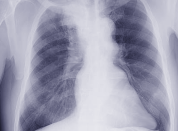
An early step in looking for a Pancoast tumor involves getting a chest x-ray. The doctor will be looking for unusual shadows or changes in the upper part of your lung, near your collarbone. This might be seen as a cloudiness, or an area that looks different than the rest of your lung. Sometimes, it can also show that the tumor has spread to the ribs. However, it can be challenging to spot these changes in the early stages due to the different body parts that lie over this area in the x-ray, like the collarbone, shoulder blade, and first rib.
If the x-ray suggests something is there, the next step is often a computed tomography (CT) scan. This type of test provides much more detailed information about the extent of the tumor and whether it has spread to nearby lymph nodes or created other tumors – factors all crucial to determine the stage of the tumor. A CT scan, however, isn’t very good at providing an accurate picture of the tumor’s local spread, though it’s excellent at identifying if the bones are involved.
In contrast, a magnetic resonance imaging (MRI) scan is much better at showing the detail of how far the cancer has spread locally because it gives a clearer view of the lung tissue, surrounding blood vessels, and spinal cord. It’s particularly good at showing if the cancer has spread to the brachial plexus, the set of nerves that run from your spinal cord to your arm. If the cancer has spread beyond certain points in these nerves, surgery may not be a suitable treatment option.
Another useful test is a positron emission tomography-computed tomography (PET-CT) scan. It helps in assessing if cancer has spread to lymph nodes or other parts of the body and accurately showing the size of the tumor. This information is crucial in planning radiation treatment.
Before and after preoperative therapy, chest CT and PET-CT scans are typically needed. However, there’s some disagreement about whether other tests, like an examination of the area between the lungs (mediastinoscopy) or ultrasound of the bronchial tubes or esophagus, are necessary.
Treatment Options for Lung Pancoast Tumor
In the late 1950s, the common treatment for certain types of lung cancer involved receiving radiation therapy before surgery. But in the 1980s, a new surgical approach was introduced. This involved reaching the front part of the chest cavity and nearby blood vessels from the neck. During the same period, this process continued to evolve.
Fast forward to the 1990s, a two-step treatment known as induction chemoradiotherapy was shown to be effective for certain advanced stages of lung cancer. This treatment involves chemotherapy (use of drugs to kill cancer cells) and radiotherapy (use of high-energy radiation to shrink tumors) followed by surgery to remove the tumor. This has since become the standard treatment for a type of lung cancer called superior sulcus tumors.
Induction chemotherapy combines potent drugs like cisplatin with other drugs like etoposide, mitomycin or vindesine. After chemotherapy, patients receive radiotherapy, with the specific amount adjusted for each individual over approximately 5 to 6 weeks. Surgery happens 3 to 5 weeks after the completion of chemoradiotherapy.
There are however certain instances where surgery is not recommended (contraindications). These include extensive invasion of the network of nerves in the shoulder (brachial plexus), involvement of the holes between the spine through which nerves pass, involvement of the soft tissue at the base of the neck, mediastinal perinodal or opposite-side neck lymph node involvement, and obstruction in veins.
There’re also cases where surgery might be reconsidered (relative contraindications) such as when the vertebral bodies (bones that make up the spinal column), the subclavian artery (a significant blood vessel) are involved, or cancer has spread to nodes on the same side of the body.
What else can Lung Pancoast Tumor be?
When trying to diagnose superior sulcus tumors, doctors consider several other possible conditions that could be causing the observed symptoms. These include:
- Mesothelioma – a type of cancer that occurs in the thin layer of tissue that covers the majority of your internal organs
- Pulmonary metastases – cancer that started in organs like the cervix, larynx, liver, or thyroid and has spread to the lungs
- Primary chest wall tumors – such as Ewing sarcoma, a rare type of cancer that occurs in bones or the soft tissue around the bones
- Adenoid cystic carcinoma – a rare type of cancer that can exist in various body sites
- Hemangiopericytoma – a rare type of tumor that can occur anywhere in the body
- Lymphoma – a group of blood malignancies that develop from lymphocytes
- Plasmacytoma – a malignant plasma cell tumor
What to expect with Lung Pancoast Tumor
The prognosis, or medical outlook, for patients with Pancoast tumor who are receiving a blend of surgery, chemotherapy, and radiotherapy, depends on several factors:
1. The T-status of the tumor – this refers to how large the tumor is and if it has spread to nearby tissues; prognosis is generally worse for T4 stage tumors (advanced stage).
2. The tumor’s response to preliminary treatment – those who see a complete response to treatment generally fare better.
3. Completeness of the tumor removal during surgery – this depends both on the tumor’s T-status and its response to the initial treatment.
In addition, those with longer duration of the disease, the existence of Horner syndrome, the spread of the tumor to the neck base, vertebral bodies, or large blood vessels, the involvement of mediastinal (middle part of the chest) lymph nodes, and wider tumor removal are generally seen to have a worse prognosis.
On the other hand, when the tumor is managed well with surgery and combination of chemotherapy and radiation, pain is significantly reduced after treatment, and the patient loses less than 5% of their weight, these signs generally suggest a better prognosis.
Possible Complications When Diagnosed with Lung Pancoast Tumor
Complications can arise both due to the disease itself and the treatments used.
Some of the complications that might affect the upper extremity due to the disease could include:
- Tumor growth into the C8 and T1 nerve roots can make the muscles of the hand weak and cause it to diminish, or instigate pain and unusual sensations in the fourth and fifth fingers and the inside part of the arm and forearm.
- If the tumor grows into the T2 nerve root, it can instigate unusual sensations and pain in the armpit and the inner area of the upper arm, potentially causing loss of the triceps reflex.
- In about 5% of patients, tumors may invade the spaces between vertebrae at the early stage of the disease, causing pressure on the spinal cord and paralysis of the lower body. Around 25% of patients eventually develop this spinal cord pressure.
- Tumors can cause issues like a phrenic or recurrent voice box nerve disorder or superior vena cava syndrome in roughly 5-10% of patients.
Potential complications from treatments may include:
- Surgical removal of lung tumors could potentially result in a medical condition where lymphatic fluid accumulates in the chest (chylothorax), paralysis of the ulnar nerve (seen in removal of the C8 nerve root), sagging of the eye and a decreased pupil size (Horner syndrome due to the removal of the stellate ganglion and sympathetic chain), leakage of brain and spinal cord fluid, as well as meningitis. The mortality rates from surgery range between 4% to 10%.
- Common side effects from radiation therapy could include localized scarring of the lung, fatigue, inflammation of the esophagus, and skin irritation. Less commonly, patients can develop lung inflammation with symptoms. Rare complications could include tightened, leathery skin with limited shoulder movement. Spinal cord inflammation and nerve inflammation in the arm may cause weakness and pain in the hand, arm, or shoulder.
- Chemotherapy also carries potential complications such as widespread inflammation and scarring of the lung tissues, kidney toxicity impairing its function, low magnesium levels, Fanconi like syndrome, etc., and damage to the nerves of the peripheral nervous system.
Preventing Lung Pancoast Tumor
Pancoast tumors are typically diagnosed quite late, and by this point, they’ve usually started to spread within the body. Because of this, patients with this type of tumor often don’t have promising outcomes, with less than 30% surviving after five years. Treatment options like combining chemotherapy and radiation therapy, known as chemoradiotherapy, haven’t shown a significant improvement in stopping local spreading of the disease or increasing long-term survival rates. Also, life after surgery can be very challenging, with many patients suffering from severe pain.
Therefore, it’s vital for patients to understand the outlook, treatment success rates, chances of the disease coming back after treatment, and potential side effects or complications. This information can help them make informed decisions about their future treatment plans.


