
Telecanthus D: Ptosis
What is Congenital Ptosis (Droopy Eyelid)?
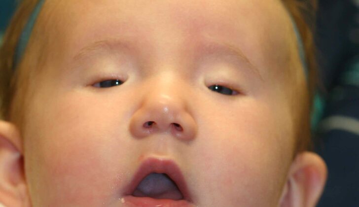
“Ptosis” is a Greek term that means “falling.” Congenital ptosis is a condition where there is an abnormal drooping of the upper eyelid from birth or within the first year of life. This condition can significantly impact a child’s function and well-being, and it can also cause distress due to its cosmetic impact for both the child and their parents.
There are two main types of ptosis:
* Real ptosis: This type of ptosis can be divided further based on when the drooping developed.
* Congenital ptosis: This occurs from birth or within the first year of life due to improper development of the muscle that lifts the upper eyelid, called the levator palpebrae superioris muscle.
* Acquired ptosis: This type of ptosis happens after the first year of life for several possible reasons. It could be due to factors affecting the nervous system, the muscles, the tendinous sheet, or mechanical issues.
* Pseudo ptosis: This appears like a droopy eyelid but it’s actually due to causes related to the eyeball and its adjacent structures.
What Causes Congenital Ptosis (Droopy Eyelid)?
Congenital ptosis refers to a condition where a child is born with a droopy eyelid. This usually happens due to some issues with the development of the muscle that lifts the upper eyelid.
Congenital ptosis can occur due to various reasons including:
* Simple congenital ptosis: This happens without any known reason.
* Congenital ptosis with weakness of the ‘superior rectus’ muscle: This is commonly known as ‘double-elevator palsy.’
* Marcus Gunn Jaw-winking ptosis: This happens when the nerve supply to the muscle that opens the jaw is wrongly connected to the muscle that lifts the eyelid. So, while chewing, the affected eyelid raises itself.
* Blepharophimosis Syndrome: This syndrome includes a narrow eye opening, congenital ptosis, a fold of skin near the inner corner of the eye, and eyes that are spaced further apart.
Less common causes of congenital ptosis include:
* Third cranial nerve palsy: Paralysis of the third cranial nerve.
* Horner Syndrome: This is identified by mild drooping of the eyelid, pupil constriction, lack of sweating on the affected side of the face, and different colors in both irises.
* Birth trauma: Injuries during birth.
* Eye tumors like plexiform neurofibromatosis (a type of nerve tissue tumor), neuroblastoma (a type of nerve cell cancer), lymphoma (a type of blood cell tumor), rhabdomyosarcoma (a type of muscle tissue tumor), neuroma leukemias can cause ptosis.
* Pseudotumor of the orbit: A condition where the eyelid drops due to an inflammation of the tissue around the eye, which in turn affects the eyelids.
Risk Factors and Frequency for Congenital Ptosis (Droopy Eyelid)
The most common type of congenital ptosis, which is an eye condition present at birth, is simple congenital ptosis according to a study by Griepentrog et al. They found that 81% of the cases were of this type, and it was usually diagnosed when the child was about 3.7 years old. This condition seems to be slightly more common in males than in females, with 57% of the cases being males and 43% females. Interestingly, 11.7% of the people with this condition had a family history of ptosis. While it can affect both eyes, this was only observed in 4% of the cases. In cases where just one eye was affected, it was typically the left eye, representing 68% of these cases.
- The most common type of congenital ptosis is simple congenital ptosis, representing 81% of cases.
- The average age of diagnosis for this condition is 3.7 years.
- Males are slightly more likely to have this condition, with a ratio of 57% males to 43% females.
- Around 11.7% of people with simple congenital ptosis have a family history of the condition.
- Although it can affect both eyes, it only does so in about 4% of cases.
- In cases where only one eye is affected, 68% of the time it’s the left eye.

slings showing the longevity of the slings
Signs and Symptoms of Congenital Ptosis (Droopy Eyelid)
Ptosis is a condition where one or both eyelids droop. There are several things healthcare providers will look at if someone is experiencing ptosis, including:
- How long the person has had ptosis
- If there are any related symptoms
- The progression or variable nature of the ptosis
A person with ptosis might bring up that they’re experiencing drooping of the eyelid(s), vision reduction, change in head posture, eye heaviness, or double vision while looking upward. They may also be concerned about how their eyes look.
The doctor will also ask about a person’s medical history, previous surgeries, birth details, and family history of ptosis to gather the necessary information.
An examination of the patient will also be done to get a better understanding of the condition. The doctor will check for things like:
- Any variation in the appearance of ptosis through old photos
- Determining visual sharpness
- Facial symmetry
- Chin elevation in case of bilateral ptosis
- Possible underlying conditions
- Signs of compensation for ptosis
- Checking for fake ptosis causes
- Determining how the eyes move
- Reflective light tests to rule out other conditions
- Personal phenomena related to the condition
- Sensitivity testing for the cornea
- Examination of tear flow
- Examining muscle function and tissue structure near the eyes
- Measurements of eyelid and gaze range
All these efforts help the healthcare professional diagnose ptosis accurately and provide appropriate treatment.
Testing for Congenital Ptosis (Droopy Eyelid)
Congenital ptosis, a condition where the upper eyelid droops, can usually be diagnosed through a clinical examination. Some of the signs that might point to this condition include mild to severe drooping of the eyelid, weakness or reduction in the function of the muscle that lifts the eyelid, a delayed response of the eyelid while looking down, a weak or absent eyelid crease, and an enlarged eyelid opening when your gaze is directed downwards.
This condition can also be graded based on the difference in distance between the upper eyelid crease and the center of the pupil in each eye. In cases when only one eye is affected, or how much it differs from what’s considered normal in cases when both eyes are involved. This grading can be categorized as:
- Mild – difference of 2mm or less
- Moderate – difference of 3mm
- Severe – difference of 4mm or more
A typical case of simple congenital ptosis often doesn’t need any additional tests for diagnosis.
Before any operation, the anesthesiologist will evaluate the patient to make sure they’re fit for anesthesia as usual. If there’s a suspicion that the ptosis is part of a syndrome, meaning it’s associated with other disorders or defects, other healthcare professionals may need to be consulted. Further tests may also be required to identify any other associated conditions.

treated with a left frontalis sling. Photographs show the improvement in the
left eyelid height and how the use of the frontalis muscle allows the left upper
eyelid to be lifted more. At rest, in unilateral cases, the ptotic corrected
eyelid will invariably be a little lower than the opposite side unless the
opposite normal levator muscle is removed and bilateral frontalis slings are
performed (the Beard procedure)
Treatment Options for Congenital Ptosis (Droopy Eyelid)
The aim of surgery to correct ptosis, or a drooping eyelid, is to restore the correct position and symmetry of the eyelids, with little to no lagophthalmos (inability to completely close the eyes) or exposure of the cornea.
The timing of the surgery depends on the individual’s situation. In cases where the child’s vision is not at risk, the surgery can be postponed until they are 3 to 4 years old. This allows for a more accurate assessment of the levator muscle, which opens the eyelid. If the ptosis is severe and interfering with the child’s vision or if both eyelids are affected, immediate surgery is recommended.
The type of surgery chosen depends mainly on the functionality of the levator muscle. A levator advancement, which adjusts the levator muscle, is suitable for mild cases of ptosis where the levator function is good. The Fasanella Servat procedure, which involves the removal of a portion of the conjunctiva (the membrane lining the inner surface of the eyelid) and the tarsal plate (a thick, stiff segment within the eyelid), is less commonly used due to the destruction of these tissues.
If the levator function is severely poor, a technique called frontalis sling surgery is often the best choice. The eyelid is essentially hitched up to the forehead muscle, which allows the person to raise their eyelid by raising their eyebrows. This is a particularly good method for severe cases of ptosis or other conditions like Marcus Gunn jaw-winking syndrome, oculomotor palsy, blepharophimosis syndrome, or if there has been a traumatic injury to the levator muscle. Various types of materials, either natural or synthetic, can be used for the sling.
If the levator function is moderate, levator resection, or removal of a section of the levator muscle, is usually the most effective solution. The surgery can be approached via the skin just above the eyelashes or via the conjunctiva.
In cases of mechanical ptosis, where there is an external force causing droopiness of the eyelid, the surgery should eliminate or fix the mechanical component that’s pushing the upper eyelid down.
In cases of Marcus Gunn Jaw-Winking Syndrome, the treatment involves the removal of the levator muscle and a frontalis suspension. This method, sometimes called the “Chicken-Beard procedure”, involves using the forehead muscle to lift the eyelid without fixing the levator muscle.

and poor levator function with over action of the frontalis muscles. Bilateral
frontalis slings corrects the eyelid position and the chin-up position
What else can Congenital Ptosis (Droopy Eyelid) be?
Congenital ptosis, which refers to a drooping of the upper eyelid from birth, must be distinguished from other instances of ptosis developed later in life. There are different types of ptosis, including:
- Neurogenic Ptosis: This can be present at birth or develop later. It’s often caused by nerve issues such as third nerve palsy, misdirection of the third nerve, Marcus Gunn jaw winking syndrome, Horner’s syndrome. Other causes include conditions like multiple sclerosis and ophthalmoplegic migraines.
- Aponeurotic Ptosis: This occurs when the levator muscle of the eye is working normally, but there is a problem with the connecting tissue (the levator aponeurosis). This can be associated with aging, injury, surgery, or a condition called blepharochalasis.
- Mechanical Ptosis: This is caused by additional weight on the upper eyelid, such as swelling, tumors, or multiple solid lumps (chalazia). Conditions that cause scarring (like trachoma or ocular pemphigoid) and the formation of hematomas can also lead to this type of ptosis.
- Myogenic Ptosis: This results from conditions affecting the muscle or nerves controlling the eyelid. This includes myasthenia gravis, myotonic dystrophy, a group of conditions known as oculopharyngeal muscular dystrophy, ocular myopathy, and injuries to the eye muscle (levator palpebrae superioris).
Ptosis must also be differentiated from pseudoptosis, which only appears to be drooping due to issues with the eye and surrounding areas.
A key way to tell the difference between ptosis and pseudoptosis is by lifting the drooping eyelid. If the other eyelid also droops, it is real ptosis. If the other eye does not change, it is pseudoptosis.
Pseudoptosis can be caused by a variety of issues, which can be grouped into ipsilateral causes (occurring on the same side of the body) and contralateral causes (occurring on the opposite side of the body).
Ipsilateral causes include:
- Shrinking of the eyeball (Phthisis bulbi)
- Sunken eyeball (Enophthalmos)
- Excessive upward deviation (Hypertropia)
- Small eyeball (Microphthalmia)
- Excess skin over the eye (Dermatochalasis)
- Complete loss of the eye (Anophthalmos)
- A hollow space in upper eye (Superior sulcus defect)
Contralateral causes include:
- Enlarged eyeball (Buphthalmos)
- Protrusion of the eyeball (Proptosis)
- Upper eyelid pulling back too far (Upper eyelid retraction)
What to expect with Congenital Ptosis (Droopy Eyelid)
The outcome for a patient with congenital ptosis, a condition characterized by drooping of the upper eyelid at birth, is influenced by various factors. These include the severity and type of ptosis, any related conditions, when the problem was first diagnosed, whether one or both eyelids are affected, the surgical method chosen, and the results of the surgery.
For accurate treatment planning, a thorough examination and precise measurements of the ptosis are essential. The type of surgery used to correct ptosis hinges on these measurements. If the measurements aren’t done correctly, it may result in the ptosis being undercorrected or overcorrected after surgery.
Possible Complications When Diagnosed with Congenital Ptosis (Droopy Eyelid)
If a serious case of congenital ptosis, a condition where the eyelid droops, isn’t diagnosed and treated promptly, it could result in serious vision problems and neck issues. Additionally, the physical appearance caused by this condition might significantly affect a person’s self-esteem and performance in daily activities.
There are several potential complications that can occur after surgery to correct ptosis. These include:
- Swelling and bruising after surgery
- Prolapse of the superior fornix, which is the top part of the area between your eye and eyelid
- Inequality in the height and shape of the eyelid in the days immediately following surgery
- Lagophthalmos, a condition where you can’t fully close your eyes
- Exposure keratopathy, a condition caused by the eye not being properly covered and protected
- Granuloma, or inflammatory mass, at the site of the stitches
- Blood collected at the surgical site, known as a hematoma
- Infection of the wound
- Pre-septal or orbital cellulitis, an infection of the tissue around the eye
- Drooping of the other eyelid following surgery on one eyelid, due to the Herrings law of equal innervation
- Under-correction or over-correction of the drooping eyelid if the extent of the ptosis and the function of the levator muscle, the muscle that elevates the eyelid, aren’t measured accurately before surgery
Recovery from Congenital Ptosis (Droopy Eyelid)
Parents naturally worry about their child’s surgery outcome, the immediate post-surgery care, the look after surgery, and the future needs and care of children with inborn droopy eyelid (also called congenital ptosis). As medical practitioners, it’s important to properly guide the parents and older children. In today’s selfie and social media age, even teens and sometimes younger kids may have concerns about their appearance post-surgery.
They may wonder if the droopy eyelid surgery needs to be repeated. As the child grows, it might be necessary to further adjust the levator muscle (the muscle that lifts the eyelid) if it functions well. In cases where temporary adjustments were made to improve vision, more permanent changes will be necessary. This involves taking a strip of tissue (the fascia lata) from the child’s leg, which can be done once the legs are big enough through a small cut just above the knee.
Parents often ask about post-surgery care. Regardless of the specific surgical procedure performed, it’s normal for the child to sleep with their eyelids a bit open. This may look dramatic at first, but it generally reduces over time. The eyelids may remain partially open for a long while or indefinitely, which is normal. After surgery, it’s crucial to apply antibiotic ointment to the incision sites three times a day for about a week, maintain clean hands, and administer any prescribed oral antibiotics. In the initial weeks, eye lubricant should be applied each time the child sleeps. After a few weeks, most kids don’t need the constant application of lubricant unless they are sick or have a cold. Most children may become more active post-surgery because they can see better. If eye patching was prescribed, it should be continued until a professional evaluation. The kids usually get back to school within three to four days and experience minimal pain that can be managed by over-the-counter pain relievers like Tylenol. The surgery progress will be monitored twice a year, and visual development and need for eye patching will be assessed by a pediatric eye team.
As for scars, when the eyelid is lifted through a direct incision, the cut is disguised in a natural eye crease. Initially, the scar may appear pink, but it nearly disappears after a few months. With frontalis slings (a method of lifting the eyelid using a sling), the incisions become almost invisible within a few months. It’s normal to feel small bumps under the skin after a sling procedure, as the sling material is attached to the forehead muscle, but these are rarely seen.
We often get asked about the possible results. We try to provide as much information as possible and often share photos of other kids with similar conditions to give you an idea of possible outcomes. However, it’s important to remember that these photos are taken months or even years after the surgery, which includes the healing process.
In cases where one eyelid has poor muscle function, we are often asked if it would be better to perform a procedure on the functional eyelid for the sake of symmetry. This is a delicate question. Some suggest interfering with the healthy muscle to create symmetrical droopy eyelids, followed by inserting slings to even out the lid heights. However, this creates a new problem where none existed before and can lead to potential complications. Therefore, many surgeons opt to insert a unilateral sling, leading to a slight difference in the eyelid heights. The decision can be made when the child is older and can provide informed consent.
Preventing Congenital Ptosis (Droopy Eyelid)
Detecting and managing ptosis, a condition where the upper eyelid droops, early can help prevent vision problems and posture issues in children. Any child who has one or both eyelids drooping should have a thorough check-up to identify the specific type of ptosis. This can range from simple inborn ptosis to more complex conditions associated with muscle dysfunction, specific syndromes, or involuntary movements. Identifying the specific type is crucial because the treatment varies for each condition.
It’s generally recommended to operate on cases of mild to moderate ptosis in one eye when the child is around 3 to 4 years old. By this age, the muscles are usually strong enough to cope with the surgery, it’s easier to accurately measure the droop in the eyelid, and close follow-up care after the surgery is possible. For severe cases affecting both eyes, surgery should be done earlier to prevent permanent vision and posture problems. Parents should take regular photos of their child to keep track of how the condition is progressing over time. If surgery is necessary but the child isn’t ready, special glasses or tapes can be used to lift the eyelid temporarily, though this is not commonly needed anymore.


