What is Subcutaneous Fat Necrosis of the Newborn?
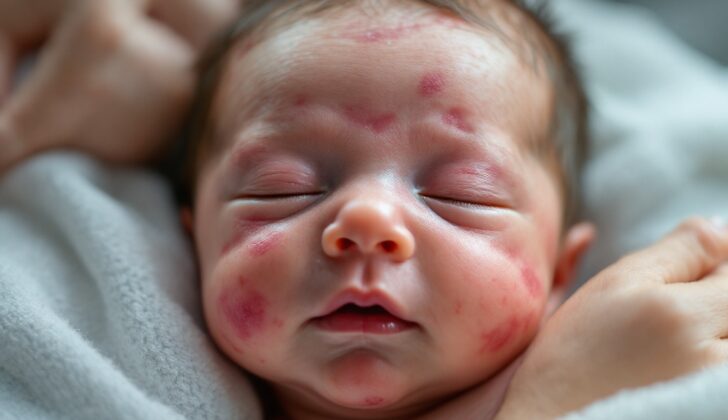
Subcutaneous fat necrosis (SCFN) is a rare condition in newborns that usually clears up by itself. This condition typically happens in babies born at term or slightly later and usually becomes noticeable within the first few weeks after birth. SCFN causes the skin to form firm, red or purple lumps and patches.
Despite mostly resolving on its own over a few weeks or months, SCFN could lead to serious changes in the body’s metabolic processes. The most common complication from SCFN is hypercalcemia, a condition where there’s too much calcium in the blood. This complication can pose a serious health risk and may even be life-threatening.
What Causes Subcutaneous Fat Necrosis of the Newborn?
Subcutaneous fat necrosis of the newborn is a condition for which the cause is unknown, but there are certain factors that seem to contribute to its development. These include stress around the time of birth such as not getting enough oxygen, being too cold, inhaling first stool, birth injuries, and certain conditions in the mother like high blood pressure during pregnancy or diabetes.
There is also evidence that being purposely cooled down as a treatment for a brain condition caused by lack of oxygen (hypoxic-ischemic encephalopathy or HIE) might trigger this skin condition in newborns. In a study revisited by researchers Del Pozzo-Magana and Ho, they found that almost all (91.1%) of the baby patients who underwent this cooling treatment for HIE developed subcutaneous fat necrosis of the newborn.
Furthermore, there’s a possibility that birth injuries might contribute to the development of this condition, although it has been observed in many babies born by Caesarean section, indicating that it isn’t just associated with traumatic births.
Risk Factors and Frequency for Subcutaneous Fat Necrosis of the Newborn
In a study looking back over 20 years, only 30 cases of the disorder being studied were found, affecting both boys and girls. All these babies were born after more than 37 weeks of pregnancy, and the average weight at birth was 3800 g. Most of them (90%) had experienced complications during their birth. In a larger study, 126 patients with the same disorder were studied. The number of boys and girls affected was roughly equal. Again, most of these babies (90%) were born after more than 37 weeks of pregnancy, with an average birth weight of 3455 g. In 20% of these cases, the babies were treated with therapeutic hypothermia, which is a controlled cooling of the body. Moreover, 86% of the babies had experienced stress while in the womb or during birth.
Signs and Symptoms of Subcutaneous Fat Necrosis of the Newborn
Subcutaneous fat necrosis of the newborn (SCFN) usually appears as skin bumps about six to ten days after birth. In fact, 92% of these cases occur within the first 28 days, with 56% in the first week. The bumps may vary, looking like small and distinct lumps under the skin that are red or purplish and mobile, or like plaques. Some can be as small as less than 1 cm, while others may be much larger, up to 8 cm in diameter. These bumps appear most commonly on arms and shoulders in about 57% of cases.
- Subcutaneous nodules (100%)
- Redness (73%)
- Pain (23%)
- Swelling (13%)
These bumps might become soft, break open, and discharge a chalk-like fatty substance. In a study of 126 infants, more than half developed high levels of calcium in the blood, mostly within the neonatal period, and 30% in the second month of life. Symptoms can range from irritability, poor feeding, excessive urination and thirst, to constipation, weak muscular tone, failure to thrive, and persistently high calcium levels in the kidneys, or even seizures.
- Irritability
- Poor feeding
- Excessive urination
- Excessive thirst
- Constipation
- Weak muscle tone
- Failure to thrive
- Persistent high calcium levels in the kidneys
- Seizures
Testing for Subcutaneous Fat Necrosis of the Newborn
If there’s a suspicion of newborn subcutaneous fat necrosis, it’s essential to identify it promptly. This condition can be accompanied by severe abnormalities that aren’t related to the skin, such as reduced platelet count, high fat levels in the blood, and excess calcium in the bloodstream. The surest way to confirm this condition is by looking at the symptoms shown by the patient and by conducting a skin biopsy, which involves taking a small sample of skin for examination.
Ordinarily, if there’s a need to sidestep the skin biopsy, an ultrasound examination and analysis of blood flow can be quite useful. These procedures can help spot subcutaneous fat necrosis in a newborn by identifying an intense signal beneath the skin, which suggests the presence of the disease, with or without calcium deposits.
Once subcutaneous fat necrosis is detected, guidelines by experts Del Pozzo-Magaña and Ho recommend monitoring the patient closely. By doing this, doctors can measure the levels of triglycerides (a type of fat), check kidney function, assess the number of platelets (cells that help the blood clot), and test blood sugar levels. If these tests come back normal, the only thing needed is to monitor the patient’s symptoms.
In patients with normal calcium levels in their blood, it’s vital to continue to measure these levels: weekly for the first month of life, then every month until the baby is six months old or until the skin’s appearance returns to normal. Additionally, a kidney ultrasound is also important, particularly in patients with high calcium levels. If the first checkup does not show any abnormalities, it can be repeated when the baby turns three months old or after the skin looks normal again.
Treatment Options for Subcutaneous Fat Necrosis of the Newborn
The treatment primarily involves relieving symptoms, as skin rashes usually heal on their own in most instances. However, if there’s an unusually high amount of calcium in the blood, this should be corrected as quickly as possible. This involves drinking plenty of fluids, taking certain types of medications known as loop diuretics via an IV drip, and eating a diet low in calcium and vitamin D. If these don’t work, other medications like bisphosphonates, which help control the amount of calcium in the blood, or systemic corticosteroids, which reduce inflammation, may be recommended.
What else can Subcutaneous Fat Necrosis of the Newborn be?
When a newborn is suspected to have subcutaneous fat necrosis, a condition affecting the fat tissue under the skin, there are a few other conditions their doctor would check for:
- Sclerema neonatorum: This is a serious condition involving hardening of the skin and underlying fat, usually seen in newborns who are very sick or premature. It is often linked to birth defects and has a high risk of causing death.
- Neonatal scleroderma: This refers to swelling of the lower parts of the body in newborns. While it is generally not harmful, it is usually followed by infections in the digestive or respiratory system.
The doctor would need to assess these possibilities before confirming that subcutaneous fat necrosis is the right diagnosis.
What to expect with Subcutaneous Fat Necrosis of the Newborn
Subcutaneous fat necrosis of the newborn is a condition where the fatty tissue under a newborn’s skin becomes damaged. It often resolves itself within a few months and likely goes undiagnosed quite frequently. However, there’s a chance it may lead to various issues related to blood and metabolism that need to be watched closely. These include low levels of platelets (thrombocytopenia), low blood sugar (hypoglycemia), high triglyceride levels (hypertriglyceridemia), and most importantly, excess calcium in the blood (hypercalcemia).
Patients with this condition require regular check-ups for at least six months and lab tests to monitor for potential complications. In one study, it took about 86 days for patients to recover from this condition. After it resolves, patients may have less fat tissue than normal (lipoatrophy). The thrombocytopenia typically resolves after the fat necrosis does.
Another study found that in 88% of the patients with hypercalcemia, the high calcium level resolved within the first 84 days of life; however, one patient still had hypercalcemia at one year. If left untreated, excess calcium in the blood can significantly damage the kidneys, which is why it’s crucial to check for kidney involvement in these patients with a renal sonogram. In fact, 83% of the patients evaluated by kidney ultrasound showed calcium deposits in their kidneys (nephrocalcinosis).
Possible Complications When Diagnosed with Subcutaneous Fat Necrosis of the Newborn
Subcutaneous fat necrosis in newborns is a condition that typically resolves on its own over several weeks. However, it’s crucial to watch the baby regularly for rare but serious complications. The most common problems include low platelet count (thrombocytopenia), low blood sugar (hypoglycemia), high levels of fat in the blood (hypertriglyceridemia), and more importantly, high levels of calcium in the blood (hypercalcemia). The reason why these complications occur is not entirely clear.
Common Complications:
- Low platelet count (Thrombocytopenia)
- Low blood sugar (Hypoglycemia)
- High levels of fat in the blood (Hypertriglyceridemia)
- High levels of calcium in the blood (Hypercalcemia)
Preventing Subcutaneous Fat Necrosis of the Newborn
It’s crucial to comfort caregivers and assure them that ‘subcutaneous fat necrosis of the newborn’ is usually harmless. This condition involves inflammation of the fat under the baby’s skin. However, it’s also important to be aware of the rare but potentially severe complications that can occur beyond the skin. For this reason, parents and caregivers must be educated about the signs and symptoms of ‘hypercalcemia’. Hypercalcemia is a condition where there is too much calcium in the blood, that can become harmful if not treated quickly. It’s equally important to emphasize the necessity of regular check-ups, to monitor the baby’s health and treat any complications right away.


